Radiation prosthetic stents are the customized oral devices fabricated for efficient administration of radiation dose to the affected areas or minimizing the unnecessary irradiation to surrounding normal tissues on head and neck neoplasms. Since the use of these stents is individualized, a close collaboration among surgeon, radiotherapist and prosthodontist is essential thereby which helps in limiting the post-therapy morbidity. In this report, two customized stents, tissue bolus and tongue depressing type were successfully applied to patients who were under RT plan due to soft palate cancer and tongue carcinoma respectively.
prosthetic oral stents, radiotherapy, maxillofacial cancer
Radiation therapy (RT) is defined as the therapeutic use of ionizing radiation in the management of neoplasms of the organs without surgery, or after surgery as an adjunctive palliative treatment, either in combination with or without chemotherapy [1]. Though RT has increasingly been conducted as an adjunctive measure in the management of head and neck cancer currently, the adverse reactions of surrounding tissues are frequently occurred such as erythema, mucositis, ulcers, xerostomia, caries, or possible osteoradionecrosis on irradiated bone [2]. These complications could diminish the quality of life, rehabilitation level, or might discourage the patient from acclimation to treatment process. Primary and supportive services from the team approach of head and neck surgeon, radiotherapist and prosthodontist are fully desirable to lessen those complications. Radiation prosthetic stents have been fabricated and tried to oral cancer patients as a tool of prevention [3]. These stents are used to adjust the position or to shield tissues or to assist in the efficient administration of irradiation to the affected spots, which limit the post therapy morbidity [4,5]. Once try-in of radiation stent is consulted from the radiotherapist, prosthodontist can actively arrange and construct the practical prostheses meeting specific conditions of RT patient. The proper control of post RT sequela can also help the future dental rehabilitation. Various types of radiation stents have been applied such as radiation source carriers, perioral cone, radiation protection/shielding, position maintaining and tissue bolus [6]. In this report, two types of customized intraoral stents (tissue bolus and tongue depressing) were respectively fabricated for patients who were planned to RT due to maxillofacial cancer with the collaborations of surgeon, radiotherapist and prosthodontist.
Case I. Tissue bolus stents
A 59-year old male patient undergone soft palate cancer (squamous cell carcinoma) with stage of T2-N2c-M0 (Figure 1) was consulted from radiotherapist. Serial concurrent chemo-radiation therapy (CCRT) with intensity-modulated 7,000 Gray (cGy) irradiations at soft palate was scheduled for 6 weeks. Prior to RT schedule, dental impression with polyvinyl siloxane was taken and polymethyl methacrylate (PMMA) stent was designed as removable type containing a relief space to place (Figure 2A) the bolus material for dose enhancement of radiation to target spot. Bolus (BOLX®, Klarity, USA) in present case was a tissue equivalent polyethylene material whose degree of radiation absorption is similar to that of soft tissues in the body (Figure 2B). After 1 year follow up, RT result was successful with stable soft tissue healing on target spot area (Figure 3) and patient is pending the prosthodontic treatment. Figure 4 represents another application of bolus combined stent specially adjusted to nearly edentulous (lonesome canine) patients with soft palate cancer.
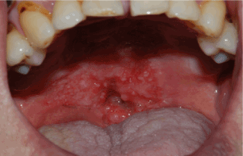
Figure 1. Pre-radiotherapy state of soft palate cancer in 69-year old male patient
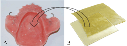
Figure 2. Transparent gel bolus (B) were packed in relief room within stent (A) to increase the radiation dose to target spot
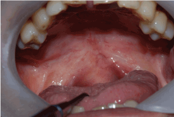
Figure 3. Postoperative healing state after 1 year follow up
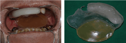
Figure 4. Application of bolus stent to nearly edentulous male patient with soft palate cancer (T2-N2c-M0)
Case II. Position maintaining (tongue depressing) stents
A 66-year old male patient undergone tongue cancer with stage of T1-N1-M0 was requested for position control device from the surgeon and radiotherapist (Figure 5A). Patient was referred to dentistry 2 weeks after surgical excision and dental impression with polyvinyl siloxane was taken. Stent was designed as tongue depressing type which could maintain the stable and repeatable position of patient during 4,800 cGy-irradiation treatments. Proper elevation of inter-occlusal vertical dimension and inferior ballooning amounts were verified by wax rim try on before resin curing stage as well as patient’s compliance (Figure 5B). Ventilation holes were made to facilitate the respiration during RT followed by PMMA stent curing (Figure 6B).
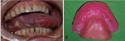
Figure 5. Right tongue cancer case with stage of T1-N1-M0 in 66-year old male patient (A), wax rim for assessment for interocclusal height and ballooning amount with co-work of radiotherapist (B)
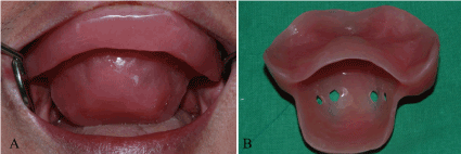
Figure 6. Try-in of position maintaining (tongue depressing) stents (A), ventilation holes for airway patency (B)
While RT is beneficial in control the oral and maxillofacial neoplasms, an increased morbidity of surrounding normal tissues can be inevitable at post-irradiation [7]. Though the needs for the oral radiation stent rely on the determination by corresponding radiotherapist, it is also big concerned to prosthodontist who will care a future dental rehabilitation.
Tissue bolus stents could help in the treatment of superficial lesions with irregular contours [6]. In case I, due to irregularities in lesion of soft palate, some areas within the field may be untreated, while others may develop isolated hotspots. BOLX® in present case is a tissue equivalent and calibrated surface gel material which compensate for surface irregularities that helps in converting perpendicular to the central axis of the ionizing beam [8]. Thereby more accurate and homogenous distribution of the radiation sources was achieved [8,9]. Other materials commonly used for bolus are tissue conditioners, water, saline, waxes and acrylic resin. A gag reflex was monitored at the initial try-in at both patients in case I. To cover the RT field, stent’s borders were extended to posterior soft palate; however, patients were adapted to devices over times before RT schedules.
Tongue depressing (position maintaining) stents can provide a patient’s position fixed and repeatable for multiple radiotherapy sessions [10] and sit usually on movable structures such as tongue, soft palate etc. In case II, PMMA balloon contoured portion depressed the tongue and this protected the parotid gland during irradiation (Figure 6A). At times, some ventilation holes in the anterior segment can be allowed to establish an airway patency during therapy (Figure 6B). It is important to assess the patient’s response or degree of comfort prior to deliver this stent due to increased inter-occlusal height and its bulky sensation under wearing. Stent should be processed as more physiologic and comfortable as possible for patient’s compliance. Therefore, wax rim try on is a critical step before a practical application of tongue depressing type.
Oral complications related to radiotherapy can be controlled with customized intraoral stents provided by prosthodontist. The team approaches with multi-center specialists should be preceded prior to initiation of surgery or radiotherapy. These cc-works could make the progress of post-radiotherapy smoother and simplify the future treatment plan including dental cares.
- Lin A (2018) Radiation therapy for oral cavity and oropharyngeal cancers. Dent Clin North Am 62(1): 99-109. [Crossref]
- Purdy JA (2008) Dose to normal tissues outside the radiation therapy patient's treated volume: a review of different radiation therapy techniques. Health Phys 95(5): 666-676. [Crossref]
- Kaanders JH, Fleming TJ, Ang KK, Maor MH, Peters LJ (1992) Devices valuable in head and neck radiotherapy. Into J Riadial Once Boil Phys 23(3): 639-645.
- Chambers MS, Tooth BB, Fleming TJ, Lemon JC (1995) Oral and dental management of the cancer patient: prevention and treatment of complications. Support Care Cancer 3: 168-175. [Crossref]
- Palates J, Gilliam KK (2008) Oral care protocols for patients undergoing cancer therapy. Gen Dent 464-478. [Crossref]
- Lee VSK, Nguyen CT, Wu J (2019) The Fabrication of an acrylic repositioning stent for use during intensity modulated radiation therapy: a feasibility study. J Prosthodont 28(6): 643-648. [Crossref]
- McCarthy D, Omer O, Nunn J, Cotter F (2005) Oral health needs of the head and neck radiotherapy Patients: epidemiology, effects of radiotherapy and role of the GDP in diagnosis. Dent update 32: 512-522. [Crossref]
- Brosky M, Lee C, Barlett T, Lo S (2000) Fabrication of radiation bolus prosthesis for the maxillectomy patient. J Prosthet Dent 83: 119-120. [Crossref]
- Miyamoto RH, Fleming TJ, Davis MG (1992) Radio therapeutic management of an orocutaneous defect with a balloon retaining stent. J Prosthet Dent 68: 115-117. [Crossref]
- Zaid M, Bajaj N, Burrows H, Mathew R, Dai A, et al. (2019) Creating customized oral stents for head and neck radiotherapy using 3D scanning and printing. Radiat Oncol 14(1): 148. [Crossref]