By facilitating the interpretation of the dentofacial complex, lateral cephalometric radiographs have become virtually indispensable to orthodontists in the treatment of patients with malocclusions. Lateral cephalograms are important in skeletal growth analysis, diagnosis, treatment planning, monitoring of therapy and evaluation of final treatment outcomes. Accurate head positioning is essential when measuring distances and angles between skeletal landmarks as part of cephalometric analysis. However, failure to position subjects ideally in natural head position (NHP) is a common operator error when taking lateral cephalograms. The purpose of this study was to determine the reliability of cephalometric measurements when natural head position (NHP) is not maintained. Here we examined the lateral cephalograms of subjects with four different head positions (with ideal positioning as control). Our results indicate that when the patient's head is tilted it is difficult to identify the proper anatomical landmarks, resulting in critical errors in cephalometric analysis including growth axis and mandibular plane angle. The deviation from the control was observed the most when the head was tilted horizontally and vertically simultaneously. In conclusion, significantly different measurements between the four head positions suggest that orientation of the head in cephalograms may affect the reliability of the measurements due to relative location of anatomic landmarks. This may negatively influence diagnosis, growth prediction, treatment planning and ultimately, assessing final outcomes.
dentofacial, cephalogram, natural head poistion
Lateral cephalograms have been widely used in orthodontic treatment planning to diagnose skeletal and dental malocclusions as well as to monitor various patterns of growth [1]. The analysis of the lateral cephalogram is done first by identifying the anatomical landmarks and subsequently performing linear and angular measurements using those identified points [2]. The results of these analyses help clinicians to properly diagnose the orthodontic case. Therefore, accurate landmark identification is essential for precise analysis and ultimately, successful treatment outcomes [3,4]. The identification of anatomical landmarks is a critical and technique sensitive procedure in cephalometric analysis and has a high potential for errors and variability [5]. These errors are most commonly a direct result of improper patient position within the radiograph unit. The standardized technique is to place the subject in a natural head position. Natural head position is defined as astandardizedand reproducible position, of the head in an upright posture,the eyes focused on a point in the distance at eye level with a horizontal visual axis [6,7] The cephalostat machine incorporates two posts which are placed in the external auditory meatus and the operator then ideally positions the patient' sagittal plane parallel to the X-ray film, the teeth in centric occlusion and the Frankfort plane horizontally. The goal of our study was to determine how positional errors may affect the outcome of cephalometricanalysis.
Two subjects were used in this study - one man and one woman (mean age 41). Both subjects gave their informed consent to participate after receiving a full explanation of the aim and the design of the study. A protractor was used to measure the angular change between different head positions of the subjects. The subjects were first positioned in the cephalostat machine ( Kodak 8000C ) at a natural head position with the Frankfort horizontal plane parallel to the floor, the sagittal plane of the face parallel to the film and patient biting on the mouthpiece (centric occlusion). This position was noted as the ideal position and used as the control. The next head position was noted as Figure 1 and Figure 4, where the subjects were directed to rotate their heads 10 degrees towards their right over the horizontal axis as measured by a protractor. The subjects were then placed once again in natural head position and then instructed to tilt their heads 10 degrees downwards across the sagittal plane into Figure 2 and Figure 5. For the final position, Figure 3 and Figure 6, the subjects were positioned with their heads tilted 10 degrees both over the vertical (downwards) and horizontal axis (to the right). One cephalogram per subject was taken at each position by the same operator on the same day to nullify inter-operator variability. All the cephalograms were then uploaded in a program called Smile stream and were traced digitally by one experienced operator only (again to diminish the inter-operator variability).
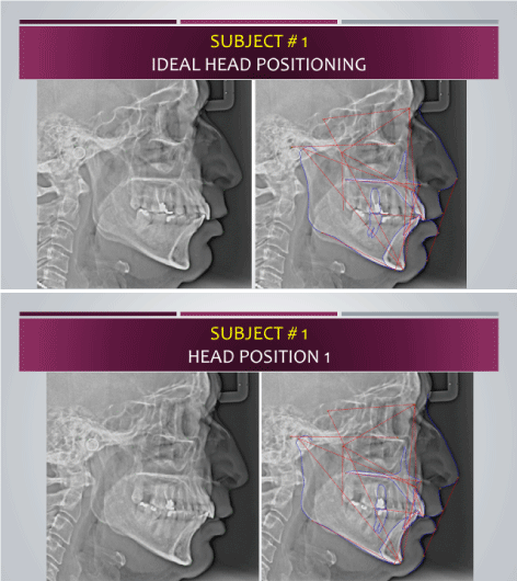
Figure 1. Where the subjects were directed to rotate their heads 10 degrees towards their right over the horizontal axis as measured by a protractor.
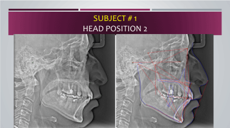
Figure 2. The subjects were then placed once again in natural head position and then instructed to tilt their heads 10 degrees downwards across the sagittal planeinto.
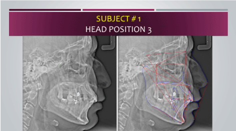
Figure 3. The subjects were positioned with their heads tilted 10 degrees both over the vertical (downwards) and horizontal axis (to the right).
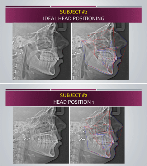
Figure 4. Where the subjects were directed to rotate their heads 10 degrees towards their right over the horizontal axis as measured by a protractor.
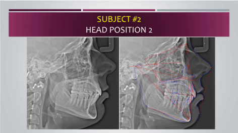
Figure 5. The subjects were then placed once again in natural head position and then instructed to tilt their heads 10 degrees downwards across the sagittal planeinto.
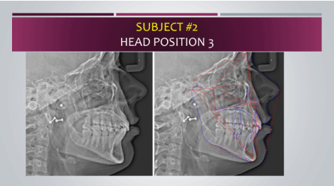
Figure 6. The subjects were positioned with their heads tilted 10 degrees both over the vertical (downwards) and horizontal axis (to the right).
All lateral cephalograms were then traced in Smile stream (Smile stream, Inc. Pvt. Ltd. 135 Columbia Suite 101, Aliso Viejo, CA 92694) using standard anatomical landmark points for the modified Steiner Analysis. When choosing points for bilateral anatomic structures or where dual images are reproduced on the cephalogram (Porion, Orbitale, Molars, and Gonion), the most superior, anterior point was selected in order to maintain consistency-- this takes into account possible magnification distortion of bilateral structures. Porion was chosen as the superior point of the external auditory meatus, Gonion as the inferior point of the ramus, central incisors oriented to the incisal edge and apex, and first molars to the mesial-occlusal surface. The results of the Modified Steiner analysis for all eight lateral cephalograms were then compared.
The data from subjects 1 and 2 were presented in Table 1 and Table 2 respectively. The mandibular plane angle was increased by 2-5 degrees at Position 3 for both subjects. The values were increased by 3-5 degrees in Position 1 and 2 for subject 2, however, it had minimal change in the same positions for subject 1. Y axis measurement showed a decrease in value in position 3 compared to ideal in both the subjects. For subject 2, the value decreased at all positions for the Y axis measurement. In case of subject 1, only the values at position 3 showed the decrease. The measurement of Maxilla to cranium became more positive with Position 1,2 and 3 as compared to the control for both of the subjects. Y axis measurement showed a decrease in value in Position 3 compared to the control in both subjects. The values of SNA, SNB, ANB and Wits did show a change in value for both the subjects in different positions compared to the control, however, no positive or negative trend could be determined. The measurement of Lower Incisor inclination based on the mandibular plane angle was shown to decrease by 5-6 degrees for both subjects in Position 3. Upper incisor inclination showed a decrease in value for all positions as compared to the ideal for subject 2 , however, such trend was not observed in case of subject 1. The maxillary height did not change significantly in different positions as compared to the ideal in subject 1, however, it showed an increase in position 3 for subject 2.
Table 1. Cephalometric analysis of the lateral cephalograms (Ideal head positioning Position 1, Position 2 and Position 3) for patient 1.
Patient 1 |
|
|
|
|
|
Ideal |
Position 1 |
Position 2 |
Position 3 |
Mand plane angle |
28.37 |
27.2 |
28.63 |
30.94 |
(ANS-PNS to Mand plane) |
|
|
|
|
Y axis (SGN-FH) |
63.89 |
64.02 |
64.65 |
62.28 |
Maxilla to Cranium |
1.45 |
1.88 |
2.09 |
4.4 |
(N perp to A point ) |
|
|
|
|
Mandible to Cranium |
-7.35 |
-10.1 |
-10.49 |
-3.52 |
( N perp to Po) |
|
|
|
|
SNA |
86.09 |
86.37 |
85.48 |
82.12 |
SNB |
80.38 |
80.91 |
79.57 |
77.47 |
ANB |
5.71 |
5.46 |
5.92 |
5.65 |
Lower incisor inclination |
101.49 |
101.87 |
103.17 |
95.9 |
L1 to MP |
|
|
|
|
Upper incisor inclination |
1.96 |
2.89 |
2.6 |
3.86 |
Upper 1 to A Vertical (to FH) |
6.77 |
7.15 |
6.61 |
9.3 |
Table 2. Cephalometric analysis of the lateral cephalograms ( Ideal head positioning, Position 1, Position 2 and Position 3) for patient 2.
Patient 2 |
|
|
|
|
|
Ideal |
Position 1 |
Position 2 |
Position 3 |
Mand plane angle |
29.01 |
31.19 |
30.04 |
34.56 |
(ANS-PNS to Mand plane) |
|
|
|
|
Y axis (SGN-FH) |
62.33 |
59.87 |
58.24 |
59.87 |
Maxilla to Cranium |
-0.18 |
-0.42 |
4.66 |
3.23 |
(N perp to A point ) |
|
|
|
|
Mandible to Cranium |
-11.57 |
-10.58 |
-3.76 |
-4.2 |
( N perp to Po) |
|
|
|
|
SNA |
76.28 |
74.23 |
78.18 |
71.3 |
SNB |
71.71 |
69.61 |
72.59 |
66.87 |
ANB |
5.09 |
4.62 |
5.59 |
4.43 |
Lower incisor inclination |
102.46 |
99.37 |
100.83 |
95.72 |
L1 to MP |
|
|
|
|
Upper incisor inclination |
1.96 |
2.89 |
2.6 |
3.86 |
Upper 1 to A Vertical (to FH) |
|
|
|
|
Cephalometric measurements are critical to orthodontic case diagnosis. Although there are specific guidelines for ideal patient positioning while taking the lateral cephalogram, quite often protocols are not properly maintained and errors occur [8]. Potential reasons for positional errors are poorly or newly trained operators, lack of time in the office, which results in rushed procedures, poor patient compliance with instructions, among others.
Although the discrepancy of positions can be varied compared to the ideal , in this study the effect of three example positions (lateral tilt, Sideways tilt and a combination of sideways and lateral tilt) was investigated. The Results clearly indicate that head positional changes ( as compared to the natural head position) have a significant effect on the lateral cephalometric analysis. Evaluation of the norms of the lateral cephalometric parameters [9] indicate that a deviation of 5-6 degrees in angular measurement or 2-4 mm in linear measurements may have significant effect on the diagnosis of a case. For example, the mandibular plane angle (ANS-PNS to MP) is considered acute when it is less than 24 degrees and considered obtuse when it is more than 33 degrees. If the measurement is 24 degrees or less, the subject is diagnosed to have a hypodivergent mandible or a future forecast of hypodivergent jaw growth ( in the case of growing patients ). Conversely, if the measurement is 33 degree or more, the subject is diagnosed to have a hyperdivergent mandible or a future forecast of hyperdivergent jaw growth ( in the case of growing patients) [10]. The treatment modalities for treating cases with hypodivergent jaws in comparison to treating cases with hyperdivergent jaws are completely different. Therefore, an erroneous measurement can affect the treatment planning as well as the future growth forecast of the subject.
For subject 1, the ideal positioning showed that the jaws have a strong hypodivergent tendency, whereas in position 3 the jaw positions looked normal. The mandibular plane angle did increase for both the subjects in position 3 as compared to the ideal position, which means that although for subject 2 in the ideal positioning the jaw analysis was normal, in position 3 the analysis showed that the jaws are hyperdivergent.
In case of Y axis norms, a measurement of 57 degree or less signifies a horizontal growth tendency of the jaws whereas a measurement of 62 degrees or more signifies a vertical jaw growth tendency [11]. In the case of subject 1, there was a similar observation of a decreased angular measurement in Position 3 when compared to the control. For subject 2 in this case, although the measurement at ideal head position shows that the subject has a vertical growth tendency the similar measurement at position 3 showed that subject 2 has normal jaw growth tendency.
Regarding linear measurements of maxilla to cranium (N perpendicular to A point) and mandible to cranium (N perpendicular to Po), an increase in values for both the subjects was observed when comparing Positions 1, 2 and 3 to the control. As per the norm, a maxilla to cranium measurement of -1 mm signifies a retruded maxilla whereas a measurement of + 3 mm signifies a protruded maxilla [12]. In case of subject 1, although ideal positioning shows an ideal maxillary position - at Position 3 the maxilla indicates a protruded position. For subject 2, while the ideal head positioning indicates a normal maxillary position, both Position 2 and Position 3 show the maxilla has a protruded position. A measurement of -4 mm mandible-to- cranium signifies a retruded mandible whereas a measurement of + 1 mm signifies a protruded mandible [13]. For subject 2, the control shows a significantly retruded mandible while in Position 1, 2 and 3 the mandibular position of subject DD are less pronounced. A similar trend in the measurements of subject 1, where Position 3 shows less mandibular protrusion as compared to the ideal positioning, was observed.
Angular measurements of SNA, SNB are also used to assess the position of the upper and lower jaw in contrast to the cranium. For SNA, an angular measurement of 83 degree or more signifies a protruded maxilla, whereas an angular measurement of 76 degree or less signifies a retruded maxilla [14]. For SNB, an angular measurement of 80 degree or above signifies a protruded mandible whereas a measurement of 75 degree or less refers to a retruded mandible [15]. For subject 2, it was observed that the value of both SNA and SNB changed the most (decreased in this case) in Position 3 when compared to the control. The values of SNA and SNB had a less substantial change for Position 1 and Position 2. For subject 1, a similar trend was observed: the most significant decrease in values occurred in Position 3. In general, the cephalometric values showed that the maxilla and mandible both became more retruded in position 3 as compared to the ideal position.
The inclination of the mandibular Lower incisors to the mandibular plane angle is a major determinant in the cephalometric analysis and subsequent orthodontic treatment planning as well as an assessment of treatment outcomes and stability. A value of 89 degrees or less refers to a retroclined position of the lower incisors whereas a value of 98 degrees or more suggests a proclined lower incisor [16]. For both subjects, the IMPA value changed in all positions (Position 1, 2 and 3) compared to the control position, however, the most substantial changes were observed in Position 3. At the ideal head position and in Position 1 and 2, the lower incisors looked proclined as per the cephalometric values, whereas in Position 3, the angulation of the lower incisors to the mandibular plane are in the normal range.
The upper incisor position is measured by the distance between the vertical plane (perpendicular to the FH plane) through A point and incisal edge of the upper incisors. The inclination of the upper incisors plays a critical role in orthodontic treatment planning specifically in evaluation of esthetic value of a patient’s profile. The normal range value of this parameter is between +2 and + 6 mm (a value less than 2 mm indicates retrusion and a value greater than 6mm indicates protrusion (17). For both subjects, the values for upper incisor position increased in Positions 1, 2 and 3 when compared to the control. For subject 1, the altered position showed protruded upper incisors while the control showed normally positioned upper incisors. For subject 2, the altered positions indicated a normal upper incisor position, whereas the control indicated retruded upper incisors.
In conclusion, we observed the following in this pilot study:
Altering subject head positioning affects the outcomes of cephalometric analysis. Both linear and angular measurements were affected by head positions.
These changes in values have significant impact on the interpretation of cephalometric analyses, thereby affecting orthodontic treatment planning as well as growth assessment.
The most substantial changes in cephalometric values occurred in Position 3where the head was tilted both in the vertical and horizontal axis.
A broader study with more subjects is needed to further substantiate the trends observed in the different head positions.
- Helal NM, Basri OA, Baeshen HA (2019) Significance of Cephalometric Radiograph in Orthodontic Treatment Plan Decision. J Contemp Dent Pract 20:789-793. [Crossref]
- Schwendicke F, Chaurasia A, Arsiwala L, Lee JH, et al (2021) Deep learning for cephalometric landmark detection: systematic review and meta-analysis. Clin Oral Investig 25:4299-4309.[Crossref]
- Doff MH, Hoekema A, Pruim GJ, Huddleston Slater JJ, Stegenga B (2010) Long-term oral-appliance therapy in obstructive sleep apnea: a cephalometric study of craniofacial changes. J Dent 38:1010-1018.
- Da Fontoura CS, Miller SF, Wehby GL, Amendt BA, Holton NE, et al (2015) Candidate Gene Analyses of Skeletal Variation in Malocclusion. J Dent Res 94:913-920.
- Major PW, Johnson DE, Hesse KL, Glover KE (1996) Effect of head orientation on posterior anterior cephalometric landmark identification. Angle Orthod 66:51-60. [Crossref]
- Cooke MS, Wei SH (1988) The reproducibility of natural head posture: a methodological study. Am J Orthod Dentofacial Orthop 93:280-288. [Crossref]
- Mehta S, Dresner R, Gandhi V, Chen PJ, Allareddy V, et al (2020) Effect of positional errors on the accuracy of cervical vertebrae maturation assessment using CBCT and lateral cephalograms. J World Fed Orthod 9:146-154.[Crossref]
- Savoldi F, Xinyue G, McGrath CP, Yang Y, Chow SC, et al (2020) Reliability of lateral cephalometric radiographs in the assessment of the upper airway in children: A retrospective study. Angle Orthod 90:47-55.[Crossref]
- Strajni� L, Sinobad DS (2012) Application of cephalometric analysis for determination of vertical dimension of occlusion--a literature review. Med Pregl 65:217-222. [Crossref]
- Ahmed M, Shaikh A, Fida M (2016) Diagnostic performance of various cephalometric parameters for the assessment of vertical growth pattern. Dental Press J Orthod 21:41-49.
- Sorin MS, Ramsey TC, Hart TC, Farrington FH (1991) Comparison of craniofacial morphology in monozygotic twins with their siblings. J Clin Pediatr Dent 15:169-173. [Crossref]
- Williams S, Leighton BC, Nielsen JH (1985) Linear evaluation of the development of sagittal jaw relationship. Am J Orthod 88:235-241.
- Carter NE (1987) Dentofacial changes in untreated Class II division 1 subjects. Br J Orthod 14:225-234.
- Alió-Sanz J, Iglesias-Conde C, Lorenzo-Pernía J, Iglesias-Linares A, Mendoza-Mendoza A, et al (2012) Effects on the maxilla and cranial base caused by cervical headgear: a longitudinal study. Med Oral Patol Oral Cir Bucal 17:e845-851. [Crossref]
- Stojanovi� Z, Nikoli� P, Nikodijevi� A, Mili� J, Duka M (2012) Analysis of variation of sagittal position of the jaw bones in skeletal Class III malocclusion. Vojnosanit Pregl 69:1039-1045.
- Irfan S, Fida M (2019) Comparison of soft and hard tissue changes between symmetric and asymmetric extraction patterns in patients undergoing orthodontic extractions. Dent Med Probl 56:257-263. [Crossref]
- Maddalone M, Losi F, Rota E, Baldoni MG (2019) Relationship between the Position of the Incisors and the Thickness of the Soft Tissues in the Upper Jaw: Cephalometric Evaluation. Int J Clin Pediatr Dent 12:391-397.[Crossref]
Editorial Information
Founding Editor-in-Chief
Shigeru Watanabe
Meikai University Japan
Editor-in-Chief
Vagner Rodrigues
Federal University of Minas Gerais
Article Type
Research Article
Publication history
Received: February 04, 2022
Accepted: February 16, 2022
Published: February 21, 2022
Copyright
©2022 Lala S. This is an open-access article distributed under the terms of the Creative Commons Attribution License, which permits unrestricted use, distribution, and reproduction in any medium, provided the original author and source are credited.
Citation
Lala S, McGann D, Dorrego D (2022) The effect of different head positions in cephalometric radiographic analysis - A pilot study. Dent Oral Maxillofac Res 8: DOI: 10.15761/DOMR.1000396

Figure 1. Where the subjects were directed to rotate their heads 10 degrees towards their right over the horizontal axis as measured by a protractor.

Figure 2. The subjects were then placed once again in natural head position and then instructed to tilt their heads 10 degrees downwards across the sagittal planeinto.

Figure 3. The subjects were positioned with their heads tilted 10 degrees both over the vertical (downwards) and horizontal axis (to the right).

Figure 4. Where the subjects were directed to rotate their heads 10 degrees towards their right over the horizontal axis as measured by a protractor.

Figure 5. The subjects were then placed once again in natural head position and then instructed to tilt their heads 10 degrees downwards across the sagittal planeinto.

Figure 6. The subjects were positioned with their heads tilted 10 degrees both over the vertical (downwards) and horizontal axis (to the right).
Table 1. Cephalometric analysis of the lateral cephalograms (Ideal head positioning Position 1, Position 2 and Position 3) for patient 1.
Patient 1 |
|
|
|
|
|
Ideal |
Position 1 |
Position 2 |
Position 3 |
Mand plane angle |
28.37 |
27.2 |
28.63 |
30.94 |
(ANS-PNS to Mand plane) |
|
|
|
|
Y axis (SGN-FH) |
63.89 |
64.02 |
64.65 |
62.28 |
Maxilla to Cranium |
1.45 |
1.88 |
2.09 |
4.4 |
(N perp to A point ) |
|
|
|
|
Mandible to Cranium |
-7.35 |
-10.1 |
-10.49 |
-3.52 |
( N perp to Po) |
|
|
|
|
SNA |
86.09 |
86.37 |
85.48 |
82.12 |
SNB |
80.38 |
80.91 |
79.57 |
77.47 |
ANB |
5.71 |
5.46 |
5.92 |
5.65 |
Lower incisor inclination |
101.49 |
101.87 |
103.17 |
95.9 |
L1 to MP |
|
|
|
|
Upper incisor inclination |
1.96 |
2.89 |
2.6 |
3.86 |
Upper 1 to A Vertical (to FH) |
6.77 |
7.15 |
6.61 |
9.3 |
Table 2. Cephalometric analysis of the lateral cephalograms ( Ideal head positioning, Position 1, Position 2 and Position 3) for patient 2.
Patient 2 |
|
|
|
|
|
Ideal |
Position 1 |
Position 2 |
Position 3 |
Mand plane angle |
29.01 |
31.19 |
30.04 |
34.56 |
(ANS-PNS to Mand plane) |
|
|
|
|
Y axis (SGN-FH) |
62.33 |
59.87 |
58.24 |
59.87 |
Maxilla to Cranium |
-0.18 |
-0.42 |
4.66 |
3.23 |
(N perp to A point ) |
|
|
|
|
Mandible to Cranium |
-11.57 |
-10.58 |
-3.76 |
-4.2 |
( N perp to Po) |
|
|
|
|
SNA |
76.28 |
74.23 |
78.18 |
71.3 |
SNB |
71.71 |
69.61 |
72.59 |
66.87 |
ANB |
5.09 |
4.62 |
5.59 |
4.43 |
Lower incisor inclination |
102.46 |
99.37 |
100.83 |
95.72 |
L1 to MP |
|
|
|
|
Upper incisor inclination |
1.96 |
2.89 |
2.6 |
3.86 |
Upper 1 to A Vertical (to FH) |
|
|
|
|


