It is about an exceptional case of endogenous endophthalmitis secondary to a liver abscess due to Klebsiella pneumonia reported in Tunisia. The patient was a 33-year-old male, who was admitted for fever and right upper quadrant abdominal pain. Abdominal computed tomography showed an abscess measured 7 cm and located in segment 4 of the liver. It was treated by antibiotics and percutaneous transhepatic drainage. Pus sample was positive for Klebsiella pneumoniae. On day 4 after admission, patient complained of a red right eye with decreased vision. The diagnosis of endogenous endophthalmitis was strongly suspected. An Early treatment was initiated with a good evolution.
The syndrome Endophthalmitis-hepatic abscess is an exceptional syndrome rarely reported in the literature. It must evoke in case of hepatic abscess caused by klebsiella pneumonia
liver, abcess, endophthalmitis
Endogenous bacterial endophthalmitis is a rare eye infection (2 to 8% of all endophthalmitis [1] and is secondary to sepsis. The portal entry is, in most cases, a deep infectious focus (cardiac, digestive, urinary). Indeed, the endocular infection is occurring by hematogenous pathway. The visual prognosis is reserved, depending on the germ in question and the treatment efficiency. We report here an exceptional association of endogenous endophthalmitis with pyogenic liver abscess caused by Klebsiella in an elderly diabetic patient.
A 70-year-old man with history insulin-dependent diabetes was admitted with a 1-week history of fever and right upper quadrant pain. On admission, body temperature was 38°C and there was a right upper quadrant abdominal tenderness. Blood investigations showed the following values: hemoglobin, 12.8 g/dl; white blood cells count, 13,600/mm3; a blood glucose level of 14 mmol/l and C reactive protein level of 250 mg/l. An abdominal computed tomography scan showed a low-density lesion that measures 7 cm suggestive of liver abscess (Figure 1). It was located in segment 4 of the liver.
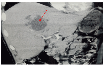
Figure 1. Abdominal computed tomography scan showing a low-density lesion in the liver suggestive of liver abscess (red arrow)
These findings led to the diagnosis of pyogenic liver abscess for which drainage was performed. Pus samples and blood cultures were positive for Klebsiella pneumoniae. The patient was treated with levofloxacin and Metronidazole. On day 4 after admission, an ophthalmic examination was performed as the patient complained of a painful red right eye with low visual acuity (Figure 2).
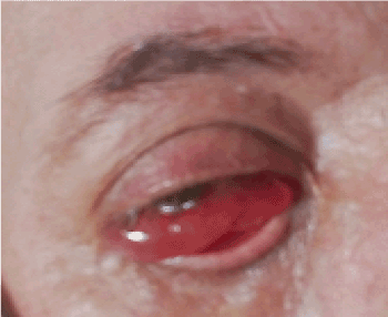
Figure 2. The right eye was erythematous with swelling and redness
A slit lamp examination revealed cells and flare in the right eye anterior chamber, posterior synechiae, an important vitritis, the fundus was barely visible. The diagnosis of endogenous endophthalmitis was strongly suspected. B-mode echo tomography showed mobile echoes in the vitreous area with total Retinal detachment (Figure 3).

Figure 3. B-mode echo tomography showed mobile echoes in the vitreous area
The patient was treated with topical ceftazidime (50 mg/ml) hourly as well as prednisolone acetate 1% fourtimes daily and atropine 1% twice daily. The evolution was favorable with an improvement in visual acuity and reduction in ocular redness.
Endogenous endophthalmitis is an exceptional intraocular infection, which results from the hematogenous spread of a microorganism from a septic focus. It accounts for 2 to 8% of all endophthalmitis, it is much more rare than exogenous endophthalmitis (post-surgical or post traumatic) [2,3]. Endogenous endophthalmitis preferentially affects immunosuppressed patients: diabetic individuals in particular. Portals entry are endocarditis (46% of cases), followed by gastrointestinal, genitourinary, dental, hepatic, meningeal and pulmonary infections. However, it remains unknown in 10% of cases [4]. In our case the portal entry was hepatic realizing the syndrome Endophthalmitis-hepatic abscess». The main germ involved is klebsiella pneumaniae and the serotype K1 is more particularly responsible for endogenous endophthalmitis. This syndrome involves the visual prognosis and sometimes vital. Well described in Asian populations, it affects Caucasian populations with an increasing number of reported cases [5].
Its description in Tunisia remains exceptional. To our knowledge, this is the 1st case reported in Tunisia. This entity must be recognized very early. Indeed, appropriate antibiotic therapy should be given to the patient in extreme emergency. The visual prognosis is involved with the risk of permanent blindness. The mortality rate varies between 3 and 42% in the literature [5].
The syndrome Endophthalmitis-hepatic abscess is an exceptional syndrome rarely reported in the literature. It must evoke in case of hepatic abscess caused by klebsiella pneumonia.
- Habib G, Badano L, Tribouilloy C, Vilacosta I, Zamorano JL, et al. (2010) Recommendations for the practice of echo-cardiography in infective endocarditis. Eur J Echocardiogr 11: 202-219.
- Jackson TL, Eykyn SJ, Graham EM, Stanford MR (2003) Endo-genous bacterial endophthalmitis: a 17-year prospective series and review of 267 reported cases. Surv Ophthalmol 48: 403-423.
- Okada AA, Johnson RP, Liles WC, D’Amico DJ, Baker AS (1994) Endogenous bacterial endophthalmitis. Report of a ten-year retrospective study. Ophthalmology 101: 832-838.
- Cornut PL, Chiquet C (2011) Endophtalmies endogènes bactériennes. J Fr Ophtalmol 34: 51-57.
- Murano T, Ooiwa Y, Takahashi K, Kodama Y, Takakura S, et al. (2013) A liver abscess deprived a healthy adult of eyesight: Endogenous endophthalmitis associated with a pyogenic liver abscess caused by serotype K1 Klebsiella pneumonia. Intern Med 52: 919-922.