Abstract
During ganglion transplantation, some neurons undergo pathological changes under the influence of ischemia, decentralization, and damage of axons, up to and including their death.The purpose of this article is to analyze the existing problem in order to prevent or reduce the death of neurons, which can lead to the death of animals in the experiment and patients in the clinic.
Keywords
ganglia, transplantation, neurons, nitricoxidecycle, nitrites, nitric oxide (NO), nitric dioxide (NO2)
Restoring the function of an organ that has been disrupted as a result of breaking its connections with the central nervous system (CNS) is one of the urgent tasks of modern biology and medicine [1-3].Breaks in connections between the organ and the central nervous system can occur as a result of a disease, injury, or during organ transplantation.Currently, the field of regenerative medicine is one of the flourishing areas of modern medicine - from cardiology and neuropathology to urology and cosmetology [3].Traditional donation cannot currently provide for all those who need organ transplants.Therefore, medicine is in dire need of such technologies that complement donor organ transplantation, and are sometimes used as an alternative to it.In this area of regenerative medicine, tissue engineering is widely used [2,3].Tissue engineering is a branch of biotechnology based on the creation of organs and tissues of the human body in the laboratory [4].Tissue engineering constructs are a biomedical cellular product that consists of cells (cell lines), a biocompatible material, and excipients [4].
John Anthony Atala (b. 1958, Peru). Currently prof. J. E. Atala – Director of the Institute of Regenerative Medicine, Wake Forest in North Carolina (United States), Head of the Department of Urology of the Medical School, Wake Forest in North Carolina (United States), Professor of Advanced Cellular Technologies, Institute of Regenerative Medicine, 1st Moscow State Medical University. One of the pioneers of regenerative medicine. In 2011, Dr. E. Atala was elected a Fellow of the Institute of Medicine of the National Academy of Sciences and named “Physician of the Year” (nomination of “Scientific American” journal) for his contributions to the study of cell, tissue and organ regeneration (Figure 1).
Figure 1.J.E. Atala – Director of the Institute of Regenerative Medicine, Wake Forest in North Carolina (United States) [4]
One of the first methods of tissue engineering structures was the method of creating a new center of local neurohumoral regulation - the gangliopexy method, based on the formation of new nerve and vascular connections [5-10].Academician of the National Academy of Sciences of Belarus D.M. Golub (1901–2001) was the founder of this method [6].Analogues of studies conducted by D.M.Golub, for a long time there was not only in our country, but also abroad.For the school D.M.Golub is characterized by a wide range of embryological, anatomical, histological, and histochemical methods using luminescent and electron microscopy methods [6-10].Employees of D.M.Golub (L.A. Davydova and other colleagues) found that during ganglion transplantation, some neurons undergo pathological changes under the influence of ischemia, decentralization, and damage to axons, up to and including their death.Often, with such pathological changes, not only neurons die, but also patients.This happens in less than 30% of cases.One of the objectives of this article is to analyze the existing problem in order to prevent or reduce the death of neurons, which can also lead to the death of animals in the experiment or patients in the clinic (Figure 2).
Figure 2.Academician of the National Academy of Sciences of Belarus David Moiseevich Golub (1901–2001) [4]
In 1998, the International Biographical Center (Cambridge, United Kingdom) included D.M. Golub among the 2000 outstanding scientists of the 20th century due to his special contribution to the field ofanatomy and embryology. Since the 1960s, scientific research has been carried out in this field in the laboratory of morphology of the Institute of Physiology of the National Academy of Sciences of the Republic ofBelarus [5].
Embryogenesis of the vegetative (autonomous) nervous system and the identification of patterns of differentiation of tissues and organs in connection with their innervation at different periods of individual development of humans and animals showed that prevertebral and organ vegetative nodes are formed as a result of dispersion (scattering) of young nerve cells from the main vegetative node (ganglion). Microgangliaappear near the main ganglion. These structures are connected to the main ganglion by nerve fibers. It turned out that microganglia can be considered as a morphological basis for the creation of new centers of local nervous regulation of internal organs by the transplantation of ganglia with the preservation of neurovascular components. Belarusian scientists developed methods for transplanting autonomic nodes into the wall of various organs (heart, bladder, prostate, etc.), as well as into the thickness of the striated muscle. On the basis of the literature and mainly data from the experiments of employees of the laboratory of morphology, it became obvious that the most favorable results were obtained with transplantation of the autonomic ganglion with preservation of the neurovascular pedicle for the rapid restoration of its interneuronal connections and blood supply [5].
Studies of the diversity of interneuronalconnections on the example of the caudal mesenteric ganglion confirm the data of morphologists and physiologists on the complexity of the structure of interneuronal relations in the gastrointestinal tract. Single and multiple synaptic structures on efferent neurons have been identified.The afferent structures of the ganglion are represented by encapsulated and non-encapsulated receptors, pericapsular plexuses.Dendrites of receptor neurons braid groups of motor neurons [5,6].
The blood supply of the transplanted caudal mesenteric ganglion is restored as a result of the appearance of collateral vessels along the course of the hypogastric nerves. Among the vessels of the transplanted node, the proportion of capillaries is 90.1%.It is somewhat higher than in the intact ganglion. It can be assumed that the survival of a large number of nerve cells during transplantation is associated with a rapid restoration of blood supply. In the connective tissue that develops around the transplanted ganglion, a large number of newly formed nerve fibers are revealed. Transplantation of the caudal mesenteric ganglion to the striated muscle with the preservation of the neurovascular "leg" favors the preservation of a large number of neurons of the transplanted ganglion, the restoration of their neuronal and vascular connections.Transplantation performed in this way deserves attention as one of the models for studying the issue of the formation of new centers of local nervous regulation of internal organs [5-7].
During transplantation of the caudal mesenteric ganglion on the neurovascular "pedicle", some of the neurons do not change.At the same time, their connections with each other and with the central nervous system coincide in structure with the intact ganglion.However, some neurons undergo pathological changes under the influence of ischemia, decentralization and damage to axons.At the same time, transneuronal atrophy, retrograde degeneration, and neuronal death are detected. The number of cases in which deaths are observed after surgery ranges from 5 to 20–30% [4,5,8,9].
Is it possible to reduce the probability of death of animals (in an animal experiment) or patients in the clinic (during operations associated with ganglion transplantation)?What factors affect the transplanted ganglion neurons, and how can they be modulated (changed, reduced) to reduce the number of deaths during operations?Is there a period of time after transplantation when the biological system in the form of a caudal mesenteric node, used as a model of tissue engineering, is in a state of bifurcation?In other words, is there a moment in time when a tissue-engineered structure, consisting of neurons and vessels, can die along with its owner, or, on the contrary, can survive and give the patient a chance to live with a transplanted ganglion?An analysis of the causes and elucidation of the mechanisms involved in neuronal damage during nerve ganglion transplantation will be presented in the discussion of these data .
An analysis of the causes and elucidation of the mechanisms involved in the above processes led us to the conclusion that almost all pathological processes occurring against the background of hypoxia/ischemia, inflammatory processes, and activation of immune/autoimmune reactions have a common component [13-18].This component is due to the occurrence of a chemical or free radical resonance between reactive nitrogen species (•NO, •NO2) and reactive oxygen species (•O2-, •OH-radicals).Chemical resonance, when •NO and •O2-, interacting with each other, create very dangerous peroxynitrites, •NO2 and •OH-radicals can become a real cause of atrophy, degeneration and death of neurons.In turn, such death of neurons can eventually lead to the death of animals in the experiment and people in the clinic [4,5, 9-11].What should be done to prevent the development of such processes?
For this it is necessary: a) to reduce hypoxia/ischemia (blood oxygenation);b) reduce the intensity of inflammatory processes (anti-inflammatory drugs);c) inhibit the activity of immune / autoimmune reactions (cytostatics, azathioprine).Do not allow excess exogenous nitrates, nitrites and NO-generating compounds to enter with water, food and drugs during the active postoperative period.During this period, it is recommended to use slightly alkaline mineral waters that inhibit the reduction of NO2- ions to NO (the nitrite reductase component of the nitric oxide cycle) (Fig. 3).This is due to the fact that heme-containing proteins in the deoxy form can reduce NO2- ions to NO.A change in pH to the acid side converts Hb-O2 into the T-conformation, in which the bond between the O2 ligand and the heme iron is weakened and the blood pigment releases the O2 molecule relatively easily.In the T-conformation, heme-containing Hb-O2 complexes can more actively reduce NO2- ions to NO (activation of the nitrite reductase component) of the nitric oxide cycle (Fig.3).The pH shift to the alkaline side ensures the preservation of Hb-O2in the R-conformation, which retains the O2 molecule well in the Hb-O2 complex.In this position (R-conformers of Hb-O2), the nitrite reductase component of the nitric oxide cycle will be inhibited, and together with it, the reproduction of NO, as well as the concentration of these molecules, will be inhibited [4,5,12] (Figure 3).
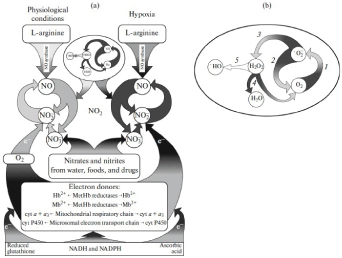
Figure 3. Cycles of nitric oxide (a) and the superoxide anion radical (b)
In the nitric oxide cycle, one can distinguish the NO-synthase component (“L-arginine - NO”), which synthesizes NO in the presence of oxygen, and the nitrite reductase component, the activity of which sharply increases under conditions of oxygen deficiency (hypoxia / ischemia). The formation of NO with the participation of the NO-synthase component is carried out as a result of the oxidation of the guanidine nitrogen of L-arginine. NO2-, ions, formed from L-arginine, can again, with the participation of nitrite reductase systems, including Hb, Mb, cyt a + a3and cyt P-450, close the chain of L-arginine → NO → NO2-/ NO3-into a cycle. Oxygen, binding to heme, inhibits the nitrite reductase activity of these proteins. During hypoxia and functional stress, when heme-containing proteins are converted into deoxy-form, NO2-ions begin to actively reduce, accepting electrons from these heme-containing proteins. An important role in the reduction of NO2– ions to NO is also played by electron donor systems that reduce Hb, Mb, cyt a + a3, and cyt P-450. In addition to these electron transport systems (chains), ascorbic acid and reduced glutathione can play an important role in the reduction of NO2-ions. In the cycle of superoxide radical anion occur: 1 - reduction of oxygen (O2) and the formation of superoxide anion radical (·O2); 2 and 3 - superoxide dismutation reactions catalyzed by superoxide dismutase; 4 - decomposition of hydrogen peroxide (H2O2) into water (H2O) and molecular oxygen (O2), carried out by the enzyme catalase; 5 - hydrogen peroxide (H2O2) 5 - hydrogen peroxide - H2O2- also decomposes to form two molecules of the ·OH-radical. The cyclic organization of reactive nitrogen and oxygen forms ensures the conversion of these reactive, highly reactive compounds into less active substances. When the cycles of nitric oxide and superoxide anion-radical are disrupted, even more active molecules of nitrogen dioxide and peroxynitrites appear, again decaying into ·NO2and ·OH-radicals, which damage the main components of living organisms [12-15].
The authors believe that the measures proposed above are fundamentally new, which can reduce the probability of NO2formation at the sites of ganglion transplantation, and thereby ensure the normal operation of the NO and •О2–cycles involved in maintaining the concentration of reactive nitrogen and oxygen species within the physiological norm.
References
- Berthiaume F, Maguire TJ, Yarmush ML (2011) Tissue engineering and regenerative medicine: history, progress, and challenges. Annu Rev Chem Biomol Eng2: 403-30. [Crossref]
- Orlando G, Wood KJ, Stratta RJ, Yoo JJ, Atala A, et al. (2011) Regenerative medicine and organ transplantation: past, present, and future. Transplantation91: 1310-7. [Crossref]
- Turner NJ, Keane TJ, Badylak SF (2013) Lessons from developmental biology for regenerative medicine. Birth Defects Res C Embryo Today99:149-59.[Crossref]
- Reutov VP, Davydova LA, Sorokina EG (2022) Tissue-engineered constructions in biophysics, neurology and other filds and branches of medicine. Biophysics67: 828–846. [Crossref]
- Davydova LA, Reutov VP (2018) Gangliopexy: creation of a new center of local neurohumoral regulation, the role of nitric oxide and specific factors for stimulating angiogenesis. The contribution of the embryologist-anatomist academician David Moiseevich Golub (1901–2001) to the development of the gangliopexy method and the current state of the problem of tissue engineering structures (TEC). Eurasian Scientific Association 37: 82-96.
- Golub DM (1963) Development of the sympathetic trunk in human embryogenesis. ArkhAnatGistolEmbriol45: 3-16.
- Golub DM, Kheĭnman FB, Novikov II, Archakova LI, Danilenko RV (1979) Reinnervation of internal organs and vessels by the neuropexy technic. ArkhAnatGistolEmbriol76:5-16. [Crossref]
- Golub DM, Bronovitskaia GM (1981) Development of the human hip joint and its innervation. Arkh. Anat. Gistol. Embriol80:47-56. [Crossref]
- Golub DM (1969) Regularities in the intrauterine development of innervation connections of internal organs and formation of new nerve pathways and centers under experimental conditions. ArkhAnatGistolEmbriol57: 3-12. [Crossref]
- Golub DM, Loyko RM, Novikov II (1979) Development of reflexogenic zone innervation of the human cardiovascular system. AnatAnz145:474-492. [Crossref]
- Golub DM, Davydova LA, Novikov II, Kheínman FB (1973) Formation of new centers of local nervous regulation of organs and tissues. ArkhAnatGistolEmbriol64:5-16. [Crossref]
- Reutov VP, Sorokina EG (2022) Causal relationship between physiological and pathological processes in the brain and in the gastrointestinal tract: the Brain–Intestinal axis. Biophysics67: 972-986. [Crossref]
- Reutov VP, Sorokina EG (1998) NO-synthase and nitrite reductase components of the nitric oxide cycle. Biochemistry 63: 874-84. [Crossref]
- Reutov VP (1999) Biochemical predetermination of the NO synthase and nitrite reductase components of the nitric oxide cycle. Biochemistry64: 528-542. [Crossref]
- Reutov VP (2002) Nitric oxide cycle in mammals and the cyclicity principle. Biochemistry67: 293-311. [Crossref]