Abstract
18F-FDG PET-CT has become the main procedure for staging and monitoring treatment response in patients with lymphoma. It can differentiate between active disease and necrosis/fibrosis in after treatment residual masses, mainly in Hodgkin Lymphoma patients. Persistence of FDG uptake is very suggestive of resistance or recurrence. However, there can be also some false positives (FP). Non necrotizing Granulomatous Lymphadenitis (NNGL) is a sarcoidosis-like inflammatory reaction and it can be a FP cause in PET-CT monitorization of HL treatment response. Here we present a case of a patient with HL who was NNGL PET positive after stem cell transplantation.
Keywords
18F-FDG; PET-CT, hodgkin lymphoma, false positive, non-necrotizing granulomatous lymphadenitis, stem cell transplantation
18F-FDG PET-CT has replaced conventional imaging techniques and become the main procedure for staging and monitoring treatment response in patients with lymphoma, emphasizing Hodgkin Lymphoma (HL) and Diffuse Large B Cell Lymphoma (DLBCL) [1,2]. PET-CT can differentiate between metabolically active disease and necrosis/fibrosis in after treatment residual masses, mainly in HL patients, which can reach a very high Negative Predictive Value (95-100%) and Positive Predictive Value of more than 90% [1,3-6]. Persistence of lymph nodes with FDG uptake after treatment is very suggestive of resistance or recurrence.
However, there can be also some false positives (FP), so starting a second line treatment without taking a previous biopsy could develop in unnecessary and potentially toxic therapies, as aggressive chemotherapy or even a stem cell transplantation (SCT). The most frequent causes of FP are infections and inflammatory-granulomatous diseases [7-10]. Non necrotizing Granolumatous Lymphadenitis (NNGL) is a sarcoidosis-like inflammatory reaction, mainly associated with lymphomas and carcinomas. Although infrequent, however, it can be a FP cause in PET-CT monitorization of HL treatment response [10]. Here we present a case of a patient with HL who was NNGL PET positive after SCT.
Case Report
Female (aged 27 years), diagnosed with nodular lymphocyte-predominant HL in 1997, Ann Arbor stage II-A, who reached complete response after treatment with 3 cycles of ABVD (adriamycin, bleomycin, vinblastine and dacarbazine) scheme followed by radiotherapy. In October 2015, aged 44 years, nodular lymphocyte-predominant HL recurrence was diagnosed, also staged II-A (bilateral axillary nodal disease). She received rescue chemo with two courses of R-ESHAP regimen [11], achieving a second complete remission, and after that, she underwent an autologous SCT in 2-2-2016.
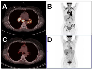
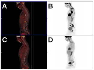
In day + 100 after SCT, a 18F-FDG PET-CT was performed. It showed focal uptake in lymph nodes (located in the mediastinum, pulmonary and liver hilum and retroperitoneum) and several locations in bone (Figures 1 and 2). Despite this result, the patient was clinically asymptomatic and blood parameters (VSG, LDH, Beta 2 microglobulin) were all normal.

Figure 1. After SCT monitoring: 18F-FDG PET-CT performed on day +100 after SCT (1A, 1B, 2A, 2B) compared with PET-CT 12 months after SCT (1C, 1D, 2C, 2D). Nodal and oseous FDG uptake were highly suggestive of malignancy; however, a mediastinoscopy confirmed NNGL diagnosis

Figure 2. After SCT monitoring: 18F-FDG PET-CT performed on day +100 after SCT (1A, 1B, 2A, 2B) compared with PET-CT 12 months after SCT (1C, 1D, 2C, 2D). Nodal and oseous FDG uptake were highly suggestive of malignancy; however, a mediastinoscopy confirmed NNGL diagnosis
Because of this clinical incongruence, a mediastinoscopy was performed. The final diagnosis was nonnecrotizing granulomatous lymphadenitis, so monitoring was decided. Finally, a new PET-CT performed 9 months after showed no metabolically active disease (Figures 1 and 2).
Discussion
18F-FDG PET-CT is standard method for staging and monitoring of treatment response in lymphomas, because of its high sensibility (>95%). However, its specificity is lower [1,2]. The most frequent causes of FP in lymphoma patients are other malignant diseases, sarcoidosis, infections (mainly Aspergillus and Mycobacterium) and other granulomatous diseases [7-9,12]. So, biopsy must be considered specially in those patients with a positive value after treatment PET-CT who show a dissociation between image result and clinical status. Despite lymph nodes and osseus foci were highly suggestive of HL relapse, her good clinical status and normal blood and biochemical parameters made it unprobeable [13].
NNGL pathogeny is still unknown. A hypothesis was made, claiming that necrotizing and degeneration of tumor residual lesions after treatment, as humoral, macrophages and lymphocytes T activation could be the cause of granuloma formation. Unlike sarcoidosis, NNGL is not accompanied by systemic symptoms and does not require any treatment. It has been suggested that it can appear in 14% of HL patients [9] and furthermore, it seems that those patients who suffer from this inflammatory reaction could have a better prognosis [14].
2021 Copyright OAT. All rights reserv
Conclusion
In conclusion, PET-CT is the best method for monitoring therapy response in patients with HL. However, new hypermetabolic foci in after treatment PET-CT are not always due to lymphoma. Our case shows that confirmation biopsy is mandatory in those patients whose recurrence or residual disease is clinically unprobeable, to avoid incorrect and potentially toxic therapies.
Disclosure
The authors state no conflict of interests.
References
- Cheson BD, Fisher RI, Barrington SF, Cavalli F, Schwartz LH, et al. (2014) Recommendations for initial evaluation, staging, and response assessment of Hodgkin and non-Hodgkin lymphoma: the Lugano classification. J Clin Oncol 32: 3059-3068.
- Barrington SF, Mikhaeel NG, Kostakoglu L, Meignan M, Hutchings M, et al. (2014) Role of imaging in the staging and response assessment of lymphoma: consensus of the International Conference on Malignant Lymphomas Imaging Working Group. J Clin Oncol 32: 3048-3058.
- Cerci JJ, Pracchia LF, Linardi CC, Pitella FA, Delbeke D, et al. (2010) 18F-FDG PET after 2 cycles of ABVD predicts event-free survival in early and advanced Hodgkin lymphoma.J Nucl Med51: 1337-1343. [Crossref]
- Engert A, Haverkamp H, Kobe C, Markova J, Renner C, et al. (2012) Reduced-intensity chemotherapy and PET-guided radiotherapy in patients with advanced stage Hodgkin's lymphoma (HD15 trial): a randomised, open-label, phase 3 non-inferiority trial. Lancet 379: 1791-1799.
- de Wit M, Bohuslavizki KH, Buchert R, Bumann D, Clausen M, et al. (2001) 18FDG-PET following treatment as valid predictor for disease-free survival in Hodgkin's lymphoma.Ann Oncol12: 29-37. [Crossref]
- Dittmann H, Sokler M, Kollmannsberger C, Dohmen BM, Baumann C, et al. (2001) Comparison of 18FDG-PET with CT scans in the evaluation of patients with residual and recurrent Hodgkin's lymphoma.Oncol Rep8: 1393-1399. [Crossref]
- Paydas S, Yavuz S, Disel U, Zeren H, Hastürk S, et al. (2002) Granulomatous reaction after chemotherapy for Hodgkin's disease.Leuk Res26: 967-970. [Crossref]
- Fallanca F, Picchio M, Crivellaro C, Mapelli P, Samanes Gajate AM, et al. (2012) Unusual presentation of sarcoid-like reaction on bone marrow level associated with mediastinal lymphadenopathy on (18)F-FDG-PET/CT resembling an early recurrence of Hodgkin's Lymphoma. Rev Esp Med Nucl Imagen Mol 31: 207-209.
- Wirk B (2010) Sarcoid Reactions after Chemotherapy for Hodgkin's Lymphoma.Clin Med Insights Case Rep3: 21-25. [Crossref]
- Cherk MH, Pham A, Haydon A (2011) 18F-fluorodeoxyglucose positron emission tomography-positive sarcoidosis after chemoradiotherapy for Hodgkin's disease: a case report. J Med Case Rep 5: 247.
- Labrador J, Cabrero-Calvo M, Pérez-López E, Mateos MV, Vázquez L, et al. (2014) ESHAP as salvage therapy for relapsed or refractory Hodgkin's lymphoma.Ann Hematol93: 1745-1753. [Crossref]
- Sanan P, Lu Y (2017) Multiorgan involvement of chemotherapy-induced sarcoidosis mimicking progression of lymphoma on FDG PET/CT. Clin Nucl Med 42: 702-703.
- Carr R, Barrington SF, Madan B, O'Doherty MJ, Saunders CA, et al. (1998) Detection of lymphoma in bone marrow by whole-body positron emission tomography.Blood91: 3340-3346. [Crossref]
- O'Connell MJ, Schimpff SC, Kirschner RH, Abt AB, Wiernik PH (1975) Epithelioid granulomas in Hodgkin disease. A favorable prognostic sign?JAMA233: 886-889. [Crossref]


