Multivessel disease is characterized by significant atherosclerotic changes in all three main epicardial coronary arteries. It is a high-risk coronary syndrome that is associated with an important increase in cardiovascular mortality. A 76-year-old female was referred to our department due to chest pain. The coronary angiography showed multivessel disease. Despite the superior diagnostic performance of nuclear imaging techniques, multivessel disease remains a challenging diagnosis. Since all three major coronary arteries had a similar degree of stenosis, the radiotracer had an homologous uptake. Pseudonormalization in MPI-gated SPECT is usually found in multivessel disease, consequently, an anatomical assessment is usually necessary.
multivessel disease, coronary artery disease, nuclear cardiology, myocardial perfusion imaging
Multivessel disease is a high-risk coronary syndrome in which there is a stenosis of 70% or more in each major epicardial coronary artery [1]. Atherosclerosis is the main mechanism responsible for such stenosis, and the main risk factors associated with this entity are hypertension, diabetes, hypercholesterolemia, and smoking. This disease carries a high risk of mortality and morbidity in both stable and unstable clinical settings; its poor prognosis may improve in patients when performing percutaneous or surgical revascularization -- depending on their clinical characteristics -- often these invasive interventions are not performed due to patient's risk underestimation [2].
A 76-year-old female was referred due to chest pain. She had hypertension, diabetes and dyslipidemia diagnosed 20 years ago. Physical examination was irrelevant and laboratory studies showed total cholesterol of 135 mg/dL and triglycerides of 155 mg/dL. A coronary angiotomography showed diffuse multivessel coronary artery disease (CAD) CAD RADS 5, with a calcium score of 2149.2 U (Figure 1). The coronary angiography showed multivessel disease with 90% stenosis of the left anterior descending artery, 80% stenosis of the first diagonal artery, presence of crystals of cholesterol in the circumflex artery and 90% stenosis of the right coronary artery (Figure 2). A transthoracic echocardiogram showed left ventricular normal wall motion and normal left ventricular systolic function.
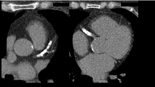
Figure 1. Coronary Calcium Score by computed tomography
An important calcification is present in the left main coronary artery, involving left anterior descending, circumflex artery (A), and right coronary artery (B). Coronary Calcium Score was 2149.2 UA
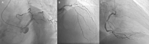
Figure 2. Coronary angiography
Panel A shows a 50% stenosis in the proximal segment of the left coronary trunk and a chronic total occlusion of the circumflex artery. Panel B shows a 90% stenosis at the proximal portion of the left anterior descending artery and an 80% stenosis in the first diagonal artery. Panel C exhibits a 90% stenosis at the mid portion of the right coronary artery
Further diagnostic tests were performed to evaluate ischemia-guided revascularization. Pharmacological stress test was positive for ischemia and perfusion study with 99m Tc-Sestamibi gated-SPECT showed a nontransmural infarction of the lateral wall and generalized ischemia of the left ventricle that caused “pseudonormalization” of perfusion, also called balanced ischemia (Figure 3).

Figure 3. MPI- gated SPECT
A small non-transmural infarction is present in the lateral wall with mild ischemia of the residual tissue (circumflex’ territory). These findings suggest pseudonormalization of the left ventricular perfusion (balanced ischemia)
Multivessel disease presents in 20-30% of individuals with obstructive (CAD) and imparts an approximately 2-fold risk of death. This entity’s definition varies in the literature, some authors define it as an angiographically visible stenosis of ≥ 50% in all three main epicardial coronary arteries, with or without involvement of the left main artery [1], and others mention that when each major epicardial coronary artery has a stenosis of ≥ 70%, the illness is diagnosed [2].
In a context of multivessel coronary artery disease, it might be difficult to recognize lesions as hemodynamically significant for a future revascularization. The use of coronary physiological testing through fractional flow reserve is beneficial in multivessel disease, especially when the findings between the coronary angiogram and the stress echocardiogram are discordant. When compared to stress test findings, positive (0.75) and negative (>0.8) fractional flow reserve values are stated to have an accuracy of >90 % [3-5].
Regarding diagnostic tests in our patient, invasive coronary angiography confirmed multivessel disease. Likewise, evidence suggestive of ischemia was found on exercise ECG (EECG); however, myocardial perfusion imaging (MPI) gated SPECT showed “pseudonormalization” of left ventricular perfusion as a result of globalized ischemia.
The diagnostic accuracy of cardiac stress tests has significantly increased as a result of the combination of exercise ECG testing with MPI- gated SPECT during the last few years [6,7].
The Coronary Artery Disease Reporting and Data System (CAD-RADS) establishes a standard categorization of CAD, it includes information regarding the degree of stenosis, plaque features, image quality, stents, and/ or coronary artery bypass grafts acquired from CCTA. Our patient was given a CAD-RADS 5, because she had a chronic total occlusion of the anterior descending coronary artery, requiring invasive coronary angiography and/or viability evaluation. For CAD-RADS classifications 4 and 5, myocardial revascularization is suggested, which imply the need for hospitalization and consultation with a cardiologist [8].
MPI-SPECT is currently one of the most sensitive diagnostic tools in the diagnosis of CAD [9]. However, the literature reports cases of patients with multivessel coronary disease confirmed through coronary angiography, who obtained normal MPI-SPECT results (false negative) and true positives in exercise electrocardiogram testing [10-13].
This case illustrates a well-known phenomenon called balanced ischemia, in which since all three major coronary arteries have a similar degree of stenosis, the radiotracer used in the study will have homologous uptake in all regions of the myocardium as a result of the globalized hypoperfusion [12,14].
Despite the superior diagnostic performance of nuclear imaging techniques, multivessel disease remains a challenging diagnosis. This disease is more frequent in patients with diabetes and is often underdiagnosed. Pseudonormalization in MPI-gated SPECT is a suggestive pattern found when multivessel disease is present. An anatomical assessment — in this case, an angiography — may help in these circumstances to guide the diagnosis and treatment options. Current guidelines recommend coronary artery bypass graft as the first-line treatment option in patients with CAD and diabetes, due to reduced mortality compared to percutaneous coronary interventions.
The authors declare no conflicts of interest.
None.
- Zhang C, Jiang L, Zhao Y, Song L, Yuan J (2018) Prognostic values of serum chloride and sodium levels in patients with three-vessel disease. J Am Coll Cardiol 71: A252.
- Arroyo-Rodríguez C, Brito-Zurita OR, Sandoval-Navarrete S, Solis-Vásquez R, Ornelas-Aguirre JM, Olea-Hernández, C., et al. (2018) Risk factors for three-vessel coronary artery disease in patients of Northwest Mexico. Arch Cardiol Mex 88: 423-431. [Crossref]
- Naji P, Sethi N, Kozman H (2015) Discordance between exercise echocardiography and fractional flow reserve in a patient with multi vessel coronary artery disease, a diagnostic dilemma for the percutaneous intervention: should we have fixed the RCA? J Am Coll Cardiol 65: A687. [Crossref]
- D'Ascenzo F, Presutti DG, Picardi E, Moretti C, Omedè P, et al. (2012) Prevalence, and non-invasive predictors of left main or three-vessel coronary disease: evidence from a collaborative international meta-analysis including 22 740 patients. Heart 98: 914-919. [Crossref]
- Mark DB, Hlatky MA, Harrell Jr, FE, Lee KL, Califf RM, et al. (1987) Exercise Treadmill Score for Predicting Prognosis in Coronary Artery Disease. Ann Intern Med 106: 793-800. [Crossref]
- Detrano R, Gianrossi R, Mulvihill D, Lehmann K, Dubach P, et al. (1989) Exercise-induced ST segment depression in the diagnosis of multivessel coronary disease: A meta-analysis. J Am Coll Cardiol 14: 1501-1508. [Crossref]
- Gianrossi R, Detrano R., Mulvihill D, Lehmann K, Dubach P, et al. (1989) Exercise-induced ST depression in the diagnosis of coronary artery disease. A meta-analysis. Circ 80: 87-98. [Crossref]
- Canan A, Ranganath P, Goerne H, Abbara S, Landeras L, et al. (2020) CAD-RADS: Pushing the Limits. Radiographics 40: 629-652. [Crossref]
- Wackers F, Russo D, Russo D, Clements J (1985) Prognostic significance of normal quantitative planar thallium-201 stress scintigraphy in patients with chest pain. J Am Coll Cardiol 6: 27-30. [Crossref]
- Madias JE, Knez P, Win MT (2000) True-positive exercise electrocardiogram/false-negative thallium-201 scintigram: a proposal of a mechanism for the paradox. Clin Cardiol 23: 625-629. [Crossref]
- Madias JE (2006) Falsely negative thallium-201 scintigram associated with truly positive exercise electrocardiogram: the case of the globally balanced myocardial ischemia. Cardiology 105: 22-24.
- Aziz E, Javed F, Alviar C, Herzog E (2011) Triple Vessel Coronary Artery Disease Presenting as a Markedly Positive Stress Electrocardiographic Test and a Negative SPECT-TL Scintigram: A Case of Balanced Ischemia. Heart Int 6: e22. [Crossref]
- Baqi A, Ahmed I, Nagher B (2020) Multi Vessel Coronary Artery Disease Presenting as a False Negative Myocardial Perfusion Imaging and True Positive Exercise Tolerance Test: A Case of Balanced Ischemia. Cureus 12: e11321. [Crossref]
- Aarnoudse W, Botman K, Pijls N (2003) False-negative myocardial scintigraphy in balanced three-vessel disease, revealed by coronary pressure measurement. JACC Cardiovasc Interv 5: 67-71. [Crossref]