Abstract
Aim: The aim of the present paper is to determine the frequency and distribution of Molar-Incisor Hypomineralization (MIH) in the mediaeval Byzantine population from Iznik in northwest Turkey.
Design: The present research was carried out on the skeletal remains of 56 individuals. Teeth were diagnosed macroscopically under a bright light, with the help of a dental probe. Criteria for the diagnosis of demarcated opacities, post-eruption breakdown, atypical restorations, and extracted permanent first molars (PFMs) due to MIH were developed by Weerheijm et al. Demarcated opacities were defined as defects of altered enamel translucency; the defective enamel is white-creamor yellow-brown in color, of normal thickness with a smooth surface, and has a distinct boundary adjacent to normal enamel.
Results: The data consisted available 68 PFMs and 13 permanent incisors. There were no demarcated opacities, or breakdowns. Many PFMs had grossly deformed cuspal architecture because of attrition however no breakdowns. Of the total number of individuals, only one PFM showed signs of MIH.
Conclusion: Our findings confirm that MIH was a rare dental anomaly in Byzantine period.
Key words
byzantine, dental hypomineralization, enamel hypomineralization, incisor, molar
Introduction
Since the late 1970s, first permanent molars (FPM) with creamy-white to yellow-brown enamel opacities, in severe cases in combination with disintegration, have been observed frequently [1]. The term molar incisor hypomineralization (MIH) was introduced in 2001 to describe the clinical appearance of enamel hypomineralization of systemic origin affecting one or more permanent first molars (PFMs) that are associated frequently with affected incisors [2]. It has also been referred to as “hypomineralized” PFMs [3], “idiopathic enamel hypomineralization” [1,4], dysmineralized” PFMs [5],“nonfluoride hypomineralization” [6,7], and “cheese molars” [8-9] . The condition is attributed to disrupted ameloblastic function during the transitional and maturational stages of amelogenesis [4,10].
Teeth are one of the most enduring physical evidences of existence of an individual after death. As such, they provide good material for palaeodental research for two reasons [11,12]. Firstly, teeth have an extremely great resistance to postmortem damages and can retain their original shape for a very long time regardless of their postmortem environment. Secondly, as dental procedures in the past (restoration and oral surgery) were non-existent or rare the epidemiology of caries can be studied in its original shape [13]. At this point it should be noted that there is only one study that MIH has been a concern in archeological populations [14].
In 13th c., Niceae, Byzantine (nowIznik, Turkey) was the third largest city of Byzantine empire, flourished under the Romans and was the scene of the ecumenical councils (A.D. 325, 787) [15]. It became an independent principality (1204–61) of the fragmented Byzantine Empire following the crusades [16]. Our skeletal samples should have lived in an urban settlement under harsh political and economical situations of 13th c. The aim of the present paper is to determine the frequency and distribution of Molar-Incisor Hypomineralization (MIH) in the mediaeval Byzantine population from Iznik in northwest Turkey. We hypothesized that modern day life factors (environmental, drugs, etc) may cause MIH, furthermore, we expected to see the prevalance of MIH in an archeological population.
Methods
The present research was carried out on the skeletal remains of 56 individuals excavated at 1984 from the archaeological site in Iznik. The samples were stored and preserved in the department of Anatomy, Medical School, Uludag University, Bursa, Turkey. All available skulls were analysed, regardless of the level of damage. The state of preservation varied from completely preserved skulls with complete mandibles, to cases where only small fragments of the mandible were preserved.
It has been detected that the present population lived through 1222-1254 during Emperor John III Ducas Vatatzes ruled the Empire of Niceae [17]. Age classification was carried out according to the criteria of Watt et al [13]. Samples were classified into following groups: 6-12 years juvenile (mixed dentition) and 26-35 years adults. Tooth loss was classified as ante- or postmortem. Teeth were considered lost postmortem if there was clear evidence of alveolar socket. Teeth were diagnosed macroscopically under a bright light, with the help of a dental probe.
Evaluation of MIH
Caries frequencies, frequencies of carious lesions on the various tooth surfaces, and the skeletal root caries index were calculated in a recent study done by our research group [18]. Thus findings were consistent with the hypothesis of caries attrition competition based on the assumption that a beneficial effect of tooth wear is to avoid development of dental caries. MIH diagnosis was carried out on digitally taken photographs by two observers (OOK and EC). The location of demarcated opacities and enamel breakdown was recorded on a specially designed patient research data sheet. The available PFMs and permanent incisors were examined for demarcated opacities, PEB, and any restorations under portable light source [19]. Criteria for the diagnosis of demarcated opacities, post-eruption breakdown, atypical restorations, and extracted PFMs due to MIH were developed by Weerheijm et al [19]. Demarcated opacities were defined as defects of altered enamel translucency; the defective enamel is white-cream or yellow-brown in color, of normal thickness with a smooth surface, and has a distinct boundary adjacent to normal enamel [3,20].
Results
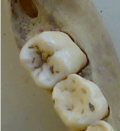
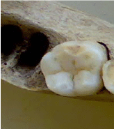
The data consisted of the skeletal remains of 56 sub-adults that had a total of 261 permanent teeth. The available 68 PFMs and 13 permanent incisors were examined regarding MIH. The inter-examiner reliability kappa statistics of the two examiners was 0.92. There were no demarcated opacities, or breakdowns (Figure1a). Many PFMs had grossly deformed cuspal architecture because of attrition however no breakdowns. Of the total number of individuals, only one PFM showed signs of MIH (Figure 1b).

Figure1a. Tooth 46 in a Byzantine child.

Figure 1b. Tooth 36 exhibits possible MIH in a Byzantine child.
Discussion
Living and dietary conditions may be a factor for MIH. Determination of aetiological factors of MIH is complicated as the child may have many medical problems in the first 3 years of life after birth. Frequent preschool age infections such as upper respiratory diseases, asthma, otitis media, tonsillitis, chickenpox, measles, and rubella, appear to be associated with MIH [3,6].
Evidence on herbal, veterinary and chemical substances used in various forms for respiratory problems of childhood such as acute otitis, acute tonsillitis and parotitis was investigated in the Byzantine medical treatizes, from the 4th to the 15th c. AD. Byzantine physicians recognized the main otolaryngological problems of childhood, comprising of acute otitis, acute tonsillitis and parotitis. DuringtheByzantineperiodtherearelimitedreferencesaboutotitisofchildhood, however, it seems that it was considered to be a common and not so much dangerous disease with milder treatment methods. The problem of asthma in childhood was also well known during the Byzantine period [21].
It has been stated that children with poor general health and systemic conditions are more likely to have developmental defects of enamel [22-23]. It has been calculated that the average life expectancy in Byzantium was about thirty-five years [24]. Also the population from Niceae analysed in the present research had a short life span, as they were found to be buried in a mass grave possibly killed during war or an attack [17] .The systemic conditions implicated in MIH to date include nutritional deficiencies, heart defects, brain injury and neurologic defects, cystic fibro sis, syndromes of epilepsy and dementia, nephritic syndrome, atopia, lead poisoning, repaired cleft lip and palate, radiation treatment, rubella embryopathy, epidermolysis bullosa, ophthalmic conditions, celiac disease, and gastrointestinal disorders [22]. In todays modern world, medical conditions give many infants to survive where we could be detecting MIH in later years. Regarding medical conditions, an animal study, Kuscu et al [25] described the range of mineral densities of enamel specimes from three groups of piglets where two groups were given different doses of amoxicillin in infancy using a X-ray micro tomography model. This study highlights some lower scores indicating hypo mineralisation regarding antibiotic usage. Archaeological, literary and documentary evidence all at test to the high rate of infant and child mortality in Byzantium, which may have approached fifty percent by age five. Although the almost universal practice of breast-feeding must have provided children with a relatively safe supply of milk and built up their antibodies, they were still pronetomal nutrition and anemia, and many no doubt died from diarrheal diseases and infections [26]. This might give us an idea it seems hard to observe MIH in the present population.
The skeletal remains studied in the present research were excavated from the site of Roman anphitheater in Iznik (Niceae) during archaeological excavations conducted at the end of 1984 by Ozbek[17]. There mains were stored in the Department of Anatomy, Uludag University Medical School since 1984. More than half of the total sample was in a state mild and good preservation. The distribution of individuals according to age groups depends on the average life span of the population. The average life span reflects numerous socio-economic factors. The harder the living conditions are, the shorter the life span. This might give us a chance to evaluate MIH easier in the present Byzantine population where age range should be determined as sub-adult. It was recently stated that eight years of age was the best time for examination of MIH [27].
In the present Byzantine population the frequency of caries was 6.8% [18].The present data guides us that this community was a mixed community that lived from hunting, fishing as well as agriculture [18,27]. Climatic factors and the lay of the land clearly influenced agricultural production in Byzantine. Not to be compared with modern western diet; the fertile low lands and river valleys of western Asia Minor and coasts of Marmara sea, possibly made it available for the present samples consume variety of food type in respect of many other societies in the 13th c. High wear in the present archeological population can be linked to the fact that the cumulative effects of attrition as a result of the Byzantine diet. The present situation also may have masked MIH, if present in Byzantine. Regarding type of diet, the empire offered a full range of typical Mediterranean diet [18]. This fact gives us the idea that extractions due to dental caries and MIH relation should be irrelevant.
Children’s developing teeth may be sensitive to environmental pollutants such as polychlorinated dibenzo-p-dioxins (PCDDs) and polychlorinated dibenzofurans (PCDFs). While MIH is a multifactorial disturbance, there might be a relationship between environmental pollutants and MIH [28]. It is obvious that environmental pollutants in 13th c would be water pollution, coal burning and mining origin rather than PCDDs and PCDFs [29]. However environmental factors in Byzantine was a dilemma where water pollution was a legal matter and even children consumed wine as main beverage.
Our findings are in contrast with Ogden et al [14] who observed group of 17th and 18th century English sub-adults exhibits a high prevalance of (MIH). This could be due to the nature of two different cultures, diets and time period.
Another point to be noticed is the evaluation criteria of MIH. Actually permanent incisors and first permanent molars would be examined wet for demarcated opacities, post-eruption breakdown (PEB), and atypical restorations according to Weerjijm et al [19]. However in the present study the samples were evaluated in dry conditions as teeth are part of an archeological collection of Uludag University and any attempt to alter the conditions of preserved data would be an ethical issue.
Future research will further investigate the formation and cause of MIH and will potentially guide us why this condition was so dramatically present or absent in different archaeological populations.
In conclusion, our findings confirm that MIH was a rare dental anomaly in Byzantine period.
References
- Koch G, Hallonsten AL, Ludvigsson N, Hansson BO, Holst A, et al (1987) Epidemiologic study of idiopathic enamel hypomineralization in permanent teeth of Swedish children. Community Dent Oral Epidemiol 15:279-285. [Crossref]
- Weerheijm KL, Jalevik B, Alaluusua S (2001) Molar-incisor hypomineralization. Caries Res 35:390-391. [Crossref]
- Jalevik B, Noren JG (2000) Enamel hypomineralization of permanent first molars: A morphological study and survey of possible aetiological factors. Int J Paediatr Dent 10:278-289. [Crossref]
- Fearne J, Anderson P, Davis GR (2004) 3D X-ray microscopic study of the extent of variations in enamel den sity in first permanent molars with idiopathic enamel hypomineralization. Br Dent J 196:634-638. [Crossref]
- Croll TP (1991) Creating the appearance of white enamel dysmineralization with bonded resins. J Esthet Dent 3:30-33. [Crossref]
- Holtta P, Kiviranta H, Leppaniemi A, Vartiainen T, Lukinmaa PL, et al (2001) Developmental dental defects in children whore side by a river polluted by dioxins and furans. Arch Environ Health 56:522-528. [Crossref]
- Leppaniemi A, Lukinmaa PL, Alaluusua S (2001) Nonfluoride hypomineralizations in the permanent first molars and their impact on the treatment need. Caries Res 35:36-40. [Crossref]
- Van Amerongen WE, Kreulen CM (1995) Cheese molars: A pilot study of the etiology of hypocalcifications in first permanent molars. J Dent Child 62:266-269. [Crossref]
- Weerheijm KL, Groen HJ, Beentjes VE, Poorterman JH (2001) Prevalence of cheese molars in 11-year-old Dutch children. J Dent Child 68:259-262. [Crossref]
- Jalevik B, Klingberg GA (2002) Dental treatment, dental fear and behavior management problems in children with severe enamel hypomineralization of their permanent first molars. Int J Paediatr Dent 12:24-32. [Crossref]
- Waldron HA (2001) Are plague pits of particular use to palaeo epidemiologists? Int J Epidemiol 30:104-108.
- Vodanovic M, Brkic H, Slaus M, Demo Z (2005) The frequency and distribution of caries in the mediaeval population of Bijelo Brdo in Croatia (10th-11th century). Arch Oral Biol 50:669-680. [Crossref]
- Watt ME, Lunt DA, Gilmour WH (1997) Caries prevalence in the permanent dentition of a mediaeval population from the south-west of Scotland. Arch Oral Biol 42:601-620. [Crossref]
- Ogden AR, Pinhasi R, White WJ (2008) Nothing new under the heavens: MIH in the past? Eur Arch Paediatr Dent 9:166-171. [Crossref]
- Angold M (1975) A Byzantine Government in Exile: Government and Society under the Laskarids of Nicaea (1204-1261), Oxford. 109-111.
- Laiou AE (2002) The Economic History of Byzantium: From the Seventh through the Fifteenth Century. Washington DC: Dumbarton Oaks Research Library and Collection. 502.
- Özbek M (1984) Roma açık hava tiyatrosundan (İznik) çıkartılan Bizans iskeletleri. Hacettepe Üni Edebi Fak Derg 2:81-89.
- Caglar E ,Kuscu OO, Sandalli N, Ari I (2007) Prevalence of dental caries and tooth wear in a Byzantine population ( 13th c. AD) from northwest Turkey. Arch Oral Biol 52:1136-1145. [Crossref]
- Weerheijm KL, Duggal M, Mejare I (2003) Judgement criteria for molar incisor hypomineralization (MIH) in epidemiologic studies: a summary of the European meeting on MIH held in Athens. Eur J Paediatr Dent 4:110-113. [Crossref]
- Commission on Oral Health Research & Epidemiology (1992) A review of the developmental defects of enamel index (DDE Index). Commission on Oral Health, Research & Epidemiology. Report of an FDI Working Group. Int Dent J 42:411-426. [Crossref]
- Ramoutsaki IA, Dimitriou H, Markaki EA, Kalmanti M (2002) Management of childhood diseases during the Byzantine period: III-respiratory diseases of childhood. Pediatr Int 44:460-462. [Crossref]
- Hall R (1989) The prevalence of developmental defects of tooth enamel (DDE) in a paediatric hospital department of dentistry population (part I). Adv Dent Res 3:114-119. [Crossref]
- Kuscu OO, Caglar E, Sandallı N (2008) The prevalence and aethiology of Molar-Incisor Hypomineralization (MIH) in a group of children, Istanbul. Eur J Ped Dent 9:139-144.
- Talbot AM (1984) Old Age in Byzantium. Byzantin ische Zeits chrift 77:267-278.
- Kuscu OO, Sandalli N, Dikmen S, Ersoy O, Tatar I, et al (2013) Association of amoxicillin use and molar incisor hypomineralization in piglets: Visual and mineral density evaluation. Arch Oral Biol 58:1422-1433. [Crossref]
- Talbot AM (2006) The Death and Burial of Children. Abstract. 2006 Spring Symposium. Dumbarton Oaks Research Library and Collection, Washington DC.
- Hobdell MH, Oliveira ER, Bautista R, Myburgh NG, Lalloo R, et al (2003) Oral diseases and socio-economic status (SES). Br Dent J 194:91-96. [Crossref]
- Kuscu OO, Çaglar E, Aslan S, Durmusoglu E, Karademir A, et al (2009) The prevalence of Molar-Incisor Hypomineralization (MIH) in a group of children in a highly polluted urban region and a wind farm-green energy island: Is there any environmental association regarding MIH? Int J Paediatr Dent 19:176-185.
- Pyatt FB, Pyatt AJ, Walker C, Sheen T, Grattanc JP (2005) The heavy metal content of skeletons from an ancient metalliferous polluted area in southern Jordan with particular reference to bioaccumulation and human health. Ecotoxicol Environ Saf 60:295-300. [Crossref]


