Abstract
The shape, size, number, and structure of dental tissues can vary considerably between primary and permanent teeth. Primary teeth tend to exhibit fewer morphological abnormalities than permanent teeth. One of the rarest anomalies in primary dentition is the presence of bi-rooted canines. This report presents a rare case of bilateral bi-rooted maxillary canines in a 6-year-old Saudi male and includes a brief review of the existing literature on this unusual finding.
Keywords
bi-rooted primary canines, dental anomalies, root morphology, primary dentition
Introduction
While permanent teeth show considerable variation in form and structure, primary teeth are generally more consistent, particularly in their root morphology [1]. A primary canine's single root has been described as the most typical form of root morphology. However, only a few instances of having a bi-rooted primary canine have been reported in Japanese, African American, Caucasian, Pueblo Indian children, and Saudis with the first reported occurring in 1941 [2-6]. In this study, the occurrence of bilateral bi-rooted canines in a Saudi male was described, along with a review of the few existing literatures on this unusual and rare anomaly.
Case presentation
A 6-year-old Saudi male patient referred from one of the Primary Health Care centers in Dammam City to the Pediatric Dental Department at Dammam Medical Complex as a case of uncooperative behavior. The patient’s medical history did not reveal any medical illnesses. Intraoral clinical examination revealed a primary dentition with multiple carious teeth and no soft tissue abnormality was detected. Radiographic intraoral examination confirmed the clinical findings of tooth decays, and interestingly has shown a maxillary canines with two roots (Figures 1 and 2).
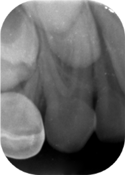
Figure 1. Periapical radiograph of the right maxillary primary canine showing a rare bifurcation of the root, consistent with a bi-rooted morphology
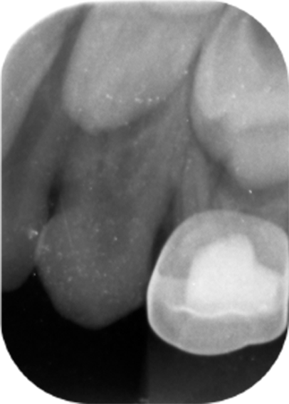
Figure 2. Periapical radiograph of the left maxillary primary canine demonstrating similar bifurcated root structure, indicating bilateral bi-rooted primary canines
Results and discussion
Additional or supernumerary roots are prevalent in the permanent dentition, with single-rooted canines and premolars typically being the most common. It is unusual for primary teeth to have extra roots. However, the published literature has documented over 18 cases of bi-rooted primary canines to date [5]. The only example from Saudi Arabia that has been documented in the literature on this uncommon finding up to date is by Assiry [7]. A 9-year-old kid who had bilateral incidence of bi-rooted canines in the maxillary arch was the subject of a case reported by Assiry [7]. Bi-rooted canines were present bilaterally in the maxillary and mandibular arches in the 6-year-old male. This is an uncommon finding that is the first of its sort to be reported in the Saudi population. Only five cases—one from Saudi Arabia—had been previously documented on the bimaxillary incidence of bi-rooted primary canines, according to a literature search. Remarkably, the bimaxillary incidences were limited to males. Based on the documented cases, the author can conclude that men are more prone to this oddity [7].
However, it has been showed that bifurcation of the bi-rooted primary canine occurs exclusively in the apical fourth of the roots and proposed that root elongation is accompanied by the possibility of producing extra/supernumerary roots [8,9]. Subsequently, it is also stated that root formation starts between 9 and 10 months after birth and may help initiate the differential growth of epithelial root sheath (Hertwig's) in teeth with multiple roots [10].
A careful examination of the primary root bifurcation and its position must be done prior to root canal treatment or extraction of multi-rooted teeth [11]. However, there is no reason and rationale to have an interim radiograph for such teeth, but it is advised to be in close evaluation during exfoliation [8,11].
Conclusion
In conclusion, it is of impertinence to note that prescribing a radiograph is needed even for the routine extraction of a primary canine. Observation of bi-rooted primary canines during development and growth may prevent problems, such as ectopic eruption or impaction or luxation of adjacent teeth during extractions. This report highlights a rare bilateral presentation of bi-rooted primary canines in a young Saudi male. Clinicians should be aware of this rare anomaly and consider radiographic assessment prior to the extraction of primary canines to avoid potential complications. Awareness and early detection of such variations can improve treatment planning and outcomes.
Conflicts of interest
The author declares no conflicts of interest.
References
- Talebi M, Parisay I, Khorakian F, Bagherian M (2010) Bi-rooted primary maxillary canines: A case report. J Dent Res Dent Clin Dent Prospects 4: 101-103. [Crossref]
- Muchizuki K, Ohtawa Y, Kubo S, Machida Y, Yakushiji M, et al. (2001) Bifurcation, birooted primary canines: A case report. Int J Paediatr Dent 11: 380-385. [Crossref]
- Atac AS, Cetinguc A (2005) Primary maxillary bi-lateral birooted canines: Report of two cases. Hacettepe Dishekimligi Fa- kultesi Dergisi 29: 24-28.
- Orhan AI, Sari S (2006) Double-rooted primary canines: A report of three cases. J Indian Soc Pedod Prev Dent 24: 204-208. [Crossref]
- Dhanpal PK, King NM (2009) Bilateral maxillary bi-rooted primary canines: Report of case. J Clin Pediater Dent 34: 113-116. [Crossref]
- Ott NV, Ball RN (1996) Birooted primary canines: Report of three cases. Paediatr Dent 18: 328-330. [Crossref]
- Assiry A (2009) Bi-rooted primary maxillary canines: A case report. J Med Case Rep 13: 261. [Crossref]
- Morrow JW, Hylin DL (1993) Supernumerary rooted primary central incisors: Report of seven cases. ASDC J Dent Child 60: 337-338. [Crossref]
- John YKL, Ricky WKW (2006) Tanaka–Johnston mixed dentition analysis for southern Chinese in Hong Kong. Angle Orthod 76: 632-636. [Crossref]
- Sicher H, Bhaskar SN (1792) Orban’s Oral Histology and Embryology, 7th edn. CV Mosby Co, St. Louis. 1792: 31-36.
- Hayutin DJ, Ralstrom CS (1992) Primary maxillary bilateral birooted canines: Report of two cases. ASDC J Dent Child 59: 235-237. [Crossref]