Abstract
Objective: The importance of dental plaque removal for prevention and control of periodontal disease and caries is well established. Most remaining plaque is in the interproximal region, and dental caries and gingivitis are more likely in this limited region. The purpose of this study was to test a novel method for evaluating interproximal accessibility of caregivers’ tooth brushing by using eyeglasses that make an artificial plaque invisible.
Study design: Twenty dental professionals participated as tooth-brushing caregivers. Participants brushed dental models of primary dentition using five different toothbrushes to compare each toothbrush’s accessibility to interproximal surfaces. Three artificial primary teeth on the left mandible were covered with a red artificial plaque. Participants were asked to brush these teeth while wearing eyeglass with red lenses that made the artificial plaque invisible. After 10 seconds of brushing, both mesial and distal surfaces of the artificial teeth were photographed and the areas of remaining artificial plaque were measured from the photographs.
Results: Significant differences of remaining artificial plaque areas were found among the five toothbrushes.
Conclusion: This novel method can effectively evaluate the accessibility of toothbrushes to interproximal surfaces.
Key words
oral hygiene, tooth brushing, toothbrush, caregiver, interproximal surface, invisibility
Introduction
The importance of dental plaque removal for prevention and control of periodontal disease and caries is well established [1]. In general, upper and lower labial surfaces have been found to harbor the least amount of plaque, while posterior teeth and interproximal surfaces have the greatest accumulation. In particular, the interproximal area is the site with the highest risk of developing both diseases, and the importance and effectiveness of interproximal plaque control has been reported [2,3].
For most people, mechanical plaque removal by manual tooth brushing remains the most common method for plaque control [4]. However, most brushing techniques commonly employed are not particularly suitable for cleaning interproximal areas because of difficulty with access [5], thus it is important to design toothbrushes to compensate and provide better access to those areas as much as possible.
To improve access to hard-to-reach areas of the mouth, modifications have been made to brush head angle, bristle material, and tuft arrangement, with several laboratory studies conducted to evaluate the effects of such toothbrush developments on simulated plaque removal, though clinical relevance has yet to be directly shown [6]. Clinical comparisons are limited, because relatively few studies have been conducted over a sufficient duration to assess the effects of such toothbrush modifications on gingival health [7,8].
Younger [9], handicapped [10], and elderly [11,12] individuals often have their teeth brushed by caregivers, as they are generally unable to adequately provide for their own oral hygiene care, especially with increasing levels of dependency [13]. Most caregivers who assist housebound individuals have not received formal training in oral care and several studies have reported poor oral hygiene in relation to dependence [13,14]. As a result, many researchers have noted that enhanced educational and training programs for this area are crucial [15-17].
For these reasons, tooth brushing provided by caregivers is an important topic in the dentistry, though it seems to have been relatively neglected. We have developed a novel method for evaluating the interproximal accessibility of tooth brushing performed by a caregiver as well as for providing training. With our method, the caregiver brushes the teeth of a dental model while wearing special “invisibility” eyeglasses that do not allow visualization of dental plaque by the wearer. The teeth of the dental model are coated with artificial plaque and the caregiver performs brushing as usual, though is unable to see the artificial plaque when wearing the eyeglasses. Upon removal of the glasses, they can see the results of their tooth brushing, i.e, remaining artificial plaque (RAP). We believe that instructions and training for oral hygiene based on this method and revealing RAP are very helpful for altering the patterns of tooth brushing by caregivers, thus making their care more effective.
Accordingly, the hypothesis of this study was that this novel method that incorporates the use of invisibility eyeglasses is effective for evaluating the interproximal accessibility of tooth brushing provided by caregivers.
Materials and methods
Study population
The participants in this study were ten right-handed female dentists and dental hygienists, respectively. Dentists and dental hygienists were selected because they represent the most-trained professional for tooth brushing. All participants were given oral and written information about the purpose and detailed procedures of the study. Subjects’ average age was 34 years, 7 months (range: 25 to 50 years of age) for the dentists and 37 years, 8 months (range: 23 to 47 years of age) for the dental hygienists. The study protocol received approval from the Ethical Committee of the Faculty of Dentistry, Niigata University, (25-R23-10-04), and each subject was given informed consent before participating in the study.
Study design and procedures
Toothbrushes Examined
Five different types of soft-bristled toothbrushes were examined in this study. The test toothbrushes were chosen to represent a variety of bristle designs mainly sold and used in Japan: Toothbrush A: wave and tapered-end, B: rippled, C, D, E: traditional flat-trim (Figures 1a, and 1b; Table 1). All toothbrushes were purchased from a retail store, except the American Dental Association (ADA) approved toothbrush (E) made in the United States, which was bought from a website.
Table 1. Size and characteristic of used toothbrushes.
Brushes |
Total Length |
Head |
Bristle |
No. of Tufts |
Length |
Width |
Height |
Length |
Diameter |
A |
143.5 |
18.1 |
10.9 |
4.5 |
8.5* |
0.203 |
30 |
B |
143.7 |
20.2 |
8 |
4.5 |
6.0-7.0** |
0.178 |
17 |
C |
136 |
17 |
8.2 |
4 |
7.5 |
0.1524 |
21 |
D |
141 |
17.5 |
8.2 |
4.5 |
6.5 |
0.1 |
17 |
E |
122.9 |
22.23 |
10.8 |
5 |
11 |
0.178 |
25 |
| |
|
|
|
|
|
|
|
|
unit:mm (data obtained from each company)
*:maximum length, **:wave-shape longest 7.0 mm, wave-shape shortest: 6.0 mm
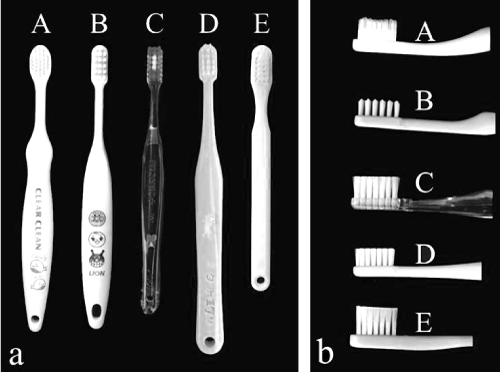
Figure 1. Five kinds of toothbrushes examined. (a) Whole shapes of the toothbrushes. (b) Head and bristle designs.
Testing procedures
Brushed teeth: In order to prepare for the tests, the mandibular left primary canine (tooth number #73 in FDI-two digit system), first primary molar (#74), and second primary molar (#75) of a D75-950 model (a maxillary and mandibular primary teeth dental model, NISSIN DENTAL PRODUCTS INC., Kyoto, Japan) were coated with a Whiteboard Marker (WBMA-40RF, PILOT CORPORATION, Tokyo, Japan) (Figure 2a). This artificial plaque is a red alcohol-based ink. The teeth were then returned to the tooth/jaw model, which was fastened to a dental mannequin (Trunk Unit Set, NISSIN DENTAL PRODUCTS INC., Kyoto, Japan). This mannequin was set on a dental chair in the supine position.
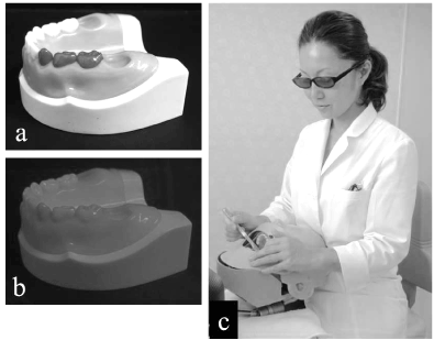
Figure 2. Brushed models and the mannequin. (a) Dental model with teeth #73, #74 and #75 coated with the artificial plaque. (b) Artificial plaque made invisible. (c) Experimental task.
“Invisibility” eyeglasses and tooth brushing: Participants were asked to brush the above-mentioned three primary teeth (#73, #74 and #75) for 10 seconds while wearing eyeglasses with red lenses that made the artificial plaque invisible. The lenses of these eyeglasses were of red plastic sheet (B5 Drip mat G-73 Transparence Red, DAISO, Higashi-Hiroshima City, Japan) (Figures 2b and 2c). No other instructions were given to each subject. This 10-second experimental tooth brushing was carried out five times by each participant. Prior to the experiment, subjects were allowed to brush these teeth for one minute to become accustomed to the experimental conditions with an arbitrary toothbrush and the mannequin, but without the artificial plaque or the eyeglasses. The order of the five toothbrushes was decided by random-sampling numbers for each participant.
Analyzing procedures
Photographing the remaining artificial plaque (RAP): Following brushing, the three teeth (#73, #74 and #75) were removed from the D75-950 model (Figure 3a). Each tooth was set on a special table to take photographs for measurement of the remaining artificial plaque (Figure 3b). The camera had a fixed bar and the table could rotate. Rotation of the table could repeatedly set the teeth in the same position relative to the camera, and also could rotate the fixed tooth to display each surface (Figure 3c). Consequently, a total of 600 photographs (i.e., 20 participants, 5 toothbrushes, 6 surfaces) were taken to measure the remaining artificial plaque areas (RAPAs).
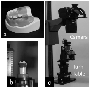
Figure 3. Taking photographs for measurement of RAPA. (a) Removal of brushed teeth from the model. (b) A tooth set on the rotating table. (c) Each surface is photographed in standardized orientation.
Definition of interproximal area template of RAP: Prior to measuring, the area of interest in the interproximal area was defined within the range between the occlusal line (Figure 4a) and the cervical line (Figure 4b) of each surface. Using Adobe Photoshop CS6 Ver. 13 (Adobe Systems Japan Inc., Tokyo, Japan), an “interproximal area template” (Figures 4c and 4d) for each mesial and distal surface of the three teeth was made and applied to the photographs, as shown in Figures 5a-5c. The RAPA was also calculated using this software.
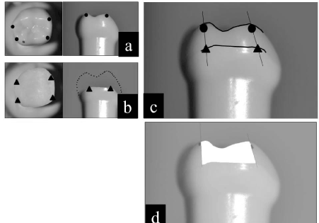
Figure 4. Definition of the interproximal area. (a) Points to define the occlusal line. (b) Points to define the cervical line. (c) Defined interproximal area. (d) Template for the interproximal area.
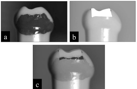
Figure 5. Definition of the remaining artificial plaque area (RAPA). (a) Remaining artificial plaque (RAP). (b) Template for the interproximal area. (c) Red area indicates the RAPA, i.e., the overlap area of both the RAP and template.
Statistical analysis: Two-way factorial ANOVA and Bonferroni statistical analyses were applied to compare the RAP areas among the 5 different toothbrushes. A p-value of 0.05 or less was considered to be statistically significant for all tests.
Results
Prior to comparing differences of the RAPAs among toothbrushes, the results for the 10 dentists and 10 dental hygienists were compared. No differences were found among the surfaces; therefore, results for both professional groups were combined.
Means and standard deviations of RAPAs on the six interproximal surfaces after tooth brushing with the five different toothbrushes are shown in Table 2. Toothbrush “A” exhibited the smallest RAPAs (in other words had the largest penetration area) on four of the six examined interproximal surfaces. The two exceptions were the distal surface of #73 and the mesial surface of #74. Toothbrush “E”, the ADA approved toothbrush, produced the smallest RAPAs for these two surfaces. With regard to the sum of the six surfaces, toothbrush “A” (mean ± SD: 8.02 ± 3.43) also had the best interproximal penetration, and toothbrush “E” (9.45 ± 3.65) had the second best. The largest total RAPA was observed with toothbrush “D” (12.55 ± 4.06).
Table 2. Measured remaining artificial plaque areas (RAPAs).
Brushes |
73 medial |
73 distal |
74 medial |
74 distal |
75 medial |
75 distal |
Sum of 6 surfaces |
A |
1.05±0.69 |
0.91±0.84 |
2.25±1.33 |
1.30±0.98 |
0.83±0.64 |
1.68±1.42 |
8.02±3.43 |
B |
1.68±1.10 |
1.13±0.81 |
2.70±1.52 |
1.40±0.91 |
1.77±1.21 |
1.82±1.08 |
10.49±3.42 |
C |
1.70±1.00 |
1.43±1.30 |
2.80±1.56 |
1.94±1.19 |
1.64±1.04 |
2.29±0.98 |
11.80±3.78 |
D |
1.51±0.69 |
1.20±1.06 |
3.01±1.38 |
2.16±1.43 |
1.86±1.66 |
2.81±1.38 |
12.55±4.06 |
E |
1.52±0.65 |
0.67±0.58 |
1.88±1.36 |
1.87±1.01 |
0.95±0.74 |
2.56±1.33 |
9.45±3.65 |
unit:mm2, mean ± SD,
A two-way ANOVA statistical evaluation of the five toothbrushes (Model A) and twenty participants (Model B) is presented in Table 3. Significant differences among the five toothbrushes were found at the distal surface of #73 (P=0.012), the mesial surface of #74 (P=0.02), the mesial (P = 0.002) and distal surfaces (P = 0.007) of #75, and also the sum of all six surfaces (P = 0.000). In addition, Bonferroni statistical pairwise comparisons (Tables 4 and 5) detected that Toothbrush “E” was significantly superior to toothbrush “C”(P=0.009) at the distal surface of #73, and also to toothbrush “D”(P=0.028) at the mesial surface of #74. At the mesial surface of #75, differences between pairs of toothbrushes “A” vs “B”(P=0.037), “A” vs “D”(P=0.015), and “E” vs “D”(P=0.047) were significant. Also, toothbrush “A” was superior to toothbrush “D”(P=0.016) at the distal surface of #75. Moreover, for the sum of the six surfaces, the differences between “A” vs “B” (P=0.037), “A” vs. “C” (P = 0.000), “A” vs. “D” (P = 0.000) and “E” vs “D” (P=0.003) were also statistically significant.
Table 3. ANOVA statistical evaluation of the RAPAs.
|
DF |
Sum of Square |
F-ratio |
P-value |
DF |
Sum of Square |
F-ratio |
P-value |
|
#73 medial surface |
#73 distal surface |
Model A
Model B |
4
19 |
5.53
20.25 |
2.195
1.693 |
0.078
0.056 |
4
19 |
6.64
49.47 |
3.477
5.451 |
0.012*
0.000** |
Error |
76 |
47.86 |
|
|
76 |
36.30 |
|
|
Total |
99 |
73.64 |
|
|
99 |
92.41 |
|
|
|
#74 medial surface |
#74 distal surface |
Model A
Model B |
4
19 |
16.76
92.53 |
3.105
3.611 |
0.020*
0.000** |
4
19 |
10.77
35.62 |
2.473
1.722 |
0.051
0.051 |
Error |
76 |
102.52 |
|
|
76 |
82.74 |
|
|
Total |
99 |
211.81 |
|
|
99 |
129.13 |
|
|
|
#75 medial surface |
#75 distal surface |
Model A
Model B |
4
19 |
18.70
44.64 |
4.782
2.404 |
0.002**
0.004** |
4
19 |
18.61
56.439 |
3.864
2.467 |
0.007**
0.003** |
Error |
95 |
74.30 |
|
|
76 |
91.49 |
|
|
Total |
99 |
137.64 |
|
|
99 |
166.54 |
|
|
|
Sum of six surface |
|
|
|
|
Model A
Model B |
4
19 |
262.42
766.69 |
9.661
5.942 |
0.000**
0.000** |
|
|
|
|
Error |
76 |
516.09 |
|
|
|
|
|
|
Total |
99 |
1545.19 |
|
|
|
|
|
|
DF: degree of freedom, * P < 0.05, ** P < 0.01
Table 4. P-values between toothbrushes based on Bonferroni statistical analysis of the #73 distal, #74 medial, #75 medial and #75 distal surface, respectively.
#73 distal |
A |
B |
C |
D |
E |
#74 medial |
A |
B |
C |
D |
E |
A |
|
1.000 |
0.214 |
1.000 |
1.000 |
A |
|
1.000 |
1.000 |
0.425 |
1.000 |
B |
|
|
1.000 |
1.000 |
0.398 |
B |
|
|
1.000 |
1.000 |
0.269 |
C |
|
|
|
1.000 |
0.009** |
C |
|
|
|
1.000 |
0.142 |
D |
|
|
|
|
0.175 |
D |
|
|
|
|
0.028* |
E |
|
|
|
|
|
E |
|
|
|
|
|
|
|
|
|
|
|
|
|
|
|
|
|
#75 medial |
A |
B |
C |
D |
E |
#75 distal |
A |
B |
C |
D |
E |
A |
|
0.037* |
0.110 |
0.015* |
1.000 |
A |
|
1.000 |
0.820 |
0.016* |
0.127 |
B |
|
|
1.000 |
1.000 |
0.107 |
B |
|
|
1.000 |
0.053 |
0.339 |
C |
|
|
|
1.000 |
0.292 |
C |
|
|
|
1.000 |
1.000 |
D |
|
|
|
|
0.047* |
D |
|
|
|
|
1.000 |
E |
|
|
|
|
|
E |
|
|
|
|
|
*P < 0.05, ** P < 0.01
Table 5. P-values between toothbrushes based on Bonferroni statistical analysis of the sum at six all surfaces.
Brushes |
A |
B |
C |
D |
E |
A |
|
0.037* |
0.000** |
0.000** |
0.877 |
B |
|
|
1.000 |
0.146 |
1.000 |
C |
|
|
|
1.000 |
0.056 |
D |
|
|
|
|
0.003** |
E |
|
|
|
|
|
* P < 0.05, ** P < 0.01
Discussion
The purpose of this study was to test the efficacy of a novel method for evaluating interproximal accessibility of caregivers’ tooth brushing by using “invisibility” eyeglasses that make an artificial plaque invisible. Differences of RAPAs among toothbrushes were found in this study. In addition, the pairwise comparisons revealed that these differences were observed at some specific surfaces. Therefore, each toothbrush might have its own characteristics for interproximal penetration.
Advantages of the method used in this study
Numerous attempts have been carried out to evaluate the penetration of interproximal surfaces both with manual toothbrushes [6,18-22] and powered toothbrushes [23-25]. Some studies used a brushing machine or robot to reproduce tooth brushing motion [6,18,21,23], compare brushing methods [21,22], compare several toothbrushes [6,18,19,21,23], and compare several interproximal surfaces [18,22]. These studies used an artificial dental model (typodont) and artificial plaque because precise evaluation of the efficacy of toothbrush penetration into interproximal surfaces requires an in vitro study. The simulated tooth morphology and the position of the teeth in the dental model used in this study provided realistic interdental embrasures, and the setting of our template enabled precise evaluation of interproximal penetration by the different toothbrushes.
On the other hand, the angles of the bristles relative to the tooth surfaces and brushing pressure are naturally subjected to some immeasurable variation because the human brain controls tooth-brushing motion. In addition, caregivers’ tooth brushing motion has not yet been clarified, therefore, these factors have to be considered under in vivo conditions.
The method in this study can evaluate the interproximal accessibility of clinical tooth brushing motion with the same precision as in vitro, therefore, this method is regarded as a semi-in vivo condition.
Although we usually increase motivation for tooth brushing and teach effective brushing methods by visualizing the plaque, brushing under such full visual control differs from usual brushing. The development of “invisibility” eyeglasses easily allows reproduction of caregivers’ usual brushing by hiding the artificial plaque from them during tooth brushing.
To our knowledge, this is the first study to evaluate interproximal penetration by caregivers’ brushing, so we selected the most-trained professional for tooth brushing (i.e., dentists and dental hygienists) as the human subjects. This semi-in vivo method could be used to compare different kinds of caregivers, and also evaluate caregivers’ abilities before and after tooth-brushing instructions in future studies.
Another advantage of this method on clinical situation is that a caregiver can share RAPAs with a dental professional after tooth brushing by taking off the eyeglasses. Dental professionals may be able to effectively instruct and train each caregiver to reduce dental plaque from the dental model.
Meaning of the results in this study
Some previous studies focused on the evaluation of characteristics among toothbrushes. Yost showed traditional flat-trim toothbrushes might be at an inherent disadvantage in penetrating interproximally because of the curvature of the teeth [6,18,19,21,23]. Volpenhein demonstrated improved interproximal penetration of rippled-pattern bristle designs compared to flat-tufted bristle designs [21,22].
The current study proved able to measure the efficacy of interproximal penetration by examined toothbrushes with a primary dentition dental model. Toothbrush “A” exhibited the smallest RAPA for the sum of all six surfaces among the five kinds of toothbrushes. Bonferroni analysis showed that toothbrush “A” was significantly superior to toothbrushes “B”,”C” and “D”, and that toothbrush “E” was significantly superior to toothbrush “D”.
The bristle designs of toothbrushes in this study included, toothbrush “A” with wave and tapered-end bristles; toothbrush “B” with a rippled bristle; and toothbrushes ”C”, ”D” and ”E” with traditional flat-trim bristles. The study’s results indicate that the ability for interproximal penetration is not determined by bristle design alone.
There were two sets of mutual interproximal surfaces in this experimental study. One set was the distal surface of #73 and the mesial surface of #74; the other set was the distal surface of #74 and the mesial surface of #75. Taking these sets into consideration, detailed observation of the RAPAs disclosed that toothbrush “E” had the smallest RAPA among the five toothbrushes for the anterior set (i.e., on the distal surface of #73 and the mesial surface of #74), and toothbrush “A” had the smallest RAPA for the posterior set (i.e., on the distal surface of #74 and the mesial surface of #75). In addition, toothbrush “A” had the smallest RAPA on the distal surface of #75.
The results for these two types of toothbrushes might suggest requirements for interproximal penetration for these two-different mutual interproximal surfaces. In other words, changes in interproximal accessibility can be complicated by not only the characteristics of the toothbrushes (e.g., form of bristles, the length and diameter of the filaments, and number of tufts), but also the environment such as the position of the tooth in the dentition and the tooth’s form.
Tooth brushing by a caregiver
The number of types of toothbrushes that can be used by caregivers for care-receivers is limited, even for younger children. Several articles have pointed out that oral care should be included in nursing work as a more highly prioritized task, and nurses’ knowledge needs to be enhanced especially in facilities for elderly and handicapped persons [26-28]. To begin with, should a caregiver brush the care-receiver’s mouth from his/her back or front? This may depend on the situation of the care-receiver or on the manner used. However, sides, directions, motions and forces of tooth brushing will be completely different for a caregiver between these two situations. Does the same toothbrush show the same efficiency in both situations?
Further studies are required to answer these questions and thereby improve oral health for care-receivers.
Conclusion
The results of this novel method for evaluating interproximal accessibility of caregivers’ tooth brushing using “invisibility” eyeglasses showed that the method can effectively evaluate the accessibility of toothbrushes to interproximal surfaces. In addition, this method also might be able to evaluate caregivers’ abilities before and after tooth brushing instructions have been given in the dental clinic and in dental school.
Conflicts of interest
The authors declare that they have no conflict of interests.
References
- Shory NL, Mitchell GE, Jamison HC (1987) A study of the effectiveness of two types of toothbrushes for removal of oral accumulations. J Am Dent Assoc 115: 717-720. [Crossref]
- Schätzle M, Löe H, Bürgin W, Anerud A, Boysen H, et al. (2003) Clinical course of chronic periodontitis. I. Role of gingivitis. J Clin Periodontol 30: 887-901. [Crossref]
- Lang WP, Ronis DL, Farghaly MM (1995) Preventive behaviors as correlates of periodontal health status. J Public Health Dent 55: 10-17. [Crossref]
- Furuichi Y, Lindhe J, Ramberg P, Volpe AR (1992) Patterns of de novo plaque formation in the human dentition. J Clin Periodontol 19: 423-433. [Crossref]
- Benson BJ, Henyon G, Grossman E (1993) Clinical plaque removal efficacy of three toothbrushes. J Clin Dent 4: 21-25. [Crossref]
- Beals D, Ngo T, Feng Y, Cook D, Grau DG, et al. (2000) Development and laboratory evaluation of a new toothbrush with a novel brush head design. Am J Dent 13: 5A-14A. [Crossref]
- Gammack JK, Pulisetty S (2009) Nursing education and improvement in oral care delivery in long-term care. J Am Med Dir Assoc 10: 658-661. [Crossref]
- Broadbent JM, Thomson WM, Boyens JV, Poulton R (2011) Dental plaque and oral health during the first 32 years of life. J Am Dent Assoc 142: 415-426. [Crossref]
- Chan SC, Tsai JS, King NM (2002) Feeding and oral hygiene habits of preschool children in Hong Kong and their caregivers' dental knowledge and attitudes. Int J Paediatr Dent 12: 322-331. [Crossref]
- Stefanovska E, Nakova M, Radojkova-Nikolovska V, Ristoska S (2010) Tooth-brushing intervention programme among children with mental handicap. Bratisl Lek Listy 111: 299-302. [Crossref]
- De Visschere L, de Baat C, Schols JM, Deschepper E, Vanobbergen J (2011) Evaluation of the implementation of an 'oral hygiene protocol' in nursing homes: A 5-year longitudinal study. Community Dent Oral Epidemiol 39: 416-425. [Crossref]
- Urrutia CG, Ormazabal FR, Santander IE, Salvo DM (2012) Oral health practices and beliefs among caregivers of the dependent elderly. Gerodontology 29: e742-747. [Crossref]
- Kandelman D, Petersen PE, Ueda H (2008) Oral health, general health, and quality of life in older people. Spec Care Dentist 28: 224-236. [Crossref]
- Stein PS, Henry RG (2009) Poor oral hygiene in long-term care. Am J Nurs 109: 44-50. [Crossref]
- Wardh I, Andersson L, Sorensen S (1997) Staff attitudes to oral health care. A comparative study of registered nurses, nursing assistants and home care aides. Gerodontology 14: 28-32. [Crossref]
- Sumi Y, Nakamura Y, Nagaosa S, Michiwaki Y, Nagaya M (2001) Attitudes to oral care among caregivers in Japanese nursing homes. Gerodontology 18: 2-6. [Crossref]
- Thean H, Wong ML, Koh H (2007) The dental awareness of nursing home staff in Singapore - a pilot study. Gerodontology 24: 58-63. [Crossref]
- Volpenhein DW, Walsh ME, Dellerman PA, Burkett TA (1994) A new method for in vitro evaluation of the interproximal penetration of manual toothbrushes. J Clin Dent 5: 27-33. [Crossref]
- Yost KG, Miluszewski KF, Chen AC (1994) Laboratory evaluations of toothbrush interproximal penetration ratios. J Clin Dent 4: 125-127. [Crossref]
- Volpenhein DW, Hartman WL (1996) A comparative evaluation of the in vitro penetration performance of the improved crest complete toothbrush versus the Colgate total toothbrush and the Oral-B Advantage toothbrush. J Clin Dent 7: 101-105. [Crossref]
- Volpenhein DW, Handel SE, Hughes TJ, Wild J (1996) A comparative evaluation of the in vitro penetration performance of the improved Crest complete toothbrush versus the Current Crest complete toothbrush, the Colgate Precision toothbrush and the Oral-B P40 toothbrush. J Clin Dent 7: 21-25. [Crossref]
- Bruun C, Ekstrand KR, Andreasen KB (1998) A new in vitro method for testing the interproximal cleaning potential of toothbrushing. J Clin Dent 9: 11-15. [Crossref]
- Driesen GM, Warren PR, Hilfinger P, Ernst CP, Willershausen B (1996) The development of the Braun Oral-B Ultra Plaque Remover: an in vitro robot study. Am J Dent 9 Spec No: S13-17. [Crossref]
- Ernst CP, Willershausen B, Driesen G, Warren PR, Hilfinger P (1997) A robot system for evaluating plaque removal efficiency of toothbrushes in vitro. Quintessence Int 28: 441-445. [Crossref]
- Heins P, Bartizek RD, Walters PA, Biesbrock AR (2002) Plaque removal efficacy of a battery-operated power toothbrush compared to two control manual toothbrushes in single use studies. Am J Dent 15 Spec No:28A-32A. Epub 2003/01/07. [Crossref]
- Adams R (1996) Qualified nurses lack adequate knowledge related to oral health, resulting in inadequate oral care of patients on medical wards. J Adv Nurs 24: 552-560. [Crossref]
- Frenkel H, Harvey I, Newcombe RG (2000) Oral health care among nursing home residents in Avon. Gerodontology 17: 33-38. [Crossref]
- Lindqvist L, Seleskog B, Wardh I, von Bultzingslowen I (2013) Oral care perspectives of professionals in nursing homes for the elderly. Int J Dent Hyg 11: 298-305. [Crossref]