We presented a case of 65- year-old man who complained of intestinal obstipation and bowel habitus. Colonoscopy and biopsy was revealed colon cancer. Laparotomy revealed triple tumoral mass in the left colon, which appeared separe. Histology of the masses revealed adenocarcinoma
colon cancer, multiple, synchronous
Multiple colon cancer(MCC) is defined as two or more cancers detected in the colon of a patient. It’s incidence is about 25%. Each tumor is independent than another tumor. The most advanced pathologically lesion is regarded as the index lesion. In some studies reported that 5year survival somewhat worse than for single colon tumors [1,2]. This report describes, a case, his medical history not revealed premalign conditions or hereditery polipoid lesions and who presented with triple colon cancer at laparotomy.
A 65-year-old man presented with abdominal pain on the left quadrant and obstipation was referred to our surgery clinic by the gastroenterologist for tumoral mass at the colon. He denied any medical history of intestinal disease. His rectal examination was unremarkable. Laboratory tests showed mild anemia. CT of abdomen revealed thickening bowel wall and tumoral mass affecting descending colon and sigmoid colon at two different levels. At colonoscopy, a tumoral mass, almost completely narrowing the lumen was found at 20 cm from the anal verge. Pathology of biopsy was reported as adenocarcinoma.
On laparatomy, a tumor was found that extending into the surrounding tissue at the upper rectum. Additionally, we identified two intraluminal tumoral masses within the left colon, with the palpation. We performed low anterior resection with left hemicolectomy.
Macroscopically triple (synchronous) colon tumor was seen (Figure 1). Microscopic examination of the specimen revealed first tumor (distal) was 3×1.3 cm in diameter and presented as ulceroinfiltrative narrowing form. A second tumor was 7.6×5×4.3 cm in diameter and third tumor was 5.7×3.5×2 cm in diameter, with a similar feature as fungating lesions. Histological section showed that three moderately differentiated adenocarcinoma with healthy colon tissue between them and three of 72 lymph nodes were positive for metastasis and 7 tumor nodule in the adipose tissue (pT3N1M0).
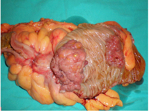
Figure 1. Specimen of left hemicolectomy showing multiple colon cancers (distal tumor in uncut bowel).
The postoperative course was uneventful. The patient denied adjuvant therapy. The patient was dead from coronary artery disease, 7 years after surgery without an apparent recurrent tumor.
Although MCC has been recognized as a clinical entity it’s clinical and pathological features is still contraversial. The concept of field cancerization was originally hypothesized to explain observations in relation to oral cancer, and is now proposed to occur in a variety of organs, including the colon. However oncologic mechanisms for this condition are still largely unknown, except for inflammation-associated cancerization. The incidence increase to 10-20% in patients with inflammatory bowel disease and polypoid diseases of the colon [3].
Obstruction of bowel in an adult without history of abdominal surgery often warrants prompt definitive diagnosis. Colonoscopy with biopsy is the diagnostic test choice for these cases. In cases with a new diagnosis of cancer, colonoscopy of the whole colon is necessary to detect another lesion. But the diagnosis of synchronous lesions can be difficult when the tumor preventing the passage of colonoscope. In this situation, CT imaging may aid in localization of the lesions and also, for staging of the cancer. If cancer diagnosed by biopsy, positron emission tomography should be used to determine the synchronous tumor/s located proximally [4].
In clinical practice, colon resection is often used to treat patients. But distribution of MCC is separated by at least one or more segment of the colon. This may be an indication of the importance of examination of the colon with manually palpation or intraoperative colonoscopy.
In the staging the cancer, index lesion, most advanced tumor histopathologically, is used to TNM classification. While some studies showed that the differently survival time for the MCC and sporadic cases, another studies reported similar time [1,2,4]. But survival time is related to the factors like tumor invasion, performance status of patients, and morbid illness etc. Case series are small and not enough to final decision.
It conclusion, all colon carcinomas require a complete pre- and per- operative examination of the large bowel. It must be remembered that more than one cancer will be found in the colon.
- Chen HS, Sheen-Chen SM (2000) Synchronous and ‘early’ metachronous colorectal adenocarcinoma. Dis Colon Rectum 43: 1093-1094.
- Kim YJ, Kim NK, Lee KY, Sohn SK, Min JS (2002) Clinicopathologic charecteristics of multiple primary colorectal cancer. J Korean Soc Coloproctol 18: 343-348.
- Yoon JW, Lee SH, Ahn BK, Baek SU (2008) Clinical characteristics of multipl primary colorectal cancers. Cancer Res Treat 40: 71-74.
2021 Copyright OAT. All rights reserv
- Yeh CC, Hsi SC, Chuu CP, Kao YH (2013) Synchronous triple carcinoma of the colon and rectum. World J Surg Oncol 11: 66.