Introduction
To illustrate a treatment of an impacted mandibular canine in a 14 years old, Kuwaiti girl.
Clinical presentation and intervention
The impacted canine was exposed through a surgical procedure then bonded with a gold chain attachment. Alignment of the impacted canine as well as treating other features of the malocclusion continued. After deboning the case, a lower fixed retainer was placed to prevent relapse of the canine.
Conclusion
The prevalence of impacted mandibular canines is lower than that of the maxillary canines. The management of impacted mandibular canine depends on several factors that were discussed in the article. These include the position of the age of the patient and the position of the tooth.
An impacted tooth can be defined as teeth are those with a delayed in eruption time or that are not expected to erupt completely based on clinical and radiographic assessment [1]. An impacted tooth in children is a major problem with potentially damaging squeal such as, damage to the adjacent teeth and cysytic formation. The prevalence of impacted maxillary canine is reported to be 1.5% [2], however, the prevalence of impaction of the mandibular canine is much lower [3,4]. In one particular study, the incidence of mandibular canine impaction was shown to be 1.29% in 5022 individuals of a Turkish population sample [5]. Clinicians should suspect impaction if the canine is not palpable in the buccal sulcus by the age of 10–11 years, hence a full clinical examination and radiographic assessment are essential in order to located the canines [6]. There are many etiological factors that can lead to the failure of eruption such as the presence of a supernumerary tooth that prevents the successful eruption of the canine. Early diagnosis of such problem will make the treatment simpler and in some cases shortens the treatment duration. The aim of this report is to illustrate a conventional orthodontic treatment of an impacted mandibular canine which was diagnosed late.
impacted canine, orthodontic treatment, surgical exposure
A 14 years old Kuwaiti girl attended the clinic complaining of “a gap between bottom teeth” (Figure 1). She was fit and well. She had a Class 1 skeletal pattern with average vertical proportions. In the intra-oral examination, it was found that, her oral hygiene was fair and required improvement prior to the orthodontic treatment (Figure 2). Furthermore, there was mild crowding in the mandibular arch and well aligned maxillary arch. The overjet was slightly increased with an average overbite and a mild central line discrepancy. The buccal segments were Class 1.
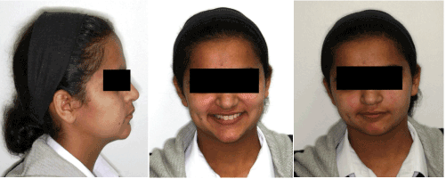
Figure 1. Pre-treatment extra-oral photographs.
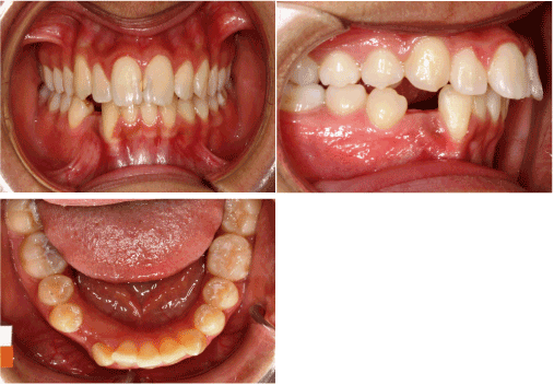
Figure 2. Pre-treatment intra-oral photographs.
All permanent dentition were present except for the lower right manbibular canine and the third molars. Further investigations revealed that the tooth was lingually displaced with no presence of any supernumerary or any other obstruction preventing its eruption (Figure 3). The treatment plan was to initiate a fixed orthodontic appliance therapy to create more space for the mandibular canine then carry on a surgical exposure procedure to uncover the tooth and enable its traction through the orthodontic appliance using a 0.22 inch slot, MBT prescription. Treatment started with leveling and alignment through 0.14 and 0.18 inch Nickel Nitatinum wires. Furthermore, the treatment continued through 0.19 x 0.25 NiTi and Stainless Steel archwires to achieve the treatment objectives (Figure 4). A retention regime was also planned which included a fixed retainer in the lower arch to minimize the risk of relapse of the impacted canine after alignment plus an upper and lower removable retainers.
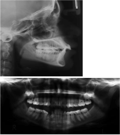
Figure 3. Pre-treatment radiographs.
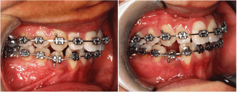
Figure 4. During the surgical exposure procedure and photographs of a following visit.
Although the impaction of a mandibular canine is not frequently occurring episode, those individuals with this problem may suffer a potentially harmful effects if left untreated. The impacted maxillary canine is extensively discussed and mentioned in the dental literature unlike the mandibular canine. The key factor here is [7] the early diagnosis, because that will allow some simpler measures such as the removal of the deciduous tooth at the appropriate time. This will enable the permanent canine to follow its course and erupt normally. However, as children grow older (i.e. beyond the age of 12-13 years), the use of this simple “interceptive” measure is no longer feasible and more comprehensive treatment should be considered. The treatment options then are categorically divided into two: 1) either to attempt to orthodontically align the tooth with or without surgical intervention, or 2) extraction of the tooth and replace it with a dental implant. The definitive treatment plan can be decided on several factors such as the age of the treatment, the age of the patient, the more likely orthodontic treatment will work and vice versa. Another factor is the position of the impacted tooth. If the tooth, as in this case report, was not far away from its original place, it is considered favorable to surgically expose it and align it orthodontically [8]. This is decided after a careful clinical and radiographic examination. Another extremely useful tool that can be used is the use of a Cone Beam Computed Tomography (CBCT) scan; however, this was available to the author at the start of treatment. It can be stated that, if the tooth was positioned deep in the bone and the surgical removal of it bears many risks, as an alternative, the tooth can be left in situ providing regular monitoring of the tooth, in order to detect any changes that may occur. Another treatment option is to auto-translate the tooth ( i.e. to extract it surgically and re-implant it in its place), nonetheless, that approach has very poor prognosis and very rarely considered as an option [9,10]. It must emphasize that for each treatment options, there are advantages and disadvantages. For instance, orthodontic treatment with surgical exposure plan was chosen (Figure 5), then, treatment duration may be long ( 2-3 years) as opposed to the extraction and the implant option which takes much less time to accomplish. However, the main advantage with the orthodontic treatment approach is that prosthesis is not required and an ideal occlusion can be achieved (Figure 6).
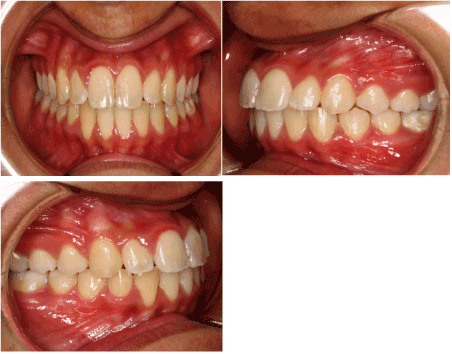
Figure 5. Post-treatment intra-oral photographs.
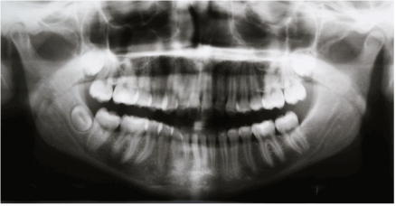
Figure 6. Post-treatment radiographs.
Impacted maxillary canines are more common than mandibular canines. The treatment plan for such teeth will be based on several factors such as the age of the patient and the position of the tooth. Treatment options include orthodontic treatment to align the tooth or removal of the tooth and replace it with a dental implant. This girl was treated successfully via surgical exposure and orthodontic alignment because the position of the impacted canine was favorable.
Permission was granted by the patient and her father to use her clinical records in this case report.
I would like to thank Dr. Joju George from the Oral Surgery Department in Bneid Al-Gar Specialty Dental Center for his great contribution in the tooth exposure stage. I would also like to extend my appreciation to patient and her father for their remarkable cooperation throughout the course of treatment. A special thank for my assistance, Deepa for her immense help.
- Richardson G, Russell KA (2000) A Review of Impacted Permanent Maxillary Cuspids: Diagnosis and Prevention. J Can Dent Assoc 66: 497-501. [Crossref]
- Ericson S, Kurol J (1987) Radiographic examination of ectopically erupting maxillary canines. Am J Orthod Dentofacial Orthop 91: 483-492. [Crossref]
- Aydin U, Yilmaz HH, Yildirim D (2004) Incidence of canine impaction and transmigration in a patient population. Dentomaxillofac Radiol 33: 164-169. [Crossref]
- Alaejos-Algarra C, Berini-Aytes L, Gay-Escoda C (1998) Transmigration of mandibular canines: report of six cases and review of the literature. Quintessence Int 29: 395-398. [Crossref]
- Yavuz MS, Aras MH, Büyükkurt MC, Tozoglu S (2007) Impacted mandibular canines. J Contemp Dent Pract 8: 78-85. [Crossref]
- The management of the palatally ectopic maxillary canine. National Clinical Guidelines. The Royal College of Surgeons of England, March 2010.
- Ericson S, Kurol J (1986) Radiographic assessment of maxillary canine eruption in children with clinical signs of eruption disturbance. Eur J Orthod 8: 133-140. [Crossref]
- McSherry PF1 (1998) The ectopic maxillary canine: a review. Br J Orthod 25: 209-216. [Crossref]
- Thomas S, Turner SR, Sandy JR (1998) Autotransplantation of teeth: is there a role? Br J Orthod 25: 275-282. [Crossref]
- Czochrowska EM, Stenvik A, Bjercke B, Zachrisson BU (2002) Outcome of tooth transplantation: survival and success rates 17-41 years posttreatment. Am J Orthod Dentofacial Orthop 121: 110-119. [Crossref]