Studies revealed that Stevia has been used throughout the world since ancient times for various purposes; for example, as a sweetener and a medicine. The present study was carried out to evaluate the wound healing potential of extract of hydroponic Stevia rebaudiana Bertoni in experimental rats. All experiments were conducted following standard procedures. The extract was administered orally in dose of 20 mg/ kg was used for evaluating the wound healing potential in excision wound model for 2 weeks. Betadine (10%) was used as standard. In conclusion, hydroponic Stevia leaf powder accelerated wound healing activity in rats.
hydroponic stevia rebaudiana, wound healing
Stevia rebaudiana (Bertoni) is a herbaceous perennial plant of the Asteraceae family, native to Paraguay (South America). Stevioside, the major sweetener present in leaf and stem tissues of stevia, was first seriously considered as a sugar substitute in the early 1970s by a Japanese consortium formed for the purpose of commercializing stevioside and stevia extracts [1]. Diterpene glycosides produced by stevia leaves are many times sweeter than sucrose. They can be utilized as a substitute to sucrose [2]; they are natural sources of non-caloric sweetener and alternatives to the synthetic sweetening agents that are now available to the diet conscious consumers. Eight diterpene glycosides with sweetening properties have been identified in leaf tissues of stevia. These are synthesized, at least in the initial stages, using the same pathway as gibberellic acid, an important plant hormone [3]. The four major sweeteners are stevioside, rebaudioside-A, rebaudioside-C and dulcoside-A. According to Kinghorn the sweetness of these compounds relative to sucrose are 210, 242, 30 and 30 times, respectively [4]. The two main glycosides are stevioside, traditionally 5-10% of the dry weight of the leaves, and rebaudioside-A 24%; these are the sweetest compounds. There are also other related compounds including minor glycosides, such as rebaudioside-B, rebaudioside-C (12%), rebaudioside- D, rebaudioside-E, rebaudioside-F, dulcoside-A, dulcoside-C and steviolbioside, as well as flavonoid glycosides, coumarins, cinnamic acids, phenylpropanoids and some essential oils [5]. Among the components of stevia, one, called rebaudioside-A, is of particular interest because it has the most desirable flavour profile [6]. Stevioside traditionally makes up the majority of the sweetener (60-70% of the total glycosides content) and is assessed as being 270 times sweeter than sugar. Rebaudioside-A is usually present as 30-40% of total sweetener and has the sweetest taste, assessed as 400 times sweeter than sugar with no bitter aftertaste (licorice taste or lingering effect). The ratio of rebaudioside-A to stevioside is the accepted measure of sweetness quality; the more rebaudioside-A the better. If rebaudioside-A is present in equal quantities to stevioside, it appears that the aftertaste is eliminated. The minor glycosides are considered to be less sweet, 30-80 times sweeter than sugar [7]. The sweetening effect of these compounds is purely taste; they are undigested and no part of the chemical is absorbed by the body. They are therefore of no nutritional value [8]. Various research data revealed that plants may worked as healing and regeneration of the tissue by multiple mechanisms. There are several reports stating that the extracts of several plants, used for wound healing properties [9,10]. Stevia rebaudiana Bertoni plant was originated from South America (Paraguay and Brazil), belongs to the family Asteraceae, claimed as a potent wound healing plant. Traditional uses and earlier reports have revealed, enhanced healing with less scarring of cuts, wounds, burns, acne, seborrhea, dermatitis, and psoriasis after topical application of aqueous Stevia extracts [11]. These are mainly comprised of stevioside, steviobioside, rebaudioside A, B, C, D, dulcoside A, B [12].
Hydroponic Stevia rebaudiana Bertoni collected from G.S.Davtyan Institute of Hydroponics Problems (Armenia), was used as a test plant for the present study. The plant was authenticated by PhD L. Hovhannisyan and Dr. M. Babakhanyan, Principal Scientist, G.S.Davtyan Institute of Hydroponics Problems and the specimen copy was preserved in the herbarium. The plant sample (leaves) was collected and oven dried at 60°C for 6 h. The dried leaves were stored at 4°C and were used for the further process.
Male albino rats weighing 200-230 g were used in wound healing model experiments. A total of 15 animals used in experiments. Stevia dose for the study was 20 mg/kg. All animals were observed for food consumption for 2 weeks.
The rats were anaesthetized prior to creation of the wounds. All the surgical interventions were carried out under sterile condition.
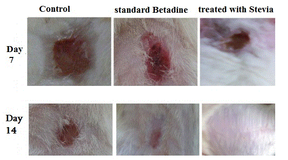
The animals were divided into 3 groups of 5 each and the following treatments were given once daily for 2 weeks. Experiments were performed at Orbeli Institute of Physiology, Neuroendocrine Relationships Lab (Figure 1 and Table 1).

Figure 1. Excision model in rat
Table 1. Biochemical, chemical, technical and radioactive composition of Stevia leaves
Indices |
Ararat valley |
Foothills |
Nagorno Karabagh Republic (NKR) (Khanabad Village)
|
Literature data |
Soil |
Hydroponics |
Extractive agents , % |
45,2-54.0 |
46,0-63,8 |
47,9-52,9 |
46,1-52,0 |
32,5-40,9 |
Stevioside , % |
8,1-8,4 |
8,9-9,2 |
8,5-8,6 |
8,0-8,5 |
4,6-8,2 |
Nitrogen, % |
3,5-4,2 |
3,7-4,6 |
3,4-3,9 |
3,5-4,3 |
|
Proteins, % |
22,4-26,2 |
23,1-28,7 |
21,2-24,3 |
21,8-26,8 |
|
Carotene, mg % |
64,3 |
66,1 |
68.4 |
65,4 |
|
Chlorophyll a+b, mg% |
119,11 |
143,1 |
131,2 |
124,4 |
|
Vitamine "C" mg% |
62,3 |
74,9 |
72,1 |
68,9 |
|
Tanning agent, % |
13,9 |
10,2 |
12,5 |
12,9 |
|
Flavonoids, % |
5,1 |
4,4 |
4,8 |
5,2 |
3,5 |
Essential oil, % |
0,1 |
0,2 |
0,2 |
0,1 |
|
Endemic microelements, mg/100 g |
|
,,_,, J |
0,8 |
8,7 |
0,6 |
0,5 |
|
,,_,, Zn |
0,9 |
1,3 |
0,7 |
0,6 |
|
,,_,, Ge |
0,00012 |
not detected |
0,0001 |
0,00016 |
not specified |
Toxic technical elements, mg/kg |
|
,,_,, Pb |
0,12 |
0,10 |
0,10 |
0,09 |
|
,,_,, As |
0,08 |
0,06 |
0,07 |
0,05 |
|
,,_,, Cd |
0,24 |
0,20 |
0,18 |
0,16 |
|
,,_,, Hg |
0,003 |
0,002 |
0,002 |
0,02 |
|
Pesticides DDT, mg/kg |
not detected |
not detected |
not detected |
not detected |
- |
Aflatoxin В1, ,,_,, |
,,_,, |
,,_,, |
,,_,, |
,,_,, |
|
Radionuclides, U· 10-6 % |
1,8 |
1,3 |
1,1 |
- |
- |
90Sr-Bq/kg |
18,6 |
14,4 |
15,8 |
- |
,,_,, |
Group I: Control (no treatment).
Group II: Standard and treated with Stevia leaf powder (20 mg/kg)
Group III: Standard Betadine 10 %
The measurements of the wound areas of the excision wound model were taken on 1st, 7th and 14th day following the initial wound using transparent paper and a permanent marker. The recorded wound areas were measured with graph paper. Progressive decrease in the wound size was monitored periodically. Wound closure time of the tissue were studied. The period of epithelialization was calculated as the number of days required for falling of the dead tissue remnants without any residual raw wound. In the excision wound model, granulation tissue formed on the wound was excised on the 14th postoperative day. There was a full thickness epidermal regeneration which covered completely the wound area. The epidermis was thick and disorganized, especially when compared with the adjacent normal skin (Figure 2 and Tables 2-5).

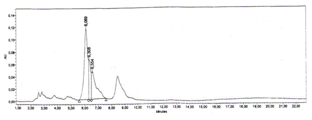
Figure 1. Thin layer chromatography of Stevia .
Table 2. Content of biologically active substances in Stevia (sample 1, standart) (thin layer chromatography was used, Silica gel 60 F 254 (Merck-Germany)
N |
Name |
Retention Time |
Area |
% Area |
Height |
1 |
Standart |
6,089 |
2061029 |
53,20 |
114769 |
2 |
Rebaudioside A |
6,308 |
662200 |
17,09 |
64659 |
3 |
Rebaudioside C |
6,554 |
1150654 |
29,70 |
45328 |
Table 3. Content of biologically active substances in Stevia (sample 2, soil)
N |
Name |
Retention Time |
Area |
% Area |
Height |
1 |
Stevioside |
6,716 |
9377124 |
77,65 |
326455 |
2 |
Rebaudioside A |
7,605 |
2698935 |
22,35 |
76589 |
Table 4. Content of biologically active substances in Stevia (sample 3, hydroponics)
N |
Name |
Retention Time |
Area |
% Area |
Height |
1 |
Stevioside |
6,279 |
13554416 |
71,37 |
566682 |
2 |
Rebaudioside A |
6,816 |
5438482 |
28,63 |
202741 |
Table 5. Content of biologically active substances in Stevia (sample 4, Nagorno Karabagh Republic, Khanabad Village). Contains diterpene glycosides about 83% and monosaccharides only 0.05%). The highest Rebaudioside A content was found in sample 4
N |
Name |
Retention Time |
Area |
% Area |
Height |
1 |
Stevioside |
6,999 |
907785 |
79,01 |
30924 |
2 |
Rebaudioside A |
7,714 |
241102 |
20,99 |
7809 |
Wound healing is a complex process of restoring cellular structures and tissue layers in damaged tissue together to its normal state and commencing in the fibroblastic stage where the area of the wound undergoes shrinkage [13]. It comprises of different phases such as contraction, granulation, epithelization and collagenation [14,15]. Wound healing can be discussed in three phases viz. Inflammatory phase, proliferative phase and maturational or remodeling phase. The 20 mg/kg Stevia was recorded similar effectiveness when compared to the control group treated with a Betadine (10%) (Figure 1). Flavonoids are known to reduce lipid peroxidation not only by preventing or slowing the onset of cell necrosis but also by improving vascularity. Studies were revealed that flavonoids are also known to promote the wound healing process mainly due to their astringent and antimicrobial properties which appear to be responsible for the wound healing and increased rate of epithelialization [16]. So the study provides a rationale for the use of hydroponic Stevia preparations in the traditional system of medicine to promote wound healing. This effect may be explained by several mechanisms such as coating the wound. Further the Stevia leaf powder did not produce any adverse effect and because of this it is possible to recommend its use in the treatment of wounds.
The study thus demonstrated the wound healing activity of hydroponic Stevia leaf powder and found to be effective in the functional recovery of the wound healing.
- Kinghorn AD and Soejarto DD (1985) Current status of stevioside as a sweetening agent for human use. In: Wagner H, Hikino H, Farnsworth NR (eds.) Economic and medical plant research (3rd edn.). Academic Press, London, UK. [Crossref]
- Megeji NW, Kumar, JK, Virendra S, Kaul VK, Ahuja PS (2005) Introducing Stevia rebaudiana, a natural zero-calorie sweetener. Current Sci 88: 801-804.
- Singh SD, Rao GP (2005) Stevia: The herbal sugar of the 21st century. Sugar Technol 7: 1724.
- Kinghorn AD (1987) Biologically active compounds from plants with reputed medical and sweetening properties. J Nat Prod 50: 1009-1024. [Crossref]
- Dacome AS, da Silva CC, da Costa CEM, Fontana JD, Adelmann J, et al. (2005) Sweet diterpenic glycosides balance of a new cultivar of Stevia rebaudiana (Bert) Bertoni: isolation and quantitative distribution by chromato- graphic, spectroscopic and electrophoretic methods. Process Biochem 44: 3587-3594.
- DuBois GE (2000) Sweeteners: non-nutritive. In: Francis FJ (ed.) Encyclopedia of food science and technology Vol.4. (2nd edn.) John Wiley and Sons, Inc., New York, NY. PP: 2245-2265. [Crossref]
- Crammer B, Ikan R (1986) Sweet glycosides from the stevia plant. Chem Britain 22: 915-916.
- Hutapea AM (1997) Digestion of stevioside, a natural sweetener, by various digestive enzymes. J Clin Biochem Nut 23: 177-186.
- Dewangan H, Bais M, Jaiswal V, Verma VK (2012) Potential wound healing activity of the ethanolic extract of Solanum xanthocarpum schrad and wendl leaves. Pak J Pharm Sci 25: 189-194. [Crossref]
- Stephen YG, Emelia K, Francis A, Kofi A, Eric W (2010) Wound healing properties and kill kinetics of Clerodendron splendens G Don, A Ghanaian wound healing plant. Pharmacognosy Res 2: 63-68. [Crossref]
- Mourey D 1992. Life with Stevia: How sweet it is. Nutritional and Medicinal Uses. https://healthfree.com/view_newsletter.php?id=153&key=b. Accessed 12 Sep 2012.
- Leung AY, Foster S (1996) Encyclopedia of common natural ingredients used in food, drugs and cosmetics (2ndedn), John Wiley and Sons Inc., New York, 478.
- Chitra S, Patil MB, Ravi K (2009) Wound healing activity of Hyptis suaveolens (L) poit (Laminiaceae). Int J Pharm Technol Res 1: 737-744.
- Wild T, Rahbarnia A, Kellner M, Sobotka L, Eberlein T (2010) Basics in nutrition and wound healing. Nutrition 26: 862- 866. [Crossref]
- Ayyanar M, Ignacimuthu S (2009) Herbal medicines for wound healing among tribal people in Southern India: ethnobotanical and scientific evidences. Int J Appl Res Nat Prod 2: 29-42.
- Tsuchiya H, Sato M, Miyazaki T, Fujiwara S, Tanigaki S, et al. (1996) Comparative study on the antibacterial activity of phytochemical flavanones against methicillin-resistant Staphylococcus aureus. J Ethnopharmacol 50: 27-34. [Crossref]
