Background: Ventilation of the middle ear cleft is essential for the success in tympanoplasty. There are well defined ventilation pathways in middle ear cleft responsible for the aeration of the middle ear. However, these can get blocked easily by disease process and disrupt the ventilation pathway resulting in chronic discharge resistant to medical therapy. Aditus ad antrum is the critical area which is most susceptible to blockage and can obstruct the ventilation pathway for the mastoid air cell system (MACS).
Objective: To find out the incidence and causes of blockage of the ventilation pathway between the mastoid air cell system and the middle ear.
Methods: 45 cases of Chronic Otitis Media (COM) Mucosal type Active (persistently discharging) despite 1 month of medication were included in this study from January 2015 to January 2018. All the patients underwent cortical mastoidectomy with tympanoplasty. Aditus patency was established by doing a free flow water test and being positive or negative. If negative the cause of the aditus blockage was determined and then removed.
Results: Out of 45 cases of chronic discharging ears the aditus was blocked in 32 cases (71.11%). The most common cause of aditus blockage in our study is granulation tissue. The incidence of blockage was directly related to the duration of the ear disease.
Conclusion: A patent aditus is a must for success of middle ear surgery and to obtain a dry ear.
aditus ad antrum, chronic suppurative otitis media, granulation tissue, water test, ventilation pathways, mastoid air cell system
The Mastoid air cell system (MACS) plays an important role in the ventilation of the middle ear [1]. The critical part is the narrowest - the aditus ad antrum. Although middle ear aeration is related to Eustachian tube function, aditus plays a vital role especially if the Eustachian tube fails in its function [2]. Aeration of middle ear is an important factor in the outcome of tympanoplasty. The lack of a well aerated mastoid and middle ear at the time of tympanoplasty may be a significant cause of failure in mucosal Chronic Otitis Media (COM) [3].
Middle ear aeration occurs from two pathways mainly: anteriorly via the Eustachian tube and posteriorly via the tympanic isthmus. The large tympanic isthmus between the medial part of the posterior incudal ligament and the tensor tendon mainly gives aeration to the epitympanic compartments. The pathway of the aeration from the Eustachian tube leads directly to the mesotympanic and hypotympanic spaces, whereas the epitympanum is only aerated through the tympanic isthmus and is away from the direct airstream [1-3]. The obstruction of the aditus ad antrum results in the pathogenesis and accentuation of the disease condition in the mucosal COM due to the low air volume of the middle ear. Also, the aeration of the mesotympanum and epitympanum is altered due to the blocked aditus and results in failure following tympanoplasty [4,5].
Many studies are there which focus on the evaluation of ventilation blocks in the isthmus; On the other hand, there is lack of articles focused on the evaluation of the aditus block. This study was done mainly to record the common causes of the blockage of aditus.
45 cases of Chronic Otitis Media (COM) Mucosal type Active (persistently discharging) despite 1 month of medication were included in this study from January 2015 to January 2018. They underwent cortical mastoidectomy and tympanoplasty. A Water test was done in all.
Inclusion criteria
- Cases of Chronic Otitis Media (COM) Mucosal type Active (persistently discharging) despite 1 month of medication
Exclusion criteria
- Cases of Chronic Otitis Media (COM) Squamousal type
- Dry cases/ Inactive mucosal cases of Chronic Otitis Media
- Cases of Chronic Otitis Media in which 1 month antibiotic therapy was not tried before surgery
All the patients underwent a thorough otorhinolaryngological examination and detailed history was elicited. Duration of the ear discharge and disease was recorded. Initial ear examination was done by otoscopy and later with operating microscope to make a proper preoperative diagnosis of safe and unsafe disease. To accurately record and document the clinical findings, otoendoscopy was also done.
All the patients underwent preoperative Tuning fork tests, pure tone audiometry and x ray of both mastoids Schuller’s view. Cortical mastoidectomy with tympanoplasty under general anaesthesia via post aural approach was done for all the patients. Aditus patency was established by doing a free flow water test. The water test was read as positive or negative. If negative the cause of the aditus blockage was determined. The Aditus patency was then established by removing the cause of the aditus blockage and obtaining a free flow of water between the mastoid cavity and the middle ear. At times the incus was removed to obtain adequate patency of the aditus. Tympanoplasty was then done after ossicular reconstruction by incus interposition or otherwise in the same surgical procedure. The patients were then assessed clinically and audiologically post operatively after 1, 3 and 6 months. A complete otological examination was carried out in a similar manner as preoperatively to compare the pre and post-operative status. The results were analysed statistically.
Written informed consent was obtained from all the patients included in this study.
INstitutional ethics committee (IEC) clearance: Study was done as per the clearance and guidelines of our IEC.
On analysing the data, we found that out of total 45 cases, 25 cases were males and 20 females. The maximum patients belonged to the age group of 26-35 years, i.e., a total of 15 cases.
Status of mastoid on X Ray mastoid Schullers view
In this study on analysing the X ray mastoid schullers view it was found that sclerosed mastoid was found in 33 cases (73.33%), diploic mastoid in 4 cases (8.89%) and pneumatised mastoid in 8 cases (17.78%)
Aditus Patency
In our study it was found that, out of 45 cases, aditus was blocked in 32 cases (71.11%) and patent in 13 cases (28.89%) (Table 2).
Table 1. Status of mastoid on x ray mastoid Schullers view.
X Ray Bilateral Mastoid Schullers view |
Number of cases |
Sclerosed mastoid |
33 (73.33%) |
Diploic mastoid |
4 (8.89%) |
Pneumatized Mastoid |
8 (17.78%) |
Total |
-
|
Table 2. Aditus Patency in a wet ear (n=45).
|
Cases |
Aditus Blocked |
32 (71.11%) |
Aditus Patent |
13 (28.89%) |
Total cases |
45 |
Table 3. Duration of the ear disease and discharge.
Duration of Discharge |
Blocked aditus (N=32) |
Patent aditus (N=13) |
> 10 yrs |
-
|
-
|
8 to 10 yrs |
-
|
-
|
6 to 8 yrs |
-
|
-
|
4 to 6 yrs |
-
|
-
|
2 to 4 yrs |
-
|
-
|
< 2 yrs |
-
|
-
|
|
-
|
-
|
Table 4. Cause of Aditus blockage (n= 32).
Causes of Aditus Blockage |
No. of Cases |
Granulation Tissue (including 2 cases of kochs) |
14 |
Hypertrophied Mucosa |
6 |
Fibrosis |
5 |
Tympanosclerosis |
5 |
Cholesteral Granuloma |
2 |
Total |
32 |
Table 5. Graft Uptake in blocked aditus.
Duration of Discharge |
Blocked aditus (N=32) |
X Ray mastoid schullers view |
Graft uptake |
> 10 yrs |
-
|
Sclerosed =11 |
Residual perforation=2
Intact graft = 9 |
8 to 10 yrs |
-
|
Sclerosed =6 |
Residual perforation=1
Intact graft = 5 |
6 to 8 yrs |
-
|
Sclerosed =6 |
Intact graft = 6 |
4 to 6 yrs |
-
|
Sclerosed =5 |
Intact graft = 5 |
2 to 4 yrs |
-
|
Sclerosed =1
Diploic = 1
|
Intact graft = 3 |
< 2 yrs |
-
|
|
Intact graft = 1 |
-
|
-
|
-
|
32 (out of these, 3 case developed residual perforation) |
Table 6. Graft Uptake in patent aditus.
Duration of Discharge |
Patent aditus (N=13) |
X Ray mastoid schullers view |
Graft uptake |
> 10 yrs |
-
|
Sclerosed =1 |
Residual perforation=1
Intact graft = 0 |
8 to 10 yrs |
-
|
Sclerosed =0 |
Intact graft = 0 |
6 to 8 yrs |
-
|
Sclerosed =2 |
Intact graft = 2 |
4 to 6 yrs |
-
|
Sclerosed =1 |
Intact graft = 1 |
2 to 4 yrs |
-
|
Diploic= 2
|
Intact graft = 4 |
< 2 yrs |
-
|
Diploic= 1
|
Intact graft = 5 |
|
-
|
-
|
13 (out of these, 1 case developed residual perforation) |
Duration of the ear disease and discharge.
The incidence of the aditus Blockage and ear discharge seems to be directly related to the period of discharge
Long standing patients had an increased incidence of aditus blockage, while patient with recent onset of disease had patent aditus
Cause of Aditus blockage
In our study it was observed that out of a total 32 patients with Aditus blockage, the cause of aditus blockage was (
- Granulation tissue (Figure 1) in 14 patients, these include 2 cases of granulation tissue due to kochs.
- Hypertrophied Mucosa (Figure 2) in 6 patients
- Fibrosis (Figure 3) in 5 patients
- Tympanosclerosis (Figure 4) in 5 patients
- Cholesterol Granuloma (Figure 5) in 2 patients
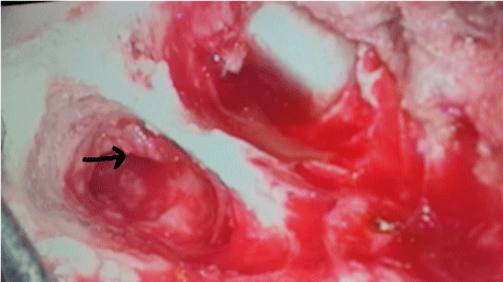
Figure 1. Granulation Tissue in aditus.
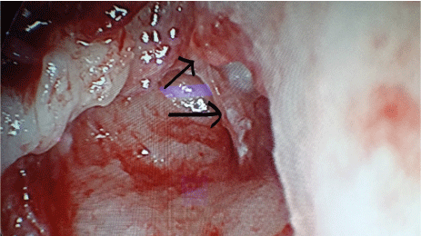
Figure 2. Hypertrophied Mucosa in aditus.
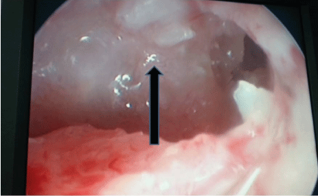
Figure 3. Fibrosis in aditus.
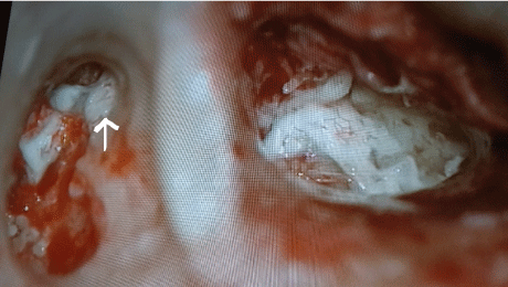
Figure 4. Tympanosclerosis blocking the aditus.
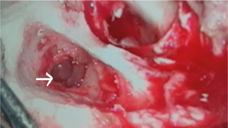
Figure 5. Cholesteral Granuloma blocking the aditus.
Graft Uptake
41 cases (91.11%) had an intact tympanic membrane post operatively at 6 months while 4 cases (8.89%) developed residual perforation 3 cases of residual perforation is with blocked aditus whereas 1 case is with patent aditus. However, all cases of residual perforation in this series are in long standing disease duration.
It is observed that longer the duration of disease, more are the chances of sclerosed mastoid and shorter the disease duration, more are the chances of a pneumatised mastoid. Moreover, sclerosed mastoids have more chances of aditus blockage as compared to pneumatised mastoids.
Statistical analysis
The data was entered into Microsoft Excel and analysed using SPSS (Statistical Package for Social Sciences) package version 21.0 (IBM inc. Chicago, USA). Results are presented in the form of tables and graphs. The Categorical variables are summarized as numbers.
Aditus ad antrum and the epitympanic diaphragm plays an important role in ventilation of mastoid air cell system [1]. Although middle ear aeration is related to Eustachian tube function, aditus also plays a vital role. Aeration of mastoid is an important factor in outcome of tympanoplasty [1]. The MACS (Mastoid Air Cell System) is now realized to be critical in the air pressure management of the middle ear. It has a large surface area in contrast to the middle ear. It has an active gas exchange system. The mucosa lining of the MACS is cuboidal close to the basilar membrane (Figure 6). It lies adjacent to submucosal capillaries. It is now considered to be the actual air pressure buffer rather than the Eustachean tube [2-4].
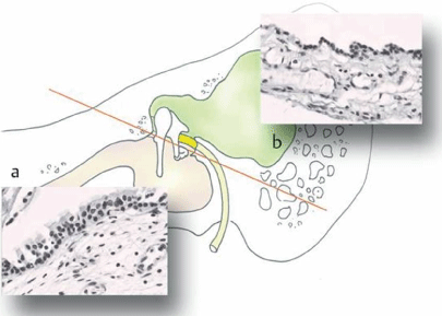
Figure 6. Mastoid Air Cell System mucosal lining for active gas exchange system.
The functions of the MACS can be considered to be acting as an air reservoir. It contributes to an active gas exchange mechanism. It also acts as an air pressure buffer. Efficiency of this will depend directly on the Surface area of the mastoid mucosa which will also vary on the extent of the pneumatization (Figure 7). Hence the pressure of the middle ear can be maintained at the ambient pressure or can be positive without the tubal openings in the healthy ears. The basic mechanism for the regulation of the middle ear pressure may be the exchange of gases over the mucosa. The release valve for the excessive positive middle ear pressure is eustachian tube and can also be released by muscular forces by equalizing the excessive negative middle ear pressure [2-5].
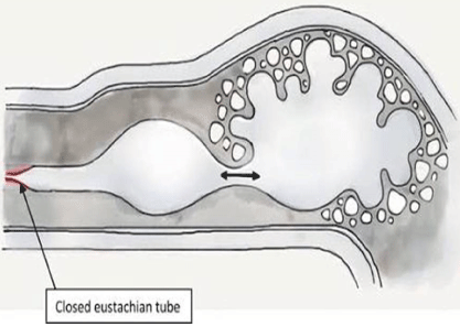
Figure 7. Mastoid Air Cell System acting as an air reservoir.
Aditus and Epitympanic blockage can cause:
- Loss of the air reservoir
- Inactivation of the buffer system
- Negative middle ear pressure due to absorption of air
- Retraction of drum
- Failure of graft in surgery
The lack of a patent aditus and an aerated mastoid at the time of tympanoplasty may be a significant cause of failure in mucosal chronic otitis media (COM) especially with a sclerosed mastoid [5]. It is now postulated that’s the middle ear aeration is from the MACS through the aditus. Failure of this route in face of a eustachian tube dysfunction will result in poor middle ear aeration which would result in tympanoplasty failure. The short process of the incus also compromises the aditus by occupying it and consequently decreasing the effective cross section area for ventilation [1,5]. This narrow space is also easily blocked due to the various causes which we listed.
The aeration of the epitympanic compartments is received through the large tympanic isthmus between the medial part of the posterior incudal ligament and the tensor tendon. The aeration pathway directly leads to the mesotympanic and hypotympanic spaces from the eustachian tube whereas the epitympanum is only aerated through the tympanic isthmus and is situated away from direct air stream [1,2]. Hence, it’s the obstruction of the aditus ad antrum that appears to contribute to the pathogenesis and accentuation of the pathological condition in the mucosal COM, Also the aeration of the epitympanum is interfered due to it and leads to failure following tympanoplasty [2].
The rate of success of the tympanoplasty depends on numerous factors, most importantly the degree of pneumatization of the mastoid air cells, the functionality of the Eustachian tube and the middle ear mucosal condition. However, in this study only one factor was considered that is role of aditus patency. Verification of the aditus patency and clearance of the cause of blocked aditus provided good hearing results after tympanoplasty in this study.
In this study it was observed that out of a total 45 patients, 32 patients had Aditus blockage while 13 patients had a patent aditus. The cause of aditus blockage was hypertrophied mucosa in 6 patients, fibrosis in 5 patients, granulation tissue including kochs in 14 patients, tympanosclerosis in 5 patients and cholesterol granuloma in 2 cases. In a study conducted by Manjunath et al. [6] 23 patients had blocked aditus and 20 patients had patent aditus. The cause of aditus blockage was myringosclerosis in 18 patients and polypoidal mucosa in 5 patients. In a study conducted by Varma et al. [7] 12 had blocked aditus while 23 had patent aditus. In a study conducted by Bhagat et al. [8], out of 50 cases, totally blocked aditus by oedematous unhealthy thickened mucosa was recorded in 10 cases (20%) while the patent aditus was recorded in other 40 cases (80%).
The duration of the disease seems to have a direct relation to the incidence of the aditus blockage, with long standing disease having a greater incidence of aditus blockage due to granulation, tympanosclerosis and fibrosis. Cases which had a disease of recent origin were more likely to have a patent aditus. This is possibly explained that these patients undergo cycles of activation and healing resulting in a persistent chronic disease and blockage- granulations and fibrosis and tympanosclerosis as end stage disease. Similar coreleation was observed in the x ray mastoid of these cases. Longer the duration of disease more were the chances of sclerosed mastoid. In this study on analysing the X ray mastoid schullers view it was found that sclerosed mastoid was found in 33 cases (73.33%), diploic mastoid in 4 cases (8.89%) and pneumatised mastoid in 8 cases (17.78%). Sclerosed mastoids were found in patients with long standing disease and with blocked aditus intraoperatively and pneumatized mastoids were found in short duration of disease with patent aditus intraoperatively in this study.
In this study it was noted that performing tympanoplasty after the removal of diseased mucosa and rendering aditus ad antrum patent, 41 cases (91.11%) had an intact tympanic membrane post operatively at 6 months while 4 cases (8.89%) developed residual perforation. In a study conducted by Sethi et al. [9], out of 50 patients 38(76%) had successful graft take up and 12(24%) had residual perforation. In a study conducted by Metin et al. [10] the patients were divided into 3 groups, poorly ventilated group, moderately ventilated group and well-ventilated group. They reported the graft uptake in poorly, moderately and well-ventilated group as 63.9%, 78.6% and 85.7% respectively.
It is important to give a well aerated middle ear space for effective hearing. This In turn depends on the MACS to function well and its function to be restored by establishing a patent aditus. The blockage of the aditus was directly related to the presence of chronic disease. This aeration through the aditus has to be established to Increase the success rate in cases of tympanoplasty. The most common cause of aditus blockage in our study is granulation tissue. Some cases required incus transposition to effectively open the aditus.
- Palva T, Ramsay H (1996) Incudal folds and epitympanic aeration. Am J Otol 17: 700-708. [Crossref]
- Marchioni D, Molteni G, Presutti L (2011) Endoscopic anatomy of the middle ear. Indian J Otolaryngol Head Neck Surg 63: 10-12. [Crossref]
- Marchioni D, Alicandri-Ciufelli M, Molteni G, Artioli FL, Genovese E, et al. (2010) Selective Epitympanic dysventilation syndrome. Laryngoscope 120: 1028-1033. [Crossref]
- Holmquist J, Bergstrom B (1978) The mastoid air cell system in ear surgery. Arch Otolaryngol 104: 127-129. [Crossref]
- Sade J (1992) The correlation between middle ear aeration and mastoid pneumatization. Eur Arch Otorhinolaryngol 249: 301-304. [Crossref]
- Manjunath M, Swarup R, Chary G, Shadab M (2011) Myringosclerosis: An Indication of A Blocked Aditus. Indian J Otolaryngol Head Neck Surg 64: 230-232. [Crossref]
- Varma A, Maithani T, Agrahari A, Pandey A, Singh V (2015) Clinical and Radiological Predictability of AditusPatency in Mucosal COM with Sclerosed Mastoid: An Analytical Study. Otorhinolaryngol Clinics Int J 7: 121-124.
- Bhagat M (2015) Patency of the aditus ad antrum in tubotympanic chronic suppurative otitis media. Otolaryngol Head Neck Surg 152: 331-335. [Crossref]
- Sethi A, Singh I, Agarwal AK, Sareen D (2005) Pneumatization correlated to myringoplasty and tubal function. Indian J Otolaryngol Head Neck Surg 57: 283-286. [Crossref]
- Metin M, Kaptan ZK, Dogan S, Yazici H, Bayraktar C, et al. (2014) Effect of preoperative mastoid ventilation on tympanoplasty success. Int J Otolaryngol 2014: 169123.