Abstract
The activation of the interventricular septum depends on the posterior fascicle of the left bundle branch but on occasions, it is due to a middle or septal fascicle. We present a peculiar case of left posterior fascicular block with septal activation preserved after ablation of the posterior fascicular ventricular tachycardia.
Key words
posterior fascicle, septal, activation
Introduction
Since Rosenbaum published his original research about the hemiblocks, the activation of the interventricular septum has been considered to depend on the posterior fascicle of the left bundle branch [1]. Some clinical and experimental studies showed that, on occasions, such activation is due to a middle or septal fascicle arising directly from the left bundle branch. We evaluated the vectocardiographic (VCG) and electrocardiographic (ECG) characteristics of the left posterior fascicular block (LPFB) produced after ablation of the posterior left ventricular papillary muscle implantation site to treat a posterior fascicular ventricular tachycardia (PFVT) [2].
Case report
A 35-year old male patient sought medical care to due to long-lasting paroxysmal episodes of rapid palpitations with sudden onset. The episodes were accompanied by dizziness and dyspnea and started three years before. He was hospitalized on several occasions due to PFVT that reverted with amiodarone or diltiazem. Doppler echocardiography did not reveal structural heart disease. The patient underwent radiofrequency catheter ablation in the area of the posterior fascicle using 3D EnSite™ NavX™ mapping navigation system. Before the procedure, the baseline ECG showed counterclockwise rotation of the QRS in the frontal plane with heart axis in +30° (Figure 1A). Continuous infusion of isoproterenol at 3 mcg/min triggered bursts of broad QRS complex tachycardia with a right bundle-branch block and left anterior fascicular block pattern (Figure 1B). Three-dimensional mapping showed that the episodes of ventricular tachycardia originated at the site of left ventricular posterior papillary muscle implantation (Figure 2). Radiofrequency energy was then applied to the site of origin of the ventricular tachycardia. The procedure was successful and the ECG after catheter ablation showed a shift in QRS rotation (clockwise rotation) in the frontal plane with right heart axis deviation (Figure 1C). The VCG showed that the first vector was orientated upwards and to the right (Figure 3, red arrow), indicating that the activation of the interventricular septum was preserved and revealing that pure LPFB without septal fascicular block directed the initial QRS forces to the right. The patient remained free of symptoms and without recurrent episodes of ventricular tachycardia for over three years.
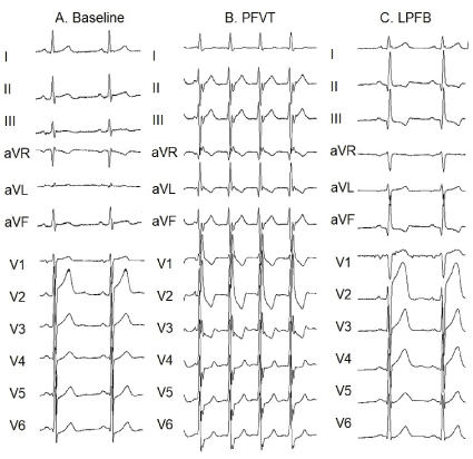
Figure 1. A) The baseline ECG shows sinus rhythm, heart axis +30°, narrow QRS complexes, with a qR pattern in lead I and Rs pattern en leads II, III and aVF, showing a counterclockwise rotation of the QRS in the frontal plane. B) Continuous isoproterenol infusion triggered a posterior fascicular vetricular tachycardia (PFVT). C) After successful ablation of the left posterior fascicle, the ECG shows right heart axis deviation with a qR pattern in leads II, III and aVF, suggesting a shift in QRS rotation (clockwise rotation) in the frontal plane. The presence of Q waves in lead I and V6 demonstrates that septal activation is preserved despite the left posterior fascicular block (LPFB).
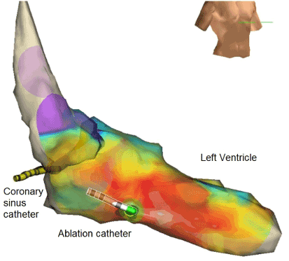
Figure 2. Three-dimensional mapping using EnSite™ NavX™ mapping system. Ventricular tachycardia originates at the site of left ventricular posterior papillary muscle implantation.
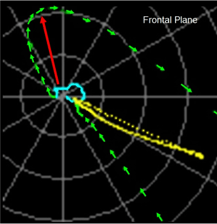
Figure 3. VCG performed after successful ablation of the left posterior fascicle. The QRS loop (in green) has clockwise rotation and the initial forces are oriented upwards and to the right (red arrow). These initial forces correspond to the normal septal activation from left to right.
Discussion and conclusion
Since the original work by Rosenbaum et al. [1], was published in 1968, the intraventricular conduction system is considered to have three well-defined anatomical and functional divisions, represented by the right bundle branch and the anterior and posterior fascicles of the left bundle branch. However, the experiments conducted by Durrer et al. [3], in human hearts showed that the activation of the interventricular septum and the sites of insertion of the left bundle branch fascicles start simultaneously.
According to the anatomical observations made by Rosenbaum et al. [1], in most human and canine hearts, a middle or septal fascicle emerges from the posterior fascicle of the left bundle branch and, less commonly, from the anterior fascicle and extends to the left subendocardial surface of the interventricular septum as a complex network of Purkinje fibers. This anatomical arrangement of the left branch divisions explains the loss of the septal Q-waves (in leads I, V5 and V6) in some cases of left posterior fascicular blocks [4,5].
Some clinical and experimental studies showed that, on occasions, the activation of the interventricular septum is due to a middle or septal fascicle arising directly from the left bundle branch. Yet, the anatomy of this fascicle is very variable and many authors dispute if this fascicle is a third independent anatomic and functional division of the left bundle branch [6-8].
In our case, the LPFB pattern produced by the ablation of the left posterior fascicle showed that the initial QRS forces are oriented upwards and to the right and not to the left because the septal activation is preserved and reveals a new variant of pure LPFB either because the site of the blockage of the left posterior fascicle is distal or because the septal activation depends on a middle fascicle that emerges independently in the left bundle branch.
References
- Rosenbaum M, Elizari M, Lázzari E (1968) Anatomy of the driving system. The hemiblocke Ed. Paidos, pp: 43-91.
- Bayés de Luna A, Pérez Riera A, Baranchuk A, Chiale P, Iturralde P, et al. (2012) Electrocardiographic manifestation of the middle fibers/septal fascicle block: a consensus report. J Electrocardiol 45: 454-460. [Crossref]
- Durrer D, van Dam RT, Freud GE, Janse MJ, Meijler FL, et al. (1970) Total excitation of the isolated human heart. Circulation 41: 899-912. [Crossref]
- Rosenbaum M, Elizari M, Lázzari E (1968) Trifascicular intraventricular blocks. The hemiblocke Ed. Paidos, pp: 155-202.
- Rosenbaum M, Elizari M, Lázzari E (1968) The anterior and posterior hemiblock. The hemiblocke Ed. Paidos, pp: 203-281.
- Perez Riera A, Ferreira C, Ferreira Filho C, Meneghini A, Uchida AH, et al. (2011) Electrovectrocardiographic diagnosis of left septal fascicular block: anatomic and clinical considerations. Ann Noninvasive Electrocardiol 16: 196-207. [Crossref]
- Demoulin JC, Kulbertus HE (1972) Histopathological examination of concept of left hemiblock. Br Heart J 34: 807-814. [Crossref]
- Elizari MV (2017) The normal variants in the left bundle branch system. J Electrocardiol 50: 389-399. [Crossref]