Abstract
Laryngocele and tracheal diverticulum are uncommon conditions that represent a bening sacculation of the upper airway with air content. Historically, x-ray studies with oral barium have been the most common technique for their detection and evaluation. However, CT has became a widely used technique for these tasks, due to its availability and extensive anatomical information. MDCT allows MPR and VR reconstructions that improve lesion detection and make easier for the clinician to understand anatomical relationships.
Keywords
multidetector computed tomography, volume rendering, laryngocele,traqueal diverticulum.
Abbreviations
CT: Computed Tomography; MDCT: Multidetector Computed Tomography; VR: Volume Rendering; MPR: Multiplanar Reconstructions.
Introduction
The airway may present evaginations with air content that depending on their anatomical location may have a different clinical presentation[1]. In the pharynx, oral contrast studies can be used to evaluate its morphology, but in the larynx and trachea the CT (computerized tomography) is the test of choice to describe its anatomy and morphology [2]. Multiplanar reconstructions may be helpful in assessing anatomical location and relationships, although it may be difficult for the clinician to correlate radiologic planes with the patient's morphostructure [3]. We present two cases, one with a symmetrical bilateral laryngocele, and the other with a tracheal diverticulum, in which a MDCT (multidetector computerized tomography) study and subsequent reconstructions significantly help the visualization of the lesions.
Case Report
The first case is a 37-year-old man referred for suspicion of submaxilitis to perform facial MRI. As an incidental finding, a right-sided paratracheal hypointense nodular lesion was observed (Figure 1), and a CT without contrast was performed to improve its anatomic definition. It was performed with a 16-row MDCT, with a cut thickness of 1.25 mm, and MPR (multiplanar reconstructions) and VR (volume rendering) reconstructions (Figure 2). An air density lesion was seen, with a thin wall but with some lobulations, with an upperright paratrachealposition, consistent with a tracheal diverticulum [4]. They are asymptomatic lesions, which in case of no complications or clinical symptoms, as was the case, do not require surgical treatment [5]. The patient has undergone other MR studies by recurrent submaxilitis, without any variations in the diverticulum.
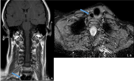
Figure 1.A)Coronal T1 TSE –MRI of the neck and B) axial STIR at the level of the
thoracic cervical junction. A hypointense lesion, well defined, right paratracheal was observed (blue arrow).
The second case is a 48-year-old man who has been referred for cervical discomfort and fluctuating mass. As a relevant fact of the story, the patient is a non-professional trumpet player. A MDCT with intravenous contrast was performed in order to characterize a potential cervical mass. It was performed with a 16-row CT, with a cut thickness of 0.625 mm, and MPR and VR reconstructions (Figure 2). A dilatation of the upper airway was observed in the pharynx, with bilateral and symmetrical sacs, which laterally extended beyond the hyoid, consistent with a bilateral laryngocele of effort [6]. VR reconstruction allows assessment of its dependence on the airway, as well as the total volume of the lesion, which justified the patient's discomfort [7]. Currently the patient is under clinical control, and no therapeutic measures have been envisaged. (Figure 3).
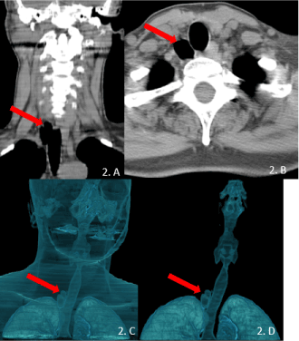
Figure 2. MDCT of the neck without intravenous contrast. A) Coronal MPR view, b) Axial View, C) VR reconstruction of soft tissue and airway and D) VR selective of airway. A and B clearly depict an hypodense lesion next to the trachea, well defined. C and D clearly depict both volume and location of the diverticulum (red arrow).
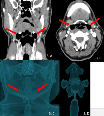
Figure 3.MDCT of the neck with intravenous contrast. A) Coronal MPR view, b) Axial View, C) VR reconstruction of soft tissue and airway and D) VR selective of upper airway demonstrate bilateral air spaces dilated during Valsalva maneuver, consistent with bilateral laryngocele (red arrows).
References
- Tanrivermis Sayit A, Elmali M, Saglam D, Celenk C (2016) The diseases of airway-tracheal diverticulum: a review of the literature. J Thorac Dis 8: E1163-1163E1167.[Crossref]
- Tao TY, Menias CO, Herman TE, McAlister WH, Balfe DM (2013) Easier to swallow: pictorial review of structural findings of the pharynx at barium pharyngography. Radiographics33:e189-208.[Crossref]
- Beigelman-Aubry C, Brillet PY, Grenier PA (2009) MDCT of the Airways: Technique and Normal Results. Radiol Clin North Am47:185-201.
- Bodet Agustí E, Martínez Vecina V, Romeu Figuerola C, Monzón Gaspà M (2007) [Tracheal diverticulum: a case report]. Acta Otorrinolaringol Esp 58: 278-279.[Crossref]
- B ÁM, E SA, Jm SC, Ji AC (2009) Universitario HC. Divertículo traqueal12:26-27.
- Suqati AA, Alherabi AZ, Marglani OA, Alaidarous TO (2016) Bilateral combined laryngocele. Saudi Med J 37: 902-903.[Crossref]
- Edmiston R, Hariri A, Karagama Y (2015) The trumpet player with a swelling in the neck. Case Reports [Internet]1:bcr2015209487-bcr2015209487.