Rationale: Isolated cardiac sarcoidosis (CS) is a life-threatening condition that can be difficult to diagnose. This case report provides an example in which 18F-FDG- and 82Rb- PET/CT studies were used to not only diagnose, but also to monitor and alter treatment for a patient with CS.
Patient concerns: A 51-year-old male presented after witnessed cardiac arrest.
Diagnosis: EKG was notable for frequent, polymorphic premature ventricular contractions and new right bundle branch block with left anterior fascicular block. TTE revealed EF of 45%. Coronary angiography was unremarkable. MRI one month later revealed EF of 20% with patchy delayed enhancement at the anterior wall and inferior septum at the mid-level, patchy right ventricular wall enhancement, and extensive transmural enhancement of the apex suggestive of CS. 18F-FDG-PET/CT showed foci of increased metabolic activity throughout the left ventricle (mainly in the inferior and anteroseptal walls). Myocardial perfusion imaging using 82Rb- PET/CT showed resting hypoperfusion in the entire inferior and mid-to-basal anteroseptal regions with a metabolism (18F-FDG) and perfusion (82Rb) mismatched pattern.
CS was diagnosed based on new conduction disease, unexplained ventricular arrhythmias and heart failure, typical cardiac MRI findings, as well as 18F-FDG uptake in the anteroinferior walls with perfusion defect. While the gold standard test is the presence of non-caseating granulomas in cardiac tissues, EMB is low yield and was not performed in our patient.
Interventions: The patient received an ICD and was treated with prednisone and mycophenolate. Repeat PET/CT one month later showed resolution of heterogeneous activity with persistent perfusion defect and the regimen was tapered. Six months later, he suffered a second cardiac arrest with an interval worsening of the inflammatory phase of CS and persistent mismatched perfusion pattern. Prednisone and mycophenolate doses were increased.
Outcomes: Most recent PET/CT was notable for resolution of inflammation with persistent perfusion defect. Therefore, the patient continued his immunosuppressive regimen and remains arrhythmia free.
Lessons: This case illustrates successful use of sequential 18F-FDG-PET/CT studies for diagnosis and monitoring of CS. Future studies are needed to determine the role of combined perfusion and metabolic PET/CT findings for guiding alterations in immunosuppressive therapy.
cardiac sarcoidosis, computed tomography, positron emission tomography
Isolated cardiac sarcoidosis (CS) is a life-threatening condition that can be difficult to diagnose. Two criteria, developed by the Japanese Ministry of Health and Welfare (JMWH) and Heart Rhythm Society (HRS), are most commonly used for CS diagnosis [1]. In addition to electrocardiogram (ECG), transthoracic echocardiography (TTE), and endomyocardial biopsy (EMB) findings, these criteria suggested adding metabolic fluorodeoxyglucose (18F-FDG-) and Rubidium (82Rb-) perfusion positron emission tomography/computed tomography (PET/CT) imaging to increase the overall sensitivity [2]. This case report provides an example in which 18F-FDG- and 82Rb- PET/CT studies were used to not only diagnose, but also to monitor and alter treatment for a patient with CS.
A 51-year-old male with a past medical history of well-controlled essential hypertension and palpitations of unknown etiology presented to an outside hospital emergency department after experiencing chest pain followed by witnessed cardiac arrest while at work. Return of spontaneous circulation was achieved after three shocks. The patient’s initial blood pressure was 100/94mmHg, and subsequent physical exam was within normal limits without evidence of murmurs, edema, or hepatomegaly. He had no known family history of sudden cardiac death or heart disease. He was physically active and used to perform as an amateur triathlete.
Myocardial infarction, ventricular arrhythmia, pulmonary embolism, and spontaneous pneumothorax were initially on the differential given the sudden onset of chest pain and cardiac arrest in an otherwise healthy patient. Laboratory values were notable for hypokalemia (2.9mmol/L), mild transaminitis, and an increase in troponin I level (0.143ng/mL). Chest x-ray was negative for acute infiltrate or pneumothorax. A 12-lead ECG showed frequent, polymorphic premature ventricular contractions, new right bundle branch block and left anterior fascicular block compared to an ECG a few years prior. Coronary angiography showed normal coronary arteries. TTE revealed a left ventricular ejection fraction (LVEF) of 45% with no valvular abnormalities or pericardial disease.
He received a single-chamber magnetic resonance imaging (MRI)-compatible implantable cardioverter-defibrillator (ICD) for secondary prevention of sudden cardiac death. Carvedilol and candesartan were prescribed for medical optimization of new heart failure. Outpatient cardiac MRI revealed LVEF of 20%, patchy delayed enhancement at the anterior wall and inferior septum at the mid-level, patchy right ventricular wall enhancement, and extensive transmural enhancement of the apex suggestive of cardiac sarcoidosis (CS).18F-FDG- PET/CT showed foci of increased metabolic activity throughout the left ventricle (mainly in the inferior and anteroseptal walls) and parts of the right ventricle, confirming the clinical suspicion of metabolically active CS (Figure 1A). Myocardial perfusion imaging using 82Rb PET/CT showed resting hypoperfusion in the entire inferior and mid-to-basal anteroseptal regions with a metabolism (18F-FDG) and perfusion (82Rb) mismatched pattern (Figure 2). There was imaging evidence of mediastinal nodal involvement, however, hilar lymph node biopsy showed no granulomatous disease. Laboratory values, including calcium, pulmonary function tests and lung appearance on chest CT were within normal limits. Based on strong clinical suspicion for CS, he was started on oral prednisone 40mg daily and mycophenolate mofetil 500mg twice daily.
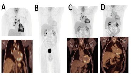
Figure 1. PET/CT Metabolic Imaging Over 16 Months. (A) 2/2017: Extensive metabolic granular activity throughout the left and right ventricles. (B) 4/2017: Interval resolution of metabolic activity in both ventricles. (C) 10/2017: Increased metabolic activity throughout the entire left ventricle in a non-CAD pattern. (D) 6/2018: Decreased interval areas of heterogenous, metabolic activity throughout the left ventricle.

Figure 2. PET/CT Perfusion Imaging with Metabolism-Perfusion Mismatch Pattern. Results between 2/2017 to 6/2018 notable for persistent hypoperfusion in the inferior and mid-to-distal lateral and apico-lateral lesions.
One month later, due to excessive steroid-related side effects (weight gain, hepatic steatosis seen on PET/CT, and severe hypertriglyceridemia), and absence of ventricular arrhythmias on ICD interrogation, prednisone was decreased to 30mg daily. A second 18F-FDG-PET/CT demonstrated interval resolution of the previously seen heterogeneous activity in both ventricles (Figure 1B), with unchanged resting hypoperfusion. The patient continued to taper prednisone by 5mg monthly, and the mycophenolate dose was decreased from 500mg to 250mg twice daily. A third 18F-FDG-PET/CT showed interval worsening of inflammatory phase of CS (Figure 1C). Subsequently, mycophenolate was increased back to 500mg twice daily while the patient remained on low dose prednisone. Six months later, the patient suffered a second cardiac arrest and ICD firing from recurrent ventricular tachycardia. He was loaded with amiodarone and maintained on high dose metoprolol. Prednisone and mycophenolate doses were increased to 40mg daily and 1.5g twice daily, respectively. Repeat 18F-FDG-PET/CT was notable for an interval decrease in myocardial inflammation (Figure 1D).
As of 5/2019, 11 months since the second cardiac arrest, the patient remained arrhythmia-free with a stable LVEF of 45-50%. He continued to take high dose mycophenolate but had been weaned off prednisone. The patient provided informed consent for publication of this case.
Isolated CS is commonly under-recognized in clinical practice due to limited understanding of disease progression. The diagnosis of CS was made based on clinical suspicion in our patient. While the gold standard test is the presence of non-caseating granulomas in cardiac tissues, EMB is low yield and was not performed in our patient. In addition to new conduction disease, unexplained ventricular arrhythmias and heart failure, our patient had typical cardiac MRI findings, as well as heterogeneous 18F-FDG uptake in the anterior and inferior walls with corresponding perfusion defect, all consistent with CS [2,3]. The combination of increased metabolic activity and decreased perfusion is associated with a four-fold increase in the annual rate of ventricular tachycardia and portends a poorer prognosis [2,4,5].
Although cardiac PET/CT imaging maybe useful in predicting life-threatening arrhythmias, its role in guiding titration of immunosuppressive therapy is unclear, and reports of its use in this capacity are scarce. This patient’s second PET/CT demonstrated an improvement in metabolic activity which prompted a decrease in immunosuppression (Figure 1B), while increased metabolic activity led to intensification of mycophenolate and prednisone dosages (Figure 1C). Serial PET/CT in this case also illustrates development of hepatic steatosis due to steroid use, prompting aggressive lipid-lowering treatment. More research is needed to determine the frequency of follow up PET/CT scans to monitor disease progression long-term, and to minimize radiation exposure and costs.
This case illustrates successful use of sequential 18F-FDG-PET/CT studies for diagnosis and monitoring of CS. Future studies are needed to determine the role of combined perfusion and metabolic PET/CT findings for guiding alterations in immunosuppressive therapy.
No funding was used for this case report.
There are no relationships with industry.
- Chareonthaitawee P, Beanlands R, Chen W, Dorbala S, Miller EJ, Murthy VL, et al. (2017) Joint SNMMI-ASNC Expert Consensus Document on the Role of 18F-FDG PET/CT in Cardiac Sarcoid Detection and Therapy Monitoring. J Nucl Med 58: 1341-1353. [Crossref]
- Blankenstein R, Osborne M, Naya M, Waller A, Kim CK, Murthy VL, et al. (2014) Cardiac positron emission tomography enhances prognostic assessments of patients with suspected cardiac sarcoidosis. J Am Coll Cardiol 63: 329-336. [Crossref]
- Youssef G, Leung E, Mylonas I, Nery P, Williams K, Wisenberg G, et al. (2012) The use of 18-F-FDG PET in the diagnosis of cardiac sarcoidosis: a systematic review and meta-analysis including the Ontario experience. J Nucl Med 53: 241-248. [Crossref]
- Bargout R, Kelly RF (2004) Sarcoid heart disease: clinical course and treatment. Int J Cardiology 97: 173-182. [Crossref]
- Skali H, Schulman A, Dorbala S (2013) 18F-FDG PET/CT for the Assessment of Myocardial Sarcoidosis. Curr Cardiol Rep 15: 352. [Crossref]