Abstract
Background: Acute abdomen is defined as a sudden onset abdominal pain that requires an urgent intervention. It is a very common presentation within an emergency department (ED). Computed tomography (CT) is a commonly used imaging modality in conjuction with clinical assessment to formulate diagnosis of an acute abdomen. The use of CT helps avoid admissions admissions for non-specific abdominal pain presentations.
Aims of the study: To compare the ED clinical working diagnosis with the CT diagnosis (CT report by a consultant radiologist) for patients with acute abdominal pain subjected to CT abdomen. We also wanted to know the final surgical diagnosis on the patients needing surgical operative intervention.
Methods: We retrospectively gathered the 6 months electronic medical records of the patients presenting to ED with acute abdomen, who had a diagnostic CT scan requested from ED. The data was obtained from their electronic records provided by Health care information technology affairs (HITA) using a designed questionnaire. The data was analyzed with counting numbers and calculating the percentages.
Results: 9124 patient case notes were retreived through HITA, who presented with acute abdominal pain and satisfied the inclusion criteria. 124 (1.3%) patients had a CT requested by the ED. Ninety seven patients (78%) who underwent CT scan had a reported finding. The CT findings of seventy-one (57%) patients matched the clinical diagnosis of ED. 19 (15%) patients had an emergency surgical operative interventation.
Conclusion: CT investigation for acute abdomen is appropriately deployed by the ED physcians and a significant number of patients’ CT findings matched the ED cliinical diagnosis.
Introduction
Acute abdomen is a common presentation seen in ED, accounting for approximately five per cent of the cases [1]. It is defined as a sudden onset of abdominal pain that requires an urgent medical or surgical intervention [2]. ED evaluation for acute abdomen can be difficult and should be assessed accurately to prevent mortality [3,4]. With the advancement in imaging techniques, ED physicians are more equipped to diagnose and manage acute abdominal emergencies [3,5].
The causes of acute abdomen can vary according to demography. It tends to be more challenging in elderly with higher mortality rate, thus requiring an urgent admission and management [3]. The most common cause of acute abdomen leading to admission is appendicitis followed by diverticulitis [6]. The prevalence of acute cholecystitis, acute diverticulitis and abdominal aortic aneurysm (AAA) rupture is increasing [7].
Diagnosis of acute abdomen includes clinical evaluation, blood tests and imaging. Whereas clinical evaluation alone is sensitive, it is less specific in diagnosing acute abdomen in comparison to imaging. Ultrasound (US) and computed tomography (CT) are the most common imaging studies used for evaluating acute abdomen in ED. CT is more sensitive (89%) in detecting acute abdomen cases compared to ultrasound (70%) [8].
CT is recommended for all patient with acute abdomen except in pregnant women, acute cholecystitis, or AAA, where US is preferred. The use of contrast enhancement with CT is indicated to detect ischemic and vascular lesions causing acute abdomen with careful consideration in patient with history of allergies, renal impairment, or taking biguanides [3].
With the increased use of CT and other imaging modalities for the diagnosis of acute abdomen in the past years, admission with a definite diagnosis instead of a nonspecific cause has increased [9]. A prospective observational study showed that 37.6% of the patients presenting to the ED with acute abdomen needed hospital admission and 36.2% of those were admitted to the surgical ward [10].
Our study is intended to find out the use of CT abdomen by physcians for patients, who present with abdominal pain in the ED and to compare the CT findings with the working diagnosis, made by the physcian prior to requesting CT.
Methods
A data request was sent to the HITA for retreiving the case records of all patients, who presented with acute abdominal pain to the ED over a 6 months retrospective period. From the 9124 patient records received, only 124 patients satisfied the inclusion criteria for our study. The clinical details of all these patients were recorded on the excel sheet including demographics, onset of pain duration, orders for imaging modalities, surgical consultation (prior or after CT), verified CT report findings, time when the verified radiology report (from consultant radiologist) was available, surgical intervention etc. The data was sent to the hospital statistician for analysis.
Inclusion Criteria
Patients with acute abdominal pain (defined as > 2 hours and < 7 days), where a CT scan was performed by the ED physcian.
Exclusion Criteria
Pregnant females.
Acute abdominal trauma. Patient with established diagnosis before arrival to ED.
Results
Demographics
Patients presented to the ED with abdominal pain between August 2020 to January 2021 were included. During that time, a total of 9124 patients were registered with acute abdominal pain. 124 patients met the inclusion criteria. Patients age ranged from 5 to 90 years with a median age of 50 years. Sixty-eight percent were female (Figure 1, Table 1). Forty-five percent had previous abdominal surgery (Table 2).
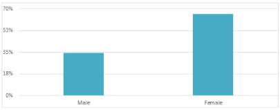
Figure 1. The Ratio of patients with/without previous abdominal surgery
Table 1. Summary of patients’ characteristics
Characteristics |
Number (percent) |
Male |
40 (32.26%) |
Female |
84 (67.74%) |
Table 2. The Ratio of patients with/without previous abdominal surgery
|
Frequency |
Percentage |
Had previous abdominal surgery |
59 |
48% |
No previous abdominal surgery |
65 |
52% |
Diagnosis and Disposition
One-hundred and twenty-four patients (1.3%) who presented to ED with abdominal pain, underwent CT within the ED, requested by an ED physcian.
The time of onset of abdominal pain ranged 6.88 days (mean 1.41 days) (Table 3).
Table 3. Time of onset of pain (in days)
Time of onset of pain |
Range |
Mean |
6.88 |
1.41 |
One hundred & seven (86%) patients had CT done with intravenous contrast (Table 7, Figure 5).
Ninety seven (78%) patients who underwent CT scan had a positive finding on the verified report by a consultant radiologist (Table 4, Figure 2).
Table 4. The patients who had positive CTs
|
Frequency |
Percentage |
Positive CT |
97 |
78.2% |
Negative CT |
27 |
21.8% |
The CT findings of seventy-one (73%) patients matched the clinical working diagnosis of ED physcian.
Twenty (16%) patients who had CT findings required surgical consultation (Table 6, Figure 4).
Ten (8.1%) consultations were made by the ED physcian, prior to the CT request (Table 5, Figure 3)
Table 5. Consultation prior to CT
|
Frequency |
Percentage |
Consultations prior to CT |
10 |
8.1% |
No consultations prior to CT |
64 |
51.6% |
NA |
50 |
40.3% |
Table 6. Patients with CT and surgical consultation
|
Frequency |
Percentage |
Surgical consultation |
20 |
16.1% |
No surgical consultation |
103 |
83.1% |
NA |
1 |
0.8% |
Table 7. CT with and without contrast
|
Frequency |
Percentage |
With contrast |
107 |
86.3% |
without contrast |
17 |
13.7% |
Table 8. Miscellaneous findings
|
Frequency |
Percentage |
“Radiology report findings” that matched the ED clinical diagnoses |
71 |
73% |
“Radiology report findings” that matched the “Emergency Diagnosis” & “surgical diagnosis |
15 |
15.4% |
Patients with positive “Radiology reports findings” who went for operative intervention |
20 |
16% |
Patients who had all three radiology investigations i-e CT, abdominal series and Ultrasound |
2 |
1.6% |
Table 9. Number of surgical cases who underwent emergency surgery
Surgical diagnosis |
Number of cases |
Acute Appendicitis |
9 |
Bowel Obstruction |
7 |
Intussusception |
1 |
Perforated gallbladder |
1 |
Incarcerated hernia |
1 |
Mesenteric Cyst |
1 |
Total |
20 |
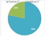
Figure 2. The patients who had positive CTs
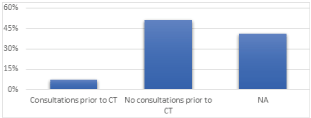
Figure 3. Consultation prior to CT
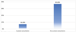
Figure 4. Patients with CT and surgical consultation
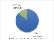
Figure 5. CT with and without contrast
Twenty patients (16%) with positive report underwent operative management (Table 9).
One hundred & four (84%) patients with findings on the CT did not require surgical consultation.
The time required for the CT scan report to be verified by a radiology consultant ranged from 60-818 min.
Discussion
Acute abdominal pain is commonly presented condition in the EDs worldwide. Evaluation of acute abdominal pain by the emergency physician is imperative. Currently with the advancement of technology, emergency physicians tend to be more skilled in diagnosing and managing acute abdominal pain. Though the conventional approach to acute abdominal pain includes history, physical examination and laboratory tests yet radiological tools ensure a non-invasive, safe and definitive diagnostic conclusion.
In our study, approx 9000 patients presented during a period of six months to the ED with a presenting symptom of acute abdominal pain. Only a selected number of patients (1.3%) underwent CT scan due to either an accurate diagnosis by ED or it turned out to be a medical cause of abdominal pain.
This also indicates how clinicians in the ED differentiate presenting symptoms to an appropriate working diagnosis, without unncecessay use of readily available radiological resources. This also complies with the basic medical principle of relying more on clinical examination and less dependence on investigations.
In 15 patients the ED diagnosis matched the surgical diagnosis, which again reinforces the clinical maturity shown by the ED physcians upfront.
There were ten consultation made by ED prior to CT, which signifies there was enough confidence in the clinical diagnosis to warrant early speciality help.
Conventional radiology (abdominal x-ray) carries its own limitation, particularly when compared to other imaging modalities. For example, plain abdominal film can show the presence of perforation or obstruction in many cases but can miss early presentation of these conditions. CT scan is almost always required to reach a definitive diagnosis in these clinically suspected cases.
CT scan also carries its own limitation. One is failure in detecting the disease at an early stage. For example, it is less accurate in detecting acute diverticulitis in presence of colon cancer. Another example is cholecystitis, as it may fail to detect gallstone when compared to US.
Only two patients (1.6%) in our study had all three radiological modalities (abdominal X-ray, US and CT abdomen) used. This could be due to demand of patients’ clinical scenario or occasssionally a recommendation comes from the radiologist e.g an US was requested to rule out cholecystitis but an incidental radiological mass seen at the same anatomical site, needed further evaluation on CT. As our institution is a large tertiary center complexity in presentation of cases is not uncommon.
Twenty patients (16%) were diagnosed as surgical conditions on the CT, which needed emergency surgery. This can also show the current trend of managing more and more surgical conditions conservatively.
Our study demonstrates the diagnostic accuracy of CT scan with sensitivity of sixty-one percent. This would be useful not only to detect the site of the disease but also the severity, which is pivotal in deciding surgical or medical management. Inflammatory process/disease is the most common presentation to the ED and our study confirms this trend (Table 2).
Majority of the CT studies were done with IV contrast, which usually enhances the quality of the CT abdomen studies (Table 7, Figure 5).
Our study findings support an introduction a diagnostic pathway for patients presenting with acute abdominal pain to the ED. This pathway will standardize the workup done in ED including a verbal discussion with a radiologist prior to placing a CT abdomen request.
Limitations
A limitation of the design of the study was that data were obtained retrospectively on the basis of the available documentation from ED and CT report. Information might have been misinterpreted or missed due to this design. This study was performed in a tertiary center in Saudi Arabia, although none of the cases described were referrals from other hospitals, the patient population may differ from other institutions.
Conclusion
In essence, our study shows that CT scan is a useful tool with high accuracy rate to detect common causes of non-traumatic acute abdomen in the ED. This method presents its potential in identifying bowel obstruction, inflammatory conditions, and perforation but it also has some limitations.
A widely used diagnostic tool to evaluate acute abdominal pain is CT scan.
With the increased use of radiological images, and in specific CT scan, admission with definitive diagnosis has increased compared to nonspecific diagnosis.
References
- Li PH, Tee YS, Fu CY, Liao CH, Wang SH, et al. (2018) The role of non-contrast CT in the evaluation of surgical abdomen patients. Am Sur 84: 1015–1021. [Crossref]
- Horton KM (2003) How We Do It. Critical Reviews in Computed Tomography 44: P. 1.
- Mayumi T, Yoshida M, Tazuma S, Furukawa A, Nishii O, et al. (2016) The practice guidelines for primary care of acute abdomen. Jpn J Radiol 34: 80–115. [Crossref]
- Yousaf A, Aziz R, Ahmed S, Ahmed I (2017) Role of pre-operative computed tomography in the assessment of acute abdomen and audit of emergency laparotomy during one year, Isra Medical Journal 9: 386-389.
- Hastings RS, Powers RD (2011) Abdominal pain in the ED: a 35 year retrospective. Am J Emerg Med 29: 711–716. [Crossref]
- Rubin GD (2014) Computed Tomography: Revolutionizing the Practice of Medicine for 40 Years. Radiology p. 273. [Crossref]
- Campbell WB, Lee EJ, Van de Sijpe K, Gooding J, Cooper MJ (2002) A 25-year study of emergency surgical admissions. Ann R Coll Surg Engl 84: 273–277. [Crossref]
- Laméris W, van Randen A, van Es HW, van Heesewijk JP, van Ramshorst B, et al. (2009) Imaging strategies for detection of urgent conditions in patients with acute abdominal pain: diagnostic accuracy study. BMJ 338: b2431. [Crossref]
- Flum DR, Morris A, Koepsell T, Dellinger EP (2001) Has misdiagnosis of appendicitis decreased over time? A population-based analysis. JAMA 286: 1748-1753. [Crossref]
- Velissaris D, Karanikolas M, Pantzaris N, Kipourgos G, Bampalis V, et al. (2017) Acute Abdominal Pain Assessment in the Emergency Department: The Experience of a Greek University Hospital. J Clin Med Res 9: 987–993. [Crossref]


