Background: Synovial sarcoma is a rare type of soft tissue sarcoma. It usually arises from areas where synovial tissue exists and can be found in almost any part of the body, especially at the extremities. Retrovesicall synovial sarcoma is a very rare tumor and represents about 1% of retrovesical tumors.
Clinical case: we report a case of a 55-year-old woman, hospitalized for acute urinary retention. Imaging of the pelvis showed a right-lateralized retrovescical mass, measuring 140x130x116 mm exerting mass effect on the bladder and the rectum. Transrectal biopsy showed a poorly differentiated tumor. Immunohistology was positive for Pancytokeratin, CD99, and BCL2, and negative for CDX2, Desmine, myogenine, and CD117, concluding to the diagnosis of synovial sarcoma. A neoadjuvant chemotherapy followed by exclusive radiotherapy was performed with good clinical response. 6 months after treatment, patient still free of recurrence.
Conclusion: Retrovesical synovial sarcoma is very rare and its management is controversial. Surgery remains the mainstay of treatment whenever possible. In case of unresectable disease, neoadjuvant chemotherapy and radiotherapy can be used with good outcomes.
Synovial sarcoma, retrovesical space, chemotherapy, radiotherapy
Synovial sarcoma is a rare type of soft tissue sarcomas [1]. The majorities of synivial sarcomas are present in the extremities and occurs most commonly in young adults [2]; however, many other sites can be affected, including the retroperitoneum, and retrovesical space [3].
We report here a rare case of unresectable synovial sarcoma arising the retrovesical space, treated by neoadjuvant chemotherapy followed by definitive radiotherapy with good outcomes.
A 55 year old Moroccan woman, presented with dysuria, pollakiuria and one episode of urinary retention, accompanied by chronic pelvic pain, she had no history of familial cancer or cigarette smoking. Digital rectal examination found a large and irregular solid masse, compressing the rectal wall. Genealogical examination was not performed because patient still virgin.
Computed tomography of the pelvis showed a large tumor measuring 140 ×130 × 115 mm, arising from the retrovesical space. This tumor had a well-defined, well-encapsulated margin. It was well demarcated from the surrounding structures and compressed the bladder anteriorly and rectum posteriorly (Figure 1). On MRI, the mass was of high-intensity on both T1-weighted and T2-weighted images. After contrast injection, the mass showed heterogeneous enhancement (Figure 2).
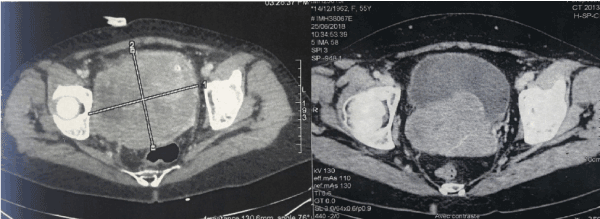
Figure 1. Computed CT scan after injection of contrast shows a large retrovesical tumor, with strong enhancement, compressing the bladder and rectum.
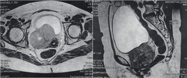
Figure 2. T2-weighted transversal and sagittal MR image shows the retrovesical mass.
Transrectal needle biopsy of the tumor was performed. Histologically, the biopsy revealed a poorly differentiated malignant tumor, consisted of short-spindle cells with rich blood vessels. On immunohistochemical examination, the tumor cells were positive for Pancytokeratin, CD99, and BCL2, and negative for CK7, CD56, CDX2, Chromogranin, synaptophysin, Desmine, myogenine, and CD117. Based on this finding, we concluded to the diagnosis of synovial sarcoma.
Patient refused categorically cystectomy, we discussed her case with the multidisciplinary board, and a neoadjuvant chemotherapy based on adriamycin- ifosfamid followed by exclusive radiotherapy was indicated. She received a total of three cycles of chemotherapy. Pelvis MRI was performed four weeks after chemotherapy, and showed a radiological response estimated equal to 60% of initial disease (Figure 3). We performed exclusive external beam radiotherapy, 66 Gy over 53 days (2 Gy daily).
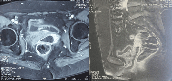
Figure 3. T1-weighted injected transversal and sagittal MR image shows the response after chemotherapy.
6 months later, results of follow-up physical and radiological examinations did not reveal any signs or symptoms of tumor recurrence.
Retrovesical tumor is defined as a tumor arising from retrovesical tissue excluding the pelvic organs (rectum, bladder, prostate or seminal vesicle) [4]. These tumors include malignant or benign lesions. The most frequent malignant tumors are liposarcoma, leiomyosarcoma, rhabdomyosarcoma and malignant lymphoma. Synovial sarcomas are unusual and only few cases have been reported [1].
Synovial sarcoma is an uncommon malignant tumor representing about 5 -10% of all primary soft tissue malignancies worldwide [1,5]. Although this tumor is seen across at any anatomical location, most synovial sarcomas (up to 95%) occur in the extremities, especially in the lower limbs [6]. Synivial sarcomas of the extraperitoneal region are extremely rare, occurring in only 0.3% of cases [2,5], for the most part arising in the abdominal retroperitoneum [6]. Only 10 cases of synovial sarcomas in the pelvic extraperitoneal region have been reported in the literature [7].
Histologically, SS is generally a biphasic tumor (with sarcomatous and epithelial components). The monophasic synovial type is rare and contain of spindle cell elements only. Immunochemical examination allows the differential diagnosis of SS from the other retroperitoneal masses as well as the presence of the characteristic chromosomal translocations (t(X; 18) (p11; q11)) [8].
Synovial sarcomas, irrespective of their location, have no specific clinical and imaging features. Clinical symptoms depend mainly on the size of the tumor, and its relationship to the surrounding organs. In our case, the patient presented urinary symptoms secondary to the bladder compression.
The radiological findings of synovial sarcoma of the rectovesical space are similar to those of tumors at other locations. On CT, we find an heterogeneous mass with an attenuation slightly lower than that of skeletal muscle, we can also identify necrosis or hemorrhage zones, and calcifications [5]. MRI present a high quality image compared to CT, and seems to be the modality of choice for diagnosis and staging. On T1- weighted MR images, SS appeared as a homogeneous rounded soft tissue mass with signal intensity similar to that of skeletal muscle. On T2-weighted images the lesion showed a prominent heterogeneity and predominant hyperintensity due to the cystic component. [9,10].
The optimal treatment of SS of the retrovesical space has not been defined; because of its rarity, there are no adequate randomized prospective studies for therapeutic standardization. The treatment of choice is an en bloc surgical resection of the tumor and adjacent affected organs including the bladder, uterus, and rectum [11]. In our case, the tumor was potentially unrespectable, and patient categorically refused cystectomy, we decide to perform in neoadjuvant chemotherapy then revaluate respectability.
The role of chemotherapy remains uncertain because we don’t have a large data. Neoadjuvant chemotherapy may be used to reduce the size of the tumor and simplifies subsequent surgery, especially in large tumors [8]. Also, the optimal regimen of chemotherapy has not been established, and the histology-tailored chemotherapy did not provide benefit over standard chemotherapy (ifosfamide, vincristine, cyclophosphamide and doxorubicin) [12]. For our patient, three cycles of adriamycin-ifosfamide was performed, with good radiological response.
Regarding radiotherapy, it may be performed in locally advanced and unresectable tumors, and it has been implied as a complementary treatment modality to surgery either before or after the excision with no difference in survival rates [13]. The benefit of radiotherapy in synovial sarcoma is less clear than for chemotherapy, despite the fact that some data in the literature suggest that neoadjuvant administration of chemotherapy combined with radiation may be beneficial [8].
Retrovesical synovial sarcoma has less satisfactory outcomes than soft tissue sarcomas at other sites. Several factors contribute to the poor outcome and high rate of recurrence as the large tumor size, hight grade, difficulties to have a complete excision surgery, and sensibility of surrounding organs to radiotherapy [14]. The 5-year survival rate is less than 30% [15].
Retrovesical synovial sarcoma is very rare tumor with poor prognostic. Surgery represents the mainstay of treatment whenever possible. In case of unresectable tumors, neoadjuvant chemotherapy and radiotherapy can be used with good outcomes.
- Spillane AJ, A'Hern R, Judson IR, Fisher C, Thomas JM (2000) Synovial sarcoma: a clinicopathologic, staging, and prognostic assessment. J Clin Oncol 18: 3794-3803. [Crossref]
- Ulusan S, Kizilkilic O, Yildirim T, Hurcan C, Bal N, et al (2005) Radiological findings of primary retroperitoneal synovial sarcoma. Br J Radiol 78: 166-169.
- Pappo AS, Fontanesi J, Luo X (1994). Synovial sarcoma in children and adolescents: the St Jude Children’s Research Hospital experience. J Clin Oncol 12: 2360-2366.
- Koji H, Yuichi T, Naotsugu I, Yasushi M, Norio N (2006) Rare case of aggressive angiomyxoma presenting as a retrovesical tumor. International Journal of Urology 13, 1012–1014.
- Murphey MD, Gibson MS, Jennings BT, Crespo-Rodríguez AM, Fanburg-Smith J, et al. (2006) From the archives of the AFIP: Imaging of synovial sarcoma with radiologic-pathologic correlation. Radiographics 26: 1543-1565. [Crossref]
- Chatzipantelis P, Kafiri G (2008) Retroperitoneal synovial sarcoma: a clinicopathological study of 6 cases. J BUON 13: 211-216. [Crossref]
- Min CK, Bum SC, Gi SH, Kil SP, Sung JK, et al (2011). Synovial Sarcoma in the Rectovesical Space: A Case Report. J Korean Soc Radiol 65: 167-170.
- Aikaterini M, Dimitrios S, Ioannis SP, George B, Nikolaos M, et al (2019). Management of primary retroperitoneal synovial sarcoma: A case report and review of literature. World J Gastrointest Surg 11: 27-33.
- Jones BC, Sundaram M, Kransdorf MJ (1993) Synovial sarcoma: MR imaging findings in 34 patients. AJR Am J Roentgenol 161: 827-830. [Crossref]
- Lucio O, Luigi B, Serena C, Carlo DB, Matteo F (2015) Monophasic Synovial Sarcoma of Prostatic Fascia: Case Report and Literature Review. Case Reports in Urology.
- Bonvalot S, Rivoire M, Castaing M, Stoeckle E, Le Cesne A, et al (2009). Primary retroperitoneal sarcomas: a multivariate analysis of surgical factors associated with local control. J Clin Oncol 27: 31-37.
- Gronchi A, Ferrari S, Quagliuolo V, Broto JM, Pousa AL, et al (2017) Histotype-tailored neoadjuvant chemotherapy versus standard chemotherapy in patients with high risk soft tissue sarcomas (ISG-STS 1001): an international, open-label, randomised, controlled, phase3, multicentre trial. Lancet Oncol 18: 812.
- Cheng H, Miura JT, Lalehzari M, Rajeev R, Donahue AE (2016). Neoadjuvant radiotherapy for retroperitoneal sarcoma: A systematic review. J Surg Oncol 113: 628-634.
- Tan MC, Brennan MF, Kuk D, Agaram NP, Antonescu CR, et al (2016). Histology-based classification predicts pattern of recurrence and improves risk stratification in primary retroperitoneal sarcoma. Ann Surg 263: 593.
- Kim DH, Joo KR, Cha JM, Shin HP, Lee JI, et al (2012). Retroperitoneal synovial sarcoma manifested by obstructive jaundice in an elderly woman: case report. Clin Endosc 45: 428-430.