The thumb carpometacarpal joint is the most common site of osteoarthritis in the hand with post-menopausal female predominance, and it can be associated with trapezial dysplasia. For treatment of osteoarthritis the use of the ArpeTM prosthesis has proven to be a useful and reliable option with a remarkable long-term survivorship and a high patient satisfaction. However, several initially undiagnosed concomitant or secondarily occurred disorders at the thumb may have negative influence on the outcome, and it can lead to a false diagnosis “failed total thumb carpometacarpal joint replacement”. In the presence of trapezial dysplasia, an exact preoperative planning is absolutely necessary to avoid postoperative dislocation of the implant.
thumb, carpometacarpal joint, osteoarthritis, trapezial dysplasia, total joint replacement, dislocation of implant
CMCJ I: thumb carpometacarpal joint; OA: osteoarthritis.
The thumb carpometacarpal joint (CMCJ I) is the most common site of osteoarthritis (OA) in the hand [1]. Biomechanically, the CMCJ I joint is best described as a "twisted saddle" with two axis for abduction-adduction and extension-flexion; and there are several volar and especially the major strong dorsal ligaments that provide joint stability [2]. Unfortunately, the great mobility that is seen in-vivo in three axis like a "cardan joint" makes that joint intrinsically unstable, and primary OA is "the price of an opposable thumb" [3,4]. This "cardan joint" is a result of evolution in hominid species as a functional adaption to stand upright, freeing the torso and upper limbs [5]. Demographic radiological studies revealed 6:1 female-to-male incidence of CMCJ I OA (mainly starting in post-menopausal females that suggests a correlation with the estrogen level), and this difference decreases with age 75 year and older with an occurrence of 40% in women and 25% in men [6,7].
Major surgical procedures (trapeziectomy with or without ligamentous reconstruction/tendon interposition/suspension, total CMCJ I replacement, CMCJ I arthrodesis) become necessary when conservative treatment (splinting, use of analgetics, intra-articular corticosteroid injections) or minor surgical procedures (denervation, debridement via arthroscopy) have been failed [8]. However, none of all procedures are free of any difficulties or complications, and maybe the clear-cut advice could be the real guiding force of one respondent in the survey by Brunton et al. [9]: "I think you should probably do what works best for you".
The "optimal therapy" for CMCJ I OA would be the in-vivo transformation of the biomechanically determined "saddle joint" to an anatomically stable "ball joint" with a third central axis for pronatoric rotation in order to perform a powerful circumduction by an endoprosthesis. Total CMCJ I replacement reduce pain, improve function, improve grip and pinch strength, and results in excellent patient satisfaction including faster re-employment if the implants have not been failed, and the Arpe prosthesis (Zimmer Biomet Holdings Inc., Warsaw, Indiana/USA), introduced in France 1991, is one type that is current in use [10-14]. However, the main problem of total CMCJ I replacement is long-term surveillance (i.e. aseptic loosening) of its cups [15,16]. To analyze the reasons for these high failure rates, it is necessary to explore the biomechanical imbalance across the long lever of the first metacarpal bone onto the small surface of the trapez bone, which is anatomically determined; and the different topographic load-bearing regions on the surface of trapez bone as well. A tip pinch of 1 kg will generate 12 kg joint compression; and for the power grip, the load may be as high as 120 kg [17].
Technical note to the Arpe prosthesis
The non-cemented ball-and-socket Arpe prosthesis has a metal-on-ultra high molecular weight polyetheylene articulation, and the design resembles a “small hip prosthesis”. The hemispherical titanium alloy and hydroxyapatite coated cup (available with 9 and 10 mm in diameter, retentive or non-retentive) with three spikes at its ground is designed for pressfit insertion providing a high primary stability (Figure 1) [18]. The disadvanatage is that the polyethylene insert is fixed to the cup, hence, if polyethylene wear occurs then the overall cup has to be removed or revised even it may not be loosened [19,20]. The titanium alloy and hydroxyapatite coated stem (available with 4 various sizes) reproduces the anatomical shape of the first metacarpal medullary space (Figure 1), and it should be inserted pressfit within the isthmus of the first metacarpal bone [21]. The intercalated cobalt-chrome head-neck components with 5 various lengths are available in form of a straight or an angled design, noted that an exchange procedure can be difficult due to a possible cold welding between the stem and the head-neck component [22]. The Arpe prosthesis (or other ball-and-socket types) can be combined with a total wrist replacement, an ulnar head prosthesis, other wrist procedures such as four corner fusion or proximal row carpectomy, and the functional outcome is improved if the total CMCJ I replacement is associated with a CMCJ I capsular release [23-26]. Noted that the Arpe prosthesis has proven to be useful as well for primary treatment of comminuted fractures at the base of the first metacarpal bone [27].
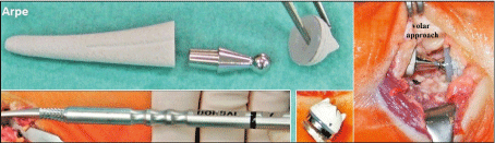
Figure 1. Design and features of the Arpe prosthesis
The problem of a ball-and-socket implants is that the central axis of cup is colinear to the axis of stem resulting in physiological load-bearing for thumb adduction only, whereas the dominant contact pattern for volar abduction and opposition is observed on the central-volar aspect of the surface of trapez bone [28]. To minimize the risk of loosening or dislocation, the Arpe cup should be placed in the centre of range of motion that is parallel to the distal articular surface of trapez bone in the posterior-anterior view, accompanied with its angulation of 7° flexion relative to the distal surface of trapez bone in the lateral view, and these results were confirmed with the use of the Maïa prosthesis (further development of the Arpe with four spikes at the ground of cup and fishscale macrostructure at metacarpal stem) [29,30]. Generally, the risk of dislocation of a total CMCJ I replacement is increased if the cup was placed with dorsal inclination, and medial or lateral inclination exceeding 20° [31]. Placement of the Arpe prosthesis in a „reversed manner“ for treatment of a failed trapeziectomy can not be recommened, however, placement of the Maïa cup into the scaphoid bone revealed first encouraging results [32,33]. A specific contraindication for the Arpe prosthesis is a collapsing trapez bone with height below 11 mm, and a Z-deformity of the thumb with closure of the first web and hyperextension in the CMCJ I which cannot be reduced by tightening the adductor muscle [34].
For placement of the cup parallel to the distal articular surface of the trapez bone it is necessary to investigate preoperatively the trapezial tilt (angle between the longitudinal axis of the second metacarpal bone and the slope of the distal articular surface of the trapez bone, also known as the Devers‘ angle) that is normally 129° ± 6°, and noted that for those cases the axis of the proximal and distal articular surfaces of the trapez bone are nearly perpendicular to the central axis of the scaphoid bone (Figure 2A-C) [35]. Trapezial dysplasia, defined as a trapezial tilt more than 135° associated with a trapezial inclination (axis between the proximal and distal articular surface of trapez bone) more than 10° (Figure 3), can be challenging and it must be considered as a potential risk factor for postoperative dislocation of the prosthesis such as described with the two following case by us [36-38].
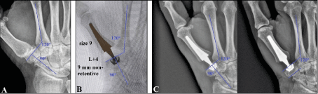
Figure 2. (A) Radiograph of a patient demonstrating advanced stage of CMCJ I OA with a normal Devers‘ angle. Note that the central axis of the scaphoid bone is perpendicular to the distal articular surface of the trapez bone. (B) Same patient intraoperative, the cup of the Arpe prosthesis was correctly inserted parallel to the distal articular surface of the trapez bone, and exatly aligned to the pre-existing normal Devers‘ angle. (C) Same patient, the postoperative radiographs demonstrating the correct placement of the Arpe prosthesis, note that in the absence of trapezial dysplasia the distal articular surface of the trapez bone runs parallel to its proximal articular surface
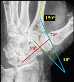
Figure 3. Radiograph of a patient with advanced stage of CMCJ I OA associated with trapezial dysplasia. The Devers‘ angle and trapezial inclination are pathologically expanded on 150° and 20°, and the central axis of scaphoid bone is only perpendicular to the proximal articular surface of the trapez bone
Case 1
A 72-year-old female presented with advanced stage of symptomatic left CMCJ I OA associated with trapezial dysplasia (trapezial tilt 150°) and subluxation of the first metacarpal bone in radial direction (Figure 4A). It was surgically treated by total CMCJ I replacement utilizing the Arpe prosthesis with a 9 mm non-retentive cup. The cup was primarily placed parallel to the distal articular surface of the trapez bone and not aligned to a normal trapezial tilt of < 135°. Two weeks after surgery, the patient reported sudden pain at her left thumb in the absence of a relevant trauma. Radiographically there was a complete dislocation of the Arpe prosthesis in radial direction (Figure 4B). For revision an exchange procedure utilizing a 10 mm non-retentive cup correctly aligned to a normal trapezial tilt of 120° became necessary (Figure 4C). At the 1-year follow-up there was unchanged correct positioning of the Arpe prosthesis radiographically, and the patient was very satisfied with her painless functional outcome (Figure 5A-B).
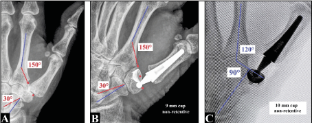
Figure 4 (Case presentation 1). (A) Radiograph showing the initial finding with advanced stage of CMCJ I OA associated with trapezial dysplasia. (B) The 9 mm cup of the Arpe prosthesis was inserted parallel to the distal articular surface of the trapez bone that resulted in early dislocation of the implant. (C) Revision surgery utilizing a 10 mm cup exactly aligned to a normal Devers‘ angle became necessary
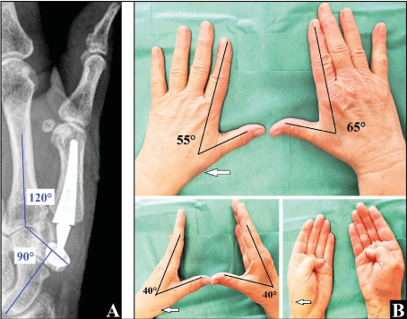
Figure 5 (Case presentation 1). (A) At the 1-year follow-up there was unchanged correct positioning of the Arpe prosthesis radiographically. (B) The patient is very satisfied with her functional outcome. The white arrows showing the uneventful healing of the dorsal surgical incision
Case 2
A 59-year-old male presented with advanced stage of symptomatic right CMCJ I OA associated with trapezial dysplasia (trapezial tilt 150°) and subluxation of the first metacarpal bone in radial direction (Figure 6A). It was surgically treated by total CMCJ I replacement utilizing the Arpe prosthesis with a 9 mm non-retentive cup. The cup was primarily placed correctly aligned to a normal trapezial tilt of 120°, and not parallel to the distal articular surface of the trapez bone (Figure 6B). The postoperative course was uneventful. At the 1-year follow-up there was unchanged correct positioning of the Arpe prosthesis radiographically, and the patient was very satisfied with his painless functional outcome (Figure 6C-D).
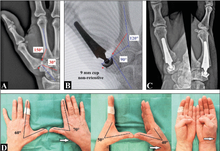
Figure 6 (Case presentation 2). (A) of CMCJ I OA associated with trapezial dysplasia. (B) The Arpe cup was placed exactly aligned to the normal Devers‘ angle that means not parallel to the distal articular surface of the trapez bone. The further course was uneventful. (C) At the 1-year follow-up there was unchanged correct positioning of the Arpe prosthesis radiographically. (D) The patient is very satisfied with his functional outcome. The white arrows showing the uneventful healing of the dorsal surgical incision
Newer studies revealed that the radiographic staging for severity of CMCJ I OA by Eaton-Littler (1973) and Eaton-Glickel (1987) alone are likely unable in decision making which treatment would be appropriate. Thus, it has been recommended to introduce a new classification that incorporates more the preoperative patient's disability; and diagnosis and treatment should be based on the surgeon's qualitative assessment combining history, physical examination, the patients claims in activities of daily living, radiographic evaluation, surgical experience of the treating physicians, and subjective patients factors [9, 39-44]. Moreover, psychological factors or disorders of the patients may have influence on the outcome after treatment of CMCJ I OA [45,46].
Compared to trapeziectomy as unchanged the most widely performed procedure worldwide, the rationale behind total CMCJ I replacement is that this procedure obtains normal length of the thumb associated with normal muscle forces resulting in a higher carpometacarpal and metacarpaophalangeal joint stability, no occurrence of painful impingement between the base of the first metacarpal bone and the scaphoid and/or trapezoid bone, and normal tendon loads in order to achieve a certain key pinch force [47-51]. Noted that total CMCJ I replacement is not generally to be considered as contraindication for patients with high claims in occupational work and leisure [52,53]. However, mid- to long-term results with the Moje ceramic prosthesis and metal-on-metal bearing types were disappointing, and for those cases the Arpe prosthesis has proven to be useful for a revision total CMCJ I replacement, and the last salvage option for a failed total CMCJ I replacement is conversion to trapeziectomy that revealed an identical outcome to those after a primary procedure [8,13,54,55]. Noted that for those cases the removal of the metacarpal stem is not required if it does not protrude beyond the resection plane at the base of the first metacarpal bone [8,13,56]. The third major procedure is the CMCJ I arthrodesis, however, due to its high complication rate it is not recommended for women aged 40 years and older, and the patient's satisfaction is only high in 88 % of cases when osseous fusion is obtained [57-59].
Four long-term outcome studies with the Arpe prosthesis revealed a remarkable survivorship after 10 to 12 years with relative portions ranging from 85% to 93.3% associated with a high patient satisfaction (Figure 7A-B), and after 15 years with 80% that is not far from the 15-year total hip survival reported from the combined Nordic Arthroplasty Registry Association in 2014 and it can be considered acceptable [60-64]. Despite these good and reliable results, the Arpe prosthesis was surprisingly withdrawn from the markektplace by the company in 2021 in the absence of any scientific evidence for it, and this is the same inexplicable situation such as with the Lateral Resurfacing Elbow system and the Maestro wrist prosthesis by the same company [65,66].
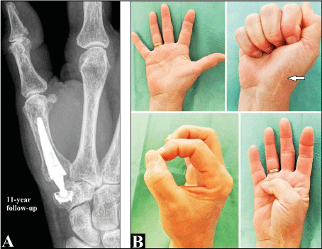
Figure 7. (A) Radiograph of a 72-year-old female (right hand) showing correct positioning of the Arpe prosthesis at the 11-year follow-up without any signs of loosening or subsidence. (B) The patient is very satisfied with her functional outcome, note the uneventful healing of the volar surgical incision (arrow)
The main postoperative complication of the Arpe implant is dislocation observed in up to 9.6% of cases which is mostly based on technical errors by the surgeons at time of insertion of implant such as primarily failed positioning of the cup or utilizing an intercalated head-neck component which is too short potentially leading to longitudinal instability [13,54,67]. The main cause of failure is aseptic loosening of the cup, however, subsidence in the absence of clinical symptoms or dislocation of the cup does not require revision surgery (Figure 8A-B). Other factors which can be associated with a poor postoperative outcome are primarily undiagnosed concomitant or secondarily occurred disorders such as trigger finger observed in 6% of cases (Figure 9A-B), carpal tunnel syndrome observed in 25% of cases (Figure 10A-D), De Quervain tenosynovitis observed in up to 21% of cases (Figure 10A-D), synovial cyst observed in 3% of cases, Dupuytren’s disease observed in 2% of cases, and pathologies around the metacarpophalangeal joint of the thumb (Figure 11A-D, Figure 12A-C) [61,68]. Noted that these disorders around the thumb can lead to a false diagnosis „failed total CMCJ I replacement“ by unexperienced surgeons (Figure 9A-B, Figure 12A-C).

Figure 8. (A) A female sustained with age of 63 years a total CMCJ I replacement (right hand) utilizing the Arpe prosthesis without any signs of subsidence of the cup at the 1-year follow-up. Marked signs of subsidence of the cup in the absence of clinical symptoms were seen at the 6-year follow-up. At the 9-year follow-up subsidence had been stabilized. (B) The patient is unchanged very satisfied with her functional outcome
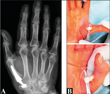
Figure 9. (A) A 67-year- old female sustained a total CMCJ I replacement (right hand) utilizing the Arpe prosthesis. (B) Six months after surgery the patient reported typical symptoms of a trigger thumb. After pulley release there was an uneventful further course. Noted that another surgeon had primarily diagnosed a failed total CMCJ I replacement
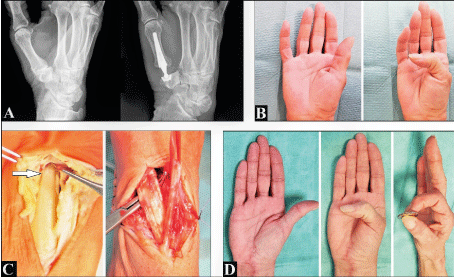
Figure 10. (A) A 59-year-old female presented with advanced stage of right CMCJ I OA sustained a total CMCJ I replacement utilizing the Arpe prosthesis. At this time the patient reported no symptoms regarding a concomitant carpal tunnel syndrom. (B) Three months after surgery there was a poor functional outcome with her thumb associated with a typical symptoms of De Quervain tenosynovitis. Surprisingly, the patient first reported about symptoms of a carpal tunnel syndrom which would have existed a long time before the total CMCJ I replacement. Neurological evaluation revealed a severe carpal tunnel syndrom. (C) Intraoperative clinical photographs showing the open decompression and neurolysis of the median nerve (white arrow: hourglass-like constriction), and opening of the first and second compartment of extensor retinaculum. (D) Six months after second surgery there was a good and painless restoration of thumb‘s functionality
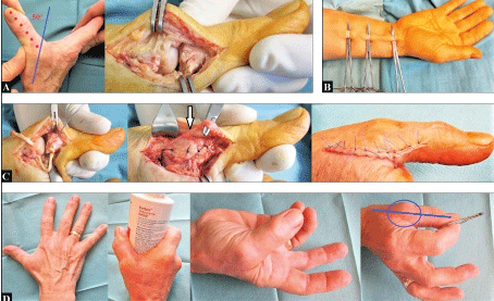
Figure 11. (A) A 59-year-old female presented with a traumatic disruption of the ulnar collateral ligament at the right metacarpophalangeal joint two years after total CMCJ I replacement utilizing the Arpe prosthesis at the same thumb. (B) Harvesting the palmaris longus tendon for ligamentous reconstruction. (C) Reconstruction of the joint capsule and the ulnar collateral ligament with the harvested palmaris longus tendon. (D) Six months after ligamentous reconstruction there ewas an excellent functional outcome. Note the complete restoration of the ulnar sided stability at the metacarpophalangeal joint when performing tip pinch (line and circle)
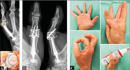
Figure 12. (A) A 58-year-old female reported increasing pain at her right thumb two years after insertion of the Arpe prosthesis. Radiographically there was a longstanding ulnar subluxation in the metacarpophalangeal joint (yellow circle) due to a traumatic disruption of the radial collateral ligament several years ago that resulted in post-traumatic OA. Noted that another surgeon had primarily diagnosed a failed total CMCJ I replacement. The intraoperative clinical photograph showing the damage of the joint cartilage at the head of the first metacarpal bone (black circle). (B) The arthrodesis of the metacarphalangeal joint was performed. (C) One year after arthrodesis the patient was very satisfied with her functional outcome
The etiology of isolated trapezial dysplasia is still widely unknown. The abnormal Devers‘ angle leads to a deviating load distribution through the CMCJ I and gradual attenuation of the surrounding ligaments and articular surface. This results in a progressive dorsoradial subluxation of the first metacarpal over the trapez bone, and later associated with development of CMCJ I OA. Symptomatic trapezial dysplasia with CMCJ I instability can lead to a disabling condition. Patients report pain with pinch, decreased range of motion, and decreased grip strength. It is most frequently seen in young female patients and in the dominant hand. The symptoms usually start during the late teens and early twenties in patients with high demands in work and leisure [37,69-76]. For early stages, osteotomies at the base of the first metacarpal bone and/or trapez bone has proven to be a useful and reliable procedure, however, it must be noted that these procedures did not always protect the CMCJ I from further progression of OA [37,69,70].
No reports in the literature could be found by us about possible difficulties for a total CMCJ I replacement (i.e. correct placement of the metacarpal stem into the intramedullary space crossing the isthmus) after a previously performed closed or open wedge osteotomy at the first metacarpal bone. With our two described cases it can be concluded that an exact preoperative planning is absolutely necessary when performing a total CMCJ I replacement in the presence of trapezial dysplasia to avoid postoperative dislocation of the implant.
The author declares no potential conflicts of interest with respect to the research, authorship, and/or publication of this article.
None.
The author would like to thank Dr. Frank Wilke and Dr. Thomas Broska including their staff from the Helios Klinikum Gotha (Germany), and Dr. Bärbel Lesser (Waltershausen, Germany) for their excellent support during writing this article.
On page 2 the reference numbers 29 and 30 were incorrectly cited „To minimize the risk of looesening or dislocation, the Arpe cup should be placed ... parallel to the distal articular surface of trapez bone ...“).
Correctly cited is: „To minimize the risk of loosening or dislocation, the Arpe cup should be placed ... parallel to the proximal articular surface of trapez bone ...“. We apologize for this failure.
To avoid a misunderstanding, with a non-dysplastic trapez bone (trapezial inclination with ≤ 10°) the distal articular surface runs parallel to the proximal surface, and both surfaces are perpendicular to the central axis of scaphoid bone (Figure 2B). Therefore, to our experience with more than 150 inserted Arpe prostheses within the last 10 years the cup should be placed parallel both to the proximal and distal surface. With a dysplastic trapez bone (trapezial inclination with > 10°) the central axis of scaphoid bone is perpendicular only to the proximal articular surface (Figure 3), therefore, for those cases the Arpe cup should be placed parallel only to the proximal articular surface.
- Giddins G (2012) Thumb arthroplasties. J Hand Surg Eur Vol 37: 603-604.
- Lin JD, Karl JW, Strauch RJ (2014) Trapeziometacarpal joint stability: the evolving importance of the dorsal ligaments. Clin Orthop Relat Res 472: 1138-1145.
- Chèze L, Doriot N, Eckert M, Rumelhart C, Comtet JJ (2001) In vivo cinematic study of the trapezometacarpal joint. Chir Main 20: 23-30.
- Turker T, Thirkannad S (2011) Trapezio-metacarpal arthritis: The price of an opposable thumb! Indian J Plast Surg 44: 308-316.
- Ladd AL, Weiss AP, Crisco JJ, Hagert E, Wolf JM, Glickel SZ, Yao J (2013) The thumb carpometacarpal joint: anatomy, hormones, and biomechanics. Instr Course Lect 62: 165-179.
- Sodha S, Ring D, Zurakowski D, Jupiter JB (2005) Prevalence of osteoarthrosis of the trapeziometacarpal joint. J Bone Joint Surg Am 87: 2614-2618.
- Van Heest AE, Kallemeier P (2008) Thumb carpal metacarpal arthritis. J Am Acad Orthop Surg 16: 140-151.
- Schmidt I (2015) Surgical Treatment Options in Thumb Carpometacarpal Osteoarthritis: A Recent Literature Overview Searching for Practice Pattern with Special Focus on Total Joint Replacement. Curr Rheumatol Rev 11: 39-46.
- Brunton LM, Wilgis EF (2010) A survey to determine practice patterns in the surgical treatment of advanced thumb carpometacarpal osteoarthrosis. Hand (NY) 5: 415-422.
- Vermeulen GM, Slijper H, Feitz R, Hovius SE, Moojen TM, Selles RW (2011) Surgical management of primary thumb carpometacarpal osteoarthritis: a systematic review. J Hand Surg Am 36: 157-169.
- Erne H, Scheiber C, Schmauss D et al. (2018) Total Endoprosthesis Versus Lundborg´s Resection Arthroplasty for the Treatment of Trapeziometacarpal Joint Osteoarthritis. Plast Reconstr Surg Glob Open 6: e1737.
- Comtet JJ. Rumelhart C (2001) Total trapezometacarpal prostheses: concepts and classification study. Chir Main 20: 48-54.
- Schmidt I (2018) Treatment Options for Thumb Carpometacarpal Joint Osteoarthritis with an Update to the ArpeTM Prosthesis. Recent Adv Arthroplast 2: 55-63.
- Lerebours A, Marin F, Bouvier S, Egles C, Rassineux A, Masquelet AC (2020) Trends in Trapeziometacarpal Implant Design: A Systematic Survey Based on Patents and Administrative Databases. J Hand Surg Am 45: 223-238.
- Krughaug Y, Lie SA, Havelin LI, Furnes O, Hove LM, Hallan G (2014) The results of 479 thumb carpometacarpal joint replacements reported in the Norwegian Arthroplasty Register. J Hand Surg Eur Vol 39: 819-825.
- Huang K, Hollevoet N, Giddins G (2015) Thumb carpometacarpal joint total arthroplasty: a systematic review. J Hand Surg Eur Vol 40: 338-350.
- Cooney WP 3rd, Chao EY (1977) Biomechanical analysis of static forces in the thumb during hand function. J Bone Joint Surg Am 59: 27-36.
- Bruyère Garnier K, Dumas R, Rumelhart C, Comtet JJ (2001) Comparison of primary trapezometacarpal cup fixation using mechanical tests. Chir Main 20: 55-62.
- Eecken SV, Vanhove W, Hollevoet N (2012) Trapeziometacarpal joint replacement with the Arpe prosthesis. Acta Orthop Belg 78: 724-729.
- Thorkildsen R, Reigstad O, Røkkum M (2021) Early dismantling of the polyethylene liner in the Arpe® trapezial cup: a report of two cases. J Hand Surg Eur Vol Apr 29: doi: 10.1177/17531934211012203. Online ahead of print.
- Apard T, Saint-Cast Y (2007) Results of a 5 years follow-up of Arpe prosthesis for the basal thumb osteoarthritis. Chir Main 26: 88-94.
- Salva-Coll G, Terrades-Cladera X (2017) Cold Welding in a Trapeziometacarpal Ball and Socket Prosthesis: A Case Report. J Hand Surg Asian Pac Vol 22: 259-261.
- Schmidt I (2017) Combined replacements of the wrist, ulnar head, and thumb carpometacarpal joint. Case report, technical note, and recent evidence to the ArpeTM prosthesis. Trauma Emerg Care 2: doi: 10.15761/TEC.1000130.
- Kandemir G, Smith S, Schmidt I, Joyce TJ (2020) Explant analysis of a Maestro™ wrist prosthesis and calculation of its lubrication regime. J Mech Behav Biomed Mat 110: 103933. doi.org/10.1016/j.jmbbm.2020.103933.
- Waitzenegger T, Leclercq C, Masmejean E, Lenoir H, Harir A, Coulet B, Chammas M (2015) Combined Treatment of Wrist and Trapeziometacarpal Joint Arthritis. J Wrist Surg 4: 301-306.
- Van Hove B , Vantilt J, Bruijnes A, Caekebeke P, Corten K, Degreef I, Duerinckx J (2020) Trapeziometacarpal total joint arthroplasty: The effect of capsular release on range of motion. Hand Surg Rehabil 39: 413-416.
- Barrera-Ochoa S, Mendez-Sanchez G, Mir-Bullo X (2017) Primary trapeziometacarpal prosthesis for complicated fracture of the base of the thumb metacarpal. J Hand Surg Eur Vol 42: 972-974.
- Goto A, Leng S, Sugamoto K, Cooney WP 3rd, Kakar S, et al (2014) In vivo pilot study evaluating the thumb carpometacarpal joint during circumduction. Clin Orthop Relat Res 472; 1106-1113.
- Duerinckx J, Caekebeke P (2001) Trapezium anatomy as a radiographic reference for optimal cup orientation in total trapeziometacarpal joint arthroplasty. J Hand Surg Eur Vol 41: 939-943.
- Caekebeke P, Duerinckx J (2018) Can surgical guidelines minimize complications after Maïa® trapeziometacarpal joint arthroplasty with unconstrained cups? J Hand Surg Eur Vol 43: 420-425.
- Brauns A, Caekebeke C, Duerinckx J (2019) The effect of cup orientation on stability of trapeziometacarpal total joint arthroplasty: a biomechanical cadaver study. J Hand Surg Eur Vol 44: 708-713.
- Goorens CK, Van Schaik DE, Goubau JF (2015) Surgical treatment after a failed trapeziectomy: A case report. Chir Main 34: 205-209.
- Chiche L, Lamarre H, Barbary S, Teissier J (2020) Scaphometacarpal arthroplasty: a report of ten cases of trapeziometacarpal prosthesis and trapeziectomy revision. J Hand Surg Eur Vol 45: 483-487.
- Teissier J, Gaudin T, Marc T (2001) Problems with the metacarpophalangeal joint in the surgical treatment of osteoarthritis by inserting an ARPE type joint prosthesis. Chir Main 20: 68-70.
- Kapandji TG, Kapandji AI (1993) New radiologic data on the trapezo-metacarpal joint: The results of 330 cases. Ann Chir 12: 263-274.
- Kapandji A, Moatti E, Raab C (1980) Specific radiography of the trapezo-metacarpal joint and its technique (author’s transl). Ann Chir 34: 719-726.
- Ropars M, Siret P, Kaila R, et al (2009) Anatomical and radiological assessement of trapezial osteotomy for trapezial dysplasia in early trapeziometacarpal joint arthritis. J Hand Surg Eur Vol 34: 264-267.
- Van Royen K, Gevers M, Vanmierlo B, Scheerlinck T, Goubau J (2021) Radiological measurement of trapezial dysplasia - variation of trapezial tilt and trapezial inclination. Hand Surg Rehabil 40: 44-50.
- Berger AJ, Momeni A, Ladd AL (2014) Intra- and interobserver reliability of the Eaton classification for trapeziometacarpal arthritis: a systematic review. Clin Orthop Relat Res 472: 1155-1159.
- Larsen SK, Østergaard AM, Hansen TB (2015) The influence of subluxation on the severity of symptoms, disability, and the results of operative treatment in TMC osteoarthritis with total joint arthroplasty. Hand (NY) 10: 593-597.
- Becker SJ, Teunis T, Ring D, Vranceanu AM (2016) The Trapeziometacarpal Arthrosis Symptoms and Disability Questionnaire: Development and Preliminary Validation. Hand (NY) 11: 197-205.
- Tsehaie J, van der Oest MJW, Poelstra R, et al. (2019) Positive experience with treatment is associated with better surgical outcome in trapeziometacarpal osteoarthritis. J Hand Surg Eur Vol 44: 714-721.
- Marks M, Grobet C, Audigé L, Herren DB (2019) Clinical thresholds of symptoms for deciding on surgery for trapeziometacarpal osteoarthritis. J Hand Surg Eur Vol 44: 937-945.
- Ottenhoff JSE, Teunis T, Janssen SJ, et al. (2020) Variation in Offer of Operative Treatment to Patients With Trapeziometacarpal Osteoarthritis. J Hand Surg Am 45: 123-130.
- Calfee R, Chu J, Sorensen A, Martens E, Elfar J (2015) What Is the Impact of Comorbidities on Self-rated Hand Function in Patients With Symptomatic Trapeziometacarpal Arthritis? Clin Orthop Relat Res 473: 3477-3483.
- Hoogendam L, van der Oest MJW, Tsehaie J, et al. (2019) Psychological factors are more strongly associated with pain than radiographic severity in non-invasively treated first carpometacarpal osteoarthritis. Disabil Rehabil 1-6.
- Degeorge B, Dagneaux L, Andrin J, Lazerges C, Coulet B, Chammas M (2018) Do trapeziometacarpal prosthesis provide better metacarpophalangeal stability than trapeziectomy and ligamentoplasty? Orthop Traumatol Surg Res 104: 1095-1100.
- Shah DS, Middleton C, Gurdezi S, Horwitz MD, Kedgley AE (2020) The Effect of Surgical Treatments for Trapeziometacarpal Osteoarthritis on Wrist Biomechanics: A Cadaver Study. J Hand Surg Am 45: 389-398.
- Hansen TB (2021) Joint replacement for trapeziometacarpal osteoarthritis: implants and outcomes. J Hand Surg Eur Vol 46: 115-119.
- Athlani L, Motte D, Martel M, Moissenet F, Mottet J, Beaulieu JY (2021) Comparison of simulated key pinch after three surgical procedures for trapeziometacarpal osteoarthritis: a cadaver study. J Hand Surg Eur Vol May 18; doi: 10.1177/17531934211015915. Online ahead of print.
- Renfree KJ, Roarke MC (2021) Thumb Metacarpal-Trapezoid Impingement as an Etiology of Pain After Trapeziectomy and Basal Joint Soft Tissue Arthroplasty: A Case Series. J Hand Surg Am Apr 9;S0363-5023(21)00126-X. doi: 10.1016/j.jhsa.2021.02.017. Online ahead of print.
- Kirkeby L, Frost P, Svendsen SW, Hansen TB (2021) Revision rates of trapeziometacarpal total joint arthroplasty in relation to occupational hand force requirements. J Hand Surg Eur Vol Mar 12;1753193421996980. doi: 10.1177/1753193421996980. Online ahead of print.
- Kirkeby L, Frost P, Hansen TB, Svendsen SW (2021) Influence of Occupational Hand Force Requirements on Return to Work, Pain, and Disability After Trapeziometacarpal Total Joint Arthroplasty: A 12-Month Follow-Up Study. J Hand Surg Eur Vol Mar 26;S0363-5023(21)00043-5. doi: 10.1016/j.jhsa.2021.01.016. Online ahead of print.
- Schmidt I (2014) Thumb CMC total exchange arthroplasty with the ARPE implant. Chir Main 33: 295-298.
- Kaszap B, Daecke W, Jung M (2013) Outcome comparision of primary trapeziectomy versus secondary trapeziectomy following failed total trapeziometacarpal joint replacement. J Hand Surg Am 38: 863-871.
- Cootjans K, Vanhaecke J, Dezillie M, Barth J, Pottel H, Stockmans F (2017) Joint Survival Analysis and Clinical Outcome of Total Joint Arthroplasties With the ARPE Implant in the Treatment of Trapeziometacarpal Osteoarthritis With a Minimal Follow-Up of 5 Years. J Hand Surg Am 42: 630-638.
- Vermeulen GM, Brink SM, Slijper H, Feitz R, Moojen TM, Hovius SE, et al. (2014) Trapeziometacarpal arthrodesis or trapeziectomy with ligament reconstruction in primary trapeziometacarpal osteoarthritis: a randomized controlled trial. J Bone Joint Surg Am 96: 726-733.
- De Smet L, Van Meir N, Verhoeven N, Degreef I (2010) Is there still a place for arthrodesis in the surgical treatment of basal joint osteoarthritis of the thumb? ActaOrthop Belg 76: 719-724.
- De Smet L, Vaes F, Van Den Broecke J (2005) Arthrodesis of the trapeziometacarpal joint for basal joint osteoarthritis of the thumb: the importance of obtaining osseous union. Chir Main 24: 222-224.
- Martin-Ferrero M (2014) Ten-year long-term results of total joint arthroplasties with ARPE® implant in the treatment of trapeziometacarpal osteoarthritis. J Hand Surg Eur Vol 39: 826-832.
- Martin-Ferrero M, Simón-Pérez C, Coco-Martín MB, Vega-Castrillo A, Aguado-Hernández H, Mayo-Iscar A (2020) Trapeziometacarpal total joint arthroplasty for osteoarthritis: 199 patients with a minimum of 10 years follow-up. J Hand Surg Eur Vol 45: 443-451.
- De Smet A, Vanhove W, Benis S, Verstraete M, Hollevoet N (2020) Ten-year outcomes of the Arpe prosthesis for the treatment of osteoarthritis of the trapeziometacarpal joint. Acta Orthop Belg 86: 131-136.
- Dumartinet-Gibaud R, Bigorre N, Raimbeau G, Jeudy J, Saint Cast Y (2020) Arpe total joint arthroplasty for trapeziometacarpal osteoarthritis: 80 thumbs in 63 patients with a minimum of 10 years follow-up. J Hand Surg Eur Vol 45: 465-469.
- Boeckstyns MEH (2020) Commentary on: Arpe total joint arthroplasty for trapeziometacarpal osteoarthritis: 80 thumbs in 63 patients with a minimum of 10 years follow-up. J Hand Surg Eur Vol 45: 470-471.
- Schmidt I (2017) A Complicated Course of a Coronal Shear Fracture Type IV of the Distal Part of Humerus Resulting in Resurfacing Radiocapitellar Joint Replacement. Open Orthop J 11: 248-254.
- Schmidt I (2018) A critical appraisal to the decision by the company Zimmer Biomet to withdraw the MaestroTM Wrist Reconstructive System from the marketplace. Trauma Emerg Care 3: doi: 10.15761/TEC.1000162.
- Mahmood I, Burton R, Choudhry MN, Coapes C (2020) Early to Mid-Term Results in Trapezio-metacarpal Joint Replacement using the ARPE Implant. Open Orthop J 14: 198-203.
- Gómez-Garrido D, Triviño-Mayoral V, Delgado-Alcala V, Cervera-Irimia J, Medina-Lorca M, et al. (2019) Five year long term result of total joint arthroplasties in the treatment of trapeziometacarpal osteoarthritis. Acta Biomed 90: 451-456.
- Tomaino MM (2000) Treatment of Eaton stage I trapeziometacarpal disease with thumb metacarpal extension osteotomy. J Hand Surg Am 25:1100-1106.
- Thuysbaert G, Goorens CK, Vanmierlo B, Mermuys K, Goubau J (2021) Addition-Subtraction Osteotomy With Ligamentoplasty for Symptomatic Trapezial Dysplasia With Metacarpal Instability: A 10-Year Follow-Up. J Hand Surg Am 46: 342.e1-342.e9.
- Wilson JN, Bossley CJ (1983) Osteotomy in the treatment of osteoarthritis of the first carpometacarpal joint. J Bone Joint Surg Br 65: 179-181.
- Kapandji AI, Heim UFA (2002) Reorientation osteotomy of the trapezial saddle. Chir Main 21: 124-133.
- Goubau JF, Kerckhove D, Berghs B (2007) Addition-subtraction osteotomy combined with ligamentoplasty for symptomatic trapezial dysplasia with instability. Chir Main 26: 26-30.
- Cheema T, Salas C, Morrell N, Lansing L, Reda Taha MM, Mercer D (2012) Opening wedge trapezial osteotomy as possible treatment for early trapeziometacarpal osteoarthritis: a biomechanical investigation of radial subluxation, contact area, and contact pressure. J Hand Surg Am 37: 699-705.
- Goorens CK, Goubau J, Van Hoonacker P, Kerckhove D, Berghs B, Scheerlinck T (2013) Addition-subtraction osteotomy without ligamentoplasty for symptomatic trapezial dysplasia with metacarpal instability. Acta Orthop Belg 79: 517-523.
- El-Mahy MM (2013) Trapezial osteotomy for the treatment of trapezial dysplasia. Ann Plast Surg 71: 485-488.