An 82-year old female patient was referred to our department for Bone Scan (BS) for differential diagnosis of soft tissue infection at amputation site. Vascular phase and whole body BS revealed focally increased thyroid uptake in the left neck. Multinodular goiter with internal micro and macrocalcifications were apparent in neck ultrasonography. 99mTc pertecnethate scintigraphy confirmed focally increased uptake with hypoactive areas probably due to cystic/necrotic field in toxic nodules. The patient’s thyroid function tests were as follows; TSH <0.003 µIU/mL, sT3 3,23 µg/mL, sT4 0,88 ng/mL. The history of patient and thyroid function tests supported diagnosis of toxic multinodular goiter.
bone scan, thyroid, toxic multinodular goitre
99mTc Methylene Diphosphonate (MDP) uptake in extraosseus areas is unexpected but it is clinically important. MDP acts as ligand adsorbing onto tissue calcium, localizing the 99mTc in the mineral phase with no significant organic subtrate interaction. The general mechanism of 99mTc MDP uptake is binding to hydroxyapatite crystals and calcium salts causing the skeletal accumulation and uptake in the other of calcium deposition in the body. In this case, we presented an 82-year old female patient who was referred to our department for Bone Scan (BS) and had focally increased thyroid uptake in the left neck at vascular phase and whole body BS.
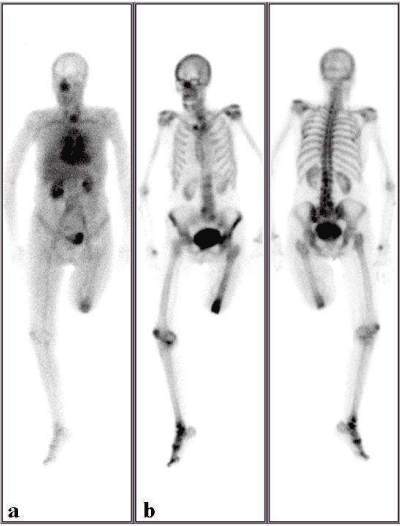
An 82-year old female patient was referred to BS for diagnosis of soft tissue infection and osteomyelitis at amputation site. After regional dynamic and blood pool images at amputation site, vascular phase of the whole-body scan has shown incidental hyperemia and whole body Bone Scan (BS) presented thyroid uptake at the left lower neck. Vascular phase and whole body BS revealed focally increased thyroid uptake in the left neck.
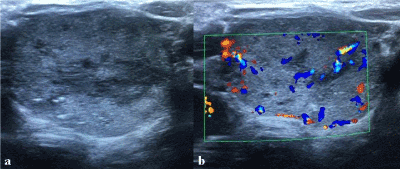
Thyroid ultrasonography showing the nodule in the left lobe which fills left lobe nearly complete (34×22 mm in size) with cystic, necrotic field with internal multiple micro and macrocalcification within highly increased vascularity of the nodules. Multinodular goiter with internal micro and macrocalcifications were apparent in neck ultrasonography. The findings were consistent with toxic multinodular goiter.
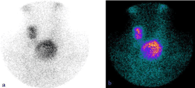
99mTc pertecnethate scintigraphy confirmed focally increased uptake with hypoactive areas probably due to cystic/necrotic field in toxic nodules. The patient’s thyroid function tests were as follows; TSH <0.003 µIU/mL, sT3 3,23 µg/mL, sT4 0,88 ng/mL. The history of patient and thyroid function tests supported diagnosis of toxic multinodular goiter.
In our case, it was the vascular phase of the whole-body scan showing focally intense hyperemia in the toxic nodules even though MDP uptake in thyroid in bone phase was also apparent. In our department, we obtain whole body scan of BS patients in vascular phase to evaluate the alterations in bone modeling profoundly and for any incidentally information about the patient that might be important without any additional radiation dose. MDP uptake in extraosseus areas is unexpected but it is clinically important. Recent research indicated that MDP acts as ligand adsorbing onto tissue calcium, localizing the 99mTc in the mineral phase with no significant organic subtrate interaction [1,2]. The general mechanism of 99mTc MDP uptake is binding to hydroxyapatite crystals and calcium salts causing the skeletal accumulation and uptake in the other of calcium deposition in the body [3,4]. In our case, increased 99mTc MDP uptake in the thyroid nodules was possibly caused by calcifications of the nodules (Figure 1). 99mTc pertecnethate scintigraphy confirmed focally increased uptake with hypoactive areas probably due to cystic/necrotic field in toxic nodules (Figure 2). Regarding Kurooka et al., classification of thyroid cartilage uptake in BS, our case has slight inhomogeneous accumulation in thyroid cartilage, which was the most common pattern in BS [5] (Figure 3).

Figure 1. A 82-year old female patient was referred to bone scan (BS) for diagnosis of soft tissue infection and osteomyelitis at amputation site. After regional dynamic and blood pool images at amputation site, vascular phase of the whole-body scan (a) has shown incidental hyperemia and whole body BS (b) presented thyroid uptake at the left lower neck.

Figure 2. Two nodules have shown focally increased radioactivity uptake on 99mTc pertechnetate thyroid scintigraphy. The largest one was at the lower pole of left thyroid lobe with hypoactive areas within probably due to cystic/necrotic field, and the other one was in the upper pole of right lobe.

Figure 3. Thyroid ultrasonography showing the nodule in the left lobe (a) which fills left lobe nearly complete (34×22 mm in size) with cystic, necrotic field with internal multiple micro and macrocalcification within highly increased vascularity of the nodules (b). The findings were consistent with toxic multinodular goiter.
Vascular phase whole body screening should be complementary to regular BS which may be very useful and give additional information about diagnosis of other diseases and allow early detection [6]. Clinician especially nuclear medicine physicians should be aware that incidental thyroid nodules may demonstrate 99mTc MDP accumulation probably due to calcification in nodular area on BS [7].
- Zuckier LS, Freeman LM (2010) Nonosseous, nonurologic uptake on bone scintigraphy: atlas and analysis. Semin Nucl Med 40:242-256. [Crossref]
- Rehm PK, Sharma S (2004) Focal thyroid uptake on bone scan due to thyroid biopsy. Clin Nucl Med 29:849-851. [Crossref]
- Wale DJ, Wong KK, Savas H (2015) Extraosseous Findings on Bone Scintigraphy Using Fusion SPECT/CT and Correlative Imaging. AJR Am J Roentgenol 205:160-172. [Crossref]
- Montes TC, Muñoz C, Rivero JI (1999) Uptake of Tc-99m sestamibi and Tc-99m MDP in anaplastic carcinoma of the thyroid (nondiagnostic CT and ultrasound scans). Clin Nucl Med 24:355-356. [Crossref]
- Kurooka H, Kawabe J, Tsumoto C (2009) Examination of pattern of RI accumulation in thyroid cartilage on bone scintigraphy. Ann Nucl Med 23:43-48. [Crossref]
- Bertagna F, Bosio G, Giubbini R (2009) Incidental thyroid Tc-99m methylene diphosphonate (MDP) uptake in a patient affected by polynodular goiter at bone scintigraphy. Nucl Med Rev Cent East Eur 12:81-82. [Crossref]
- Mulazimoglu M, Karyagar S, Saglampinar S (2008) Does radioiodine treatment change the methylene diphosphonate uptake in a toxic thyroid nodule on bone scintigraphy? Clin Nucl Med 33:731-733. [Crossref]


