Mesonephric-like adenocarcinomas are rare tumors occurring in the uterine body and ovaries and show histological similarities to mesonephric carcinoma of the uterine cervix and can be identified immunohistochemical expression of mesonephric markers. The occurrence of mesonephric carcinomas in the cervical serosa where mesonephric remnants occur is well recognized, however, it is controversial as to whether these carcinomas of the ovary and uterine body originate from mesonephric remnants.
Here we report on the histomorphological and immunohistochemical characteristics of 3 cases of mesonephric-like adenocarcinomas occurring within the uterine body and the ovaries and present a comparative analysis with mesonephric remnants.
None of the three cases, one case of uterine body, and 2 cases of the ovaries, displayed any characteristic histological findings applicable other gynecological cancers. Additionally, an eosinophilic colloid-like material was observed in some glands within the tumor, which appeared to be similar to mesonephric carcinoma originating in the mesonephric remnants and mesonephric carcinoma.
Immunohistochemical analysis revealed GATA3, TTF-1 reactivity in all cases with positive calretinin reactivity in 2 cases with varying frequency with these markers being considered critical for the diagnosis of mesonephric-like adenocarcinoma similar to mesonephric carcinoma. Importantly, there was negative for ER and PgR, which are frequently expressed in gynecological tumors. In mesonephric remnant, GATA3, AR, were positive and ER, PgR were negative. However, TTF-1 was negative. The implication of expression of TTF-1 in mesonephric remnant was unclear. k-ras mutations were confirmed, in all 3 cases and were considered to be important factors for tumor development.
mesonephric-like adenocarcinoma, pathology, immunohistochemistry, K-ras
The mesonephric (Wolffian) ducts are the primordial origin of the male genitalia and undergo regression in females during development [1]. However, vestiges of the mesonephric (Wolffian) ducts may be found on the lateral walls of the cervix as well as the uterine appendages in adult women. In some cases, these remnants have been confirmed as the source of hyperplastic and neoplastic lesions.
Mesonephric remnant and mesonephric hyperplasia are often found in incidentally resected uterine material and observed on the lateral serous membrane side of the cervix (in the 3 and 9 o’clock positions).
Mesonephric and mesonephric-like carcinomas show pathologically mixture of glandular ducts, solid nests, and cord-like structures [1]. It is difficult to apply any of the other tissue type epithelial malignancies of other female genital organs (uterus and ovaries) from the varied histological pattern. From the lack of its frequency and histological characteristics, it may often be mistaken for other tissue types such as endometrial cancer and serous adenocarcinoma. However, some of these tumors, glandular ducts containing eosinophilic colloids as seen in the mesonephric remnant is confirmed [1-3].
In recent years, GATA3 and TTF1 have been reported to be useful immunohistochemical markers for mesonephric carcinoma and mesonephric-like adenocarcinomas, and in combination with calretinin, have also been reported as useful for their diagnosis [1-5]. Mesonephric remnants may be observed near the mesonephric carcinoma and is considered to be of true mesonephric origin. Mesonephric-like adenocarcinoma that develops in the uterine corpus and ovaries displays morphological similarities with mesonephric carcinoma [1], notwithstanding as there is poor evidence to indicate mesonephric ductal origin is often referred to as mesonephric-like adenocarcinoma.
KRAS mutations have been reported in mesenteric ductal carcinoma and mesenteric duct-like adenocarcinoma, suggesting factors associated with tumorigenesis. [3-6]
In this study, the histopathological and immunohistochemical characteristics and genetic changes of three cases of mesonephric ductal adenocarcinoma arising in the uterine corpus and ovary were examined in detail, and the similarities with mesonephric ductal remnant tissue reviewed.
Case
There were 3 cases diagnosed as mesonephric-like adenocarcinoma and 1 case of mesonephric remnant discovered during surgery at Tokai University Hospital.
Immunohistochemistry
Immunohistochemistry was performed using an automated immunostaining system (Leica Bond max and Ventana Benchmark XT). In addition to antibodies used for pathological diagnosis of generally gynecological tumors, antibodies used as markers of the mesonephric duct were used for immunohistochemical analysis and included vimentin, cytokeratin 7 (CK7), cytokeratin 20 (CK20), calretinin, GATA3, Pax8, TTF-1, Thyroglobulin (TG), CD10, androgen receptor (AR), and estrogen receptor (ER), Progesterone receptor (PgR), WT-1, HNF- 1 beta, Napsin A, p.16, p.53 and Ki-67.
Genetic extraction
DNA was extracted from tissues using Maxwell®16 AS2000 (Promega), i-densyTMIS-5320 Quenching Probe (Arkray), the QP method using KRAS exon 2 (codons 12/13), KRAS exon 3 (codons 59/61), KRAS exon 4 (codon 117) , KRAS exon 4 (codon 146), NRAS exon 2 (codons 12/13), NRAS exon 3 (codons 59/61), BRAF (V600E) mutation analysis was conducted and analysis was performed using the genetic analysis assistance system MEQNET iDia.
Case 1
A woman in her 70’s with a history of 2 pregnancies and 2 deliveries and menopause at 55 years old and no particular events in her family history presented with atypical genital bleeding. Examination revealed a mass in the lateral left endometrium.
No abnormalities were observed for tumor markers CEA, CA19-9 and uterine and bilateral salpingo‑oophorectomy and pelvic lymphadenectomy were performed based on the suspicion of uterine sarcoma.
CT findings indicated metastases of the pelvic and paraaortic lymph nodes however, lymphadenectomy was not performed. Chemotherapy (TC therapy) was carried out post-surgery.
Pathological findings (Figure 1)
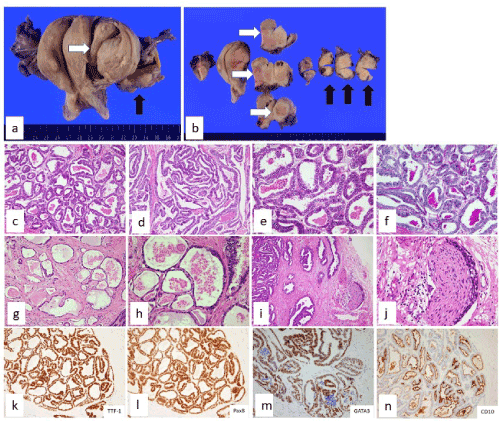
Figure 1. Pathological findings of Case 1
A tumor measuring 5 cm was found spanning from the left endometrial layer of the uterine corpus to the parametrium and a 3 cm mass was also found within the left ovary. The tumors of the uterus and left appendages were histologically similar.
Tumors consisted of atypical cells with basophilic cytoplasm and enlarged hyperchromatic nuclei and complex tubular structures which display papillary or cord-like structures. Glandular structures consisting of invasive and atrophied tumor cells and containing PAS-positive amorphous materials were observed. There was no involvement the endometrium. Perineural invasion was focally noted. Lymphatic invasion of tumor cells was observed. Immunohistochemical analysis showed that the tumor cells were positive for CK7 (focal), Pax8 and TTF-1CK20 (high frequency/diffuse), and negative for CK20. Some tumor cells were positive for GATA3, HNF-1βand focalized luminal CD10 positivity was also observed. Tumor cells were negative for ER, PgR and p53 overexpression was not observed. The MIB-1 positive rate was approximately 60%. Genetic analysis identified KRAS (codon12/13) mutation whereas no mutation was observed for NRAS, BRAF. While mesonephric remnants are commonly observed on the lateral side of the uterine wall and are indicative of uterine origin and ovarian metastasis, no such tissue was observed around the tumor.
Case 2
A woman in her 50’s with no previous pregnancies or births and an unremarkable family medical history presented with a main complaint of leukorrhea. No abnormalities were observed during cytological examination of the cervix and endometrium; however, a tumorous lesion was detected during an MRI. While CA19-9 was mildly elevated (57.9 U/ml compared to the normal value of 30 and below), other tumor markers, CEA, CA125 were within the normal range. Uterine and bilateral salpingo-oophorectomy, omentectomy, pelvic and para-aortic lymphadenectomy were performed based on suspicion of right ovarian cancer. Intraoperative cytological examination revealed an adenocarcinoma. The patient is currently undergoing postoperative chemotherapy (TC therapy).
Pathological findings (Figure 2)
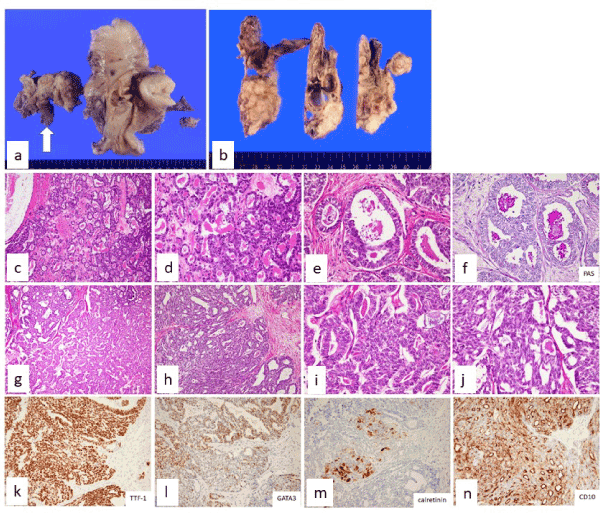
Figure 2. Pathological findings of Case 2
A tumor mass of 5 cm was found in the right ovary and the cut surface revealed a solid-cystic composition. Histological examination revealed atypical cells with basophilic cytoplasm and enlarged hyperchromatic nuclei. A dense ductal and papillary structure constituted the solid composition of the tumor. Presence of a PAS positive eosinophilic amorphous material was found within some tumor glands. In addition, a part of the tumor also showed a short-spindle-shaped tumor cell. Immunohistochemical analysis identified Pax-8 positive tumor cells, a high frequency of CK7 positive cells and an absence of cells positive for CK20. Additionally, there was a high frequency of TTF-1 positive cells, whereas GATA3, calretinin and HNF-1βpositivity was detected only in some cells and CD10 positivity was observed only in specific regions in the luminal surface of some tumor glands. The tissue was negative for ER, PgR, AR as well as p.53. A positive reaction of approximately 60% was observed for MIB-1. Genetic analysis identified KRAS (codon12/13) mutation whereas no mutation was observed for NRAS, BRAF. Additionally, no mesonephric remnants were associated with or the surrounding of the tumor. No neoplasia was found in the right fallopian tubes or the left appendages. Multiple leiomyomas and adenomyosis were identified in the uterus and endometriosis observed in the omentum. No metastases were found in the dissected lymph nodes.
Case 3
A woman in her 50’s who had experienced 2 pregnancies and 2 deliveries with an unremarkable family medical history. Detailed investigation of pollakiuria and examination of a lower abdominal mass by transvaginal ultrasound revealed an ovarian neoplastic lesion. The tumor marker CA125, was mildly elevated (49.6 U/ml compared to the normal range of below 35 U/ml) whereas CA19-9 levels were within the normal range. The patient underwent hysterectomy, bilateral salpingo-oophorectomy, and pelvic and para-aortic lymphadenectomy. The patient is undergoing postoperative chemotherapy (TC therapy).
Pathological findings (Figure 3)
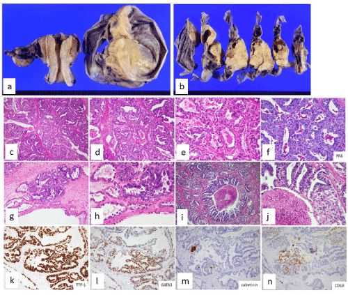
Figure 3. Pathological findings of Case 3
A tumor mass of 5 cm was found in the left ovary and the cut surface revealed a solid-cystic composition. Histological examination revealed atypical cells with basophilic cytoplasm and enlarged hyperchromatic nuclei. The solid components consisted of dense glandular ducts and papillary structures of tumor cells and a mixture of solid nests. PAS positive eosinophilic amorphous material was focally observed within a region of the ductal structure. The cystic component has been coated on the single layered tumor cells, including atrophic tumor cells, mucin laden tumor cells or hobnail-like nuclear protruded tumor cells. Comedo-like necrosis was focally seen.
Immunohistochemical analysis of the tumor cells identified a high frequency of Pax-8 and CK7 positive cells. However, these cells were negative for CK20. Additionally, there was a high frequency of TTF-1 positive cells, whereas GATA3 positivity was detected only in some cells and CD10 and calretinin positivity was observed only in specific regions in the lining cells of tubules/cysts and cells were negative for HNF1β. The cells were also negative for ER, PgR, and AR. There was a negative reaction for p.53. A 60% positivity rate was observed for MIB-1. Gene mutation analysis identified the KRAS (codon 12/13) but no mutations were observed for NRAS, and BRAF. There were no mesonephric remnants were associated with or surrounding the tumor. No neoplasias were found in the right fallopian tubes, uterus or omentum.
No neoplastic lesions were observed in the uterus and right adnexa.
Tumor confirmation was done using intraoperative peritoneal lavage cytology.
Summary of histopathology and immunohistochemistry of mesonephric-like adenocarcinoma
Composition of all the cases of mesonephric-like adenocarcinoma consisted mainly of varied glandular structures with partial expression of papillary, tubular and solid cystic structures. Histologically there is a different characterization from other gynecological epithelial malignant tissue types (endometrioid, serous, clear cell, and mucinous carcinomas). However, a part of the tumor reveals solid nests, mucin laden tumor cells or hobnail like tumor cells, and these areas required distinction from other tissue types of gynecological tumors. Immunohistochemically, all cases displayed a high frequency of TTF-1 positivity, GATA3 positivity, and intraluminal ductal CD-10 positivity. All cases were immunohistochemically negative for ER, PgR, and AR. While calretinin positivity was observed in 2 of 3 cases, expression was partial. All cases were immunohistochemically negative for thyroglobulin and expressed wild type p.53. Focal expression of HNF1beta (nucleus-positive pattern) and Napsin A positivity was observed in 2 of the 3 cases (Table. 1).
Gene mutation testing in mesonephric-like adenocarcinoma
Analysis of the KRAS (codon12/13, codon59/61, codon117, codon146), NRAS (codon12/13, codon59/61), BRAF(V600E) genetic mutations in mesonephric ductal adenocarcinoma, identified mutations in KRAS (codon12/13) and the absence of NRAS, BRAF mutations in all cases (Figure. 4).
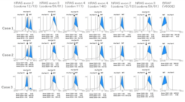
Figure 4. Results of gene analysis in three cases of mesonephric-like adenocarcinoma
Clinical course and pathological findings in mesonephric remnants (Reference case)
(Figure 5)
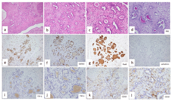
Figure 5. Mesonephric remnant (Reference case)
A patient in their 40s who underwent surgery for a bicornate uterus and uterine leiomyoma. Histopathological characterization identified small clusters of non-atypical small glands and intraluminal presence of eosinophilic colloid-like material.
Immunohistochemical characterization of the ductal cells identified high frequency of Pax-8, GATA3, AR positivity. These cells were negative for TTF-1, CD10, ER, PgR, thyroglobulin (Table. 1).
Table 1. Result of immunohistochemistry
| |
Mesonephric like carcinoma |
Mesonephric remnant |
| |
Case 1 |
Case 2 |
Case 3 |
Reference case |
Vimentin |
+ |
++ |
- |
++ |
CK7 |
+ |
++ |
++ |
++ |
CK20 |
- |
-- |
-- |
- |
Pax8 |
++ |
++ |
++ |
++ |
GATA3 |
+ |
+ |
+ |
++ |
TTF-1 |
++ |
++ |
++ |
- |
TG |
- |
- |
- |
- |
CD10 |
+luminal |
+luminal |
+luminal |
- |
Calretinin |
- |
+focal |
+focal |
- |
AR |
- |
- |
- |
+ |
ER |
- |
- |
- |
- |
PgR |
- |
- |
- |
- |
PTEN loss |
- |
- |
- |
- |
WT-1 |
- |
- |
- |
- |
HNF-1β(muclear staining) |
+focal |
+focal |
- |
- |
Napsin A |
+focal |
+focal |
- |
- |
p16 |
+occasional |
+occasional |
+occasional |
- |
p53 over expression |
- |
- |
- |
- |
Ki-67 labelling index |
60% |
60% |
60% |
less than 5% |
++: Positive rate above 50%; +: Positive rate of 50% and under; +focal: positive in only a small part of the tumor; +occasional: Sparsely positive within the tumor
Histological findings
Condensed nests of small gland ducts are seen (a). The ducts are round or slightly irregular, with eosinophilic substances inside (b). Unstructured colloidal eosinophilic substances are found in the glandular cavities [high power view] (c). The colloidal substance in the gland duct is positive for PAS reaction (d).
Immunohistochemistry
AR (e), GATA3 (f) and Pax8 (g) are positive, but calretinin (h) is negative. TTF-1 (i weak enlargement, j strong enlargement) was negative. CD10 (k weak enlargement, l strong enlargement) was negative for the mesonephric remnant tissue but surrounding stromal cells are positive.
Here we presented 3 cases of mesonephric-like adenocarcinoma, a rare tumor of the uterus and ovaries. Histological findings of the mesonephric-like adenocarcinoma demonstrated histological similarities to those of the uterine cervix and an eosinophilic colloid-like deposit was observed in ductal lumens. Additionally, importantly for diagnosis, immunohistochemical analysis identified TTF-1 and GATA3 positivity as well as the presence of the CD10 in the luminal surface of a part of the duct and staining similarities with mesonephric carcinoma of uterine cervix. [1-5].
GATA3 is recognized as a specific marker of the urothelium and its expression is confirmed in gynecological malignant tumors such as Brenner tumors [7], and has been frequently observed in mesonephric remnants and mesonephric carcinoma, and is thought to play an important role as a marker of the mesonephric duct [1,6,8] GATA3 expression was confirmed in the uterine corpus and ovarian mesonephros-like adenocarcinoma cases observed and it was determined to be an important marker for the diagnosis of this tumor.
While generally known as a marker for thyroid cancer and lung adenocarcinoma, the expression of TTF-1 in mesonephric carcinoma and mesonephric duct-like adenocarcinoma is also recognized. However, there was a lack of TTF-1 expression in the mesonephric remnant of the cases presented here and thus may not be a true mesonephric marker [1,2]. Moreover, thyroglobulin expression, which is observed in the follicular epithelium of the thyroid is absent in mesonephric carcinoma and mesonephric-like adenocarcinoma [9].
Similarly, gynecological malignancies including endometrial stromal tumors, some endometroid carcinomas, cervical mesonephric remnants also express CD10 [10]. However, CD10-positivity was focal and mesonephric origins could not be excluded by its absence. No luminal expression of CD10 was observed in the remnant tissue of the mesonephros shown in our presentation.
The association between p16 expression and HPV related cervical cancer is well recognized, however its expression in uterine and ovarian serous carcinoma is unknown. There are no reports of HPV involvement in mesonephric carcinoma and HPV expression is only either negative or when positive, sporadically scattered [11].
Recently, genomic analysis of mesonephric carcinoma and mesonephric-like adenocarcinoma has confirmed a high frequency of activating mutations of k-ras, whereas no such mutations have been found for PTEN, ARID1A, and p53. [3,5,6] Activation of k-ras mutation was confirmed in all 3 cases using the QP method suggesting that k-ras mutation may be involved in tumor development.
Immunohistochemistry and simple genetic analysis using QP method of these tumors were considered important in confirming the character of these tumors.
English Language Editing, Translation with Editing, and Manuscript Formatting were supported by Editage/Cactus communications Inc.
- Howitt BE, Nucci MR (2018) Mesonephric proliferations of the female genital tract. Pathology 50: 141-150.
- Pors J, Cheng A, Leo JM, Kinloch MA, Gilks B, et al. (2018) A comparison of GATA3, TTF1, CD10, and calretinin in identifying mesonephric and mesonephric-like carcinomas of the gynecologic tract. Am J Surg Pathol 42: 1596-1606.
- Kiyong NA, Kim HS (2019) Clinicopathologic and molecular characteristics of mesonephric adenocarcinoma arising from the uterine body. Am J Surg Pathol 43: 12-25.
- Horn LC, Höhn AK, Krücken I, Stiller M, Obeck U, et al. (2020) Mesonephric-like adenocarcinomas of the uterine corpus: Report of a case series and review of the literature indicating poor prognosis for this subtype of endometrial adenocarcinoma. J Cancer Res Clin Oncol 146: 971-983.
- Mirkovic J, McFarland M, Garcia E, Sholl LM, Lindeman N, et al. (2018) Targeted genomic profiling reveals recurrent K-ras mutations in mesonephric-like adenocarcinomas of the female genital tract. Am J Surg Pathol 42: 227-233.
- Mirkovic J, Sholl LM, Garcia E, Lindeman N, MacConaill L, et al. (2015) Targeted genomic profiling reveals recurrent KRAS mutations and gain of chromosome 1q in mesonephric carcinomas of the female genital tract. Modern Pathology 28: 1504-1514.
- Esheba GE, Longacre TA, Atkins KA, Higgins JP (2009) Expression of the urothelial differentiation markers GATA3 and Placental S100 (S100P) in female genital tract transitional cell proliferations. Am J Surg Pathol 33: 347-353.
- Howitt BE, Emori MM, Drapkin R, Gaspar C, Barletta JA, et al. (2015) GATA3 is a sensitive and specific marker of benign and malignant mesonephric lesions in the lower female genital tract Am J Surg Pathol 39: 1411-1419.
- McFarland M, Quick CM, McCluggage WG (2016) Hormone receptor-negative, thyroid transcription factor 1‐positive uterine and ovarian adenocarcinomas: report of a series of mesonephric‐like adenocarcinomas. Histopathology 68: 1013-1020.
- McCluggage WG, Oliva E, Herrington CS, McBride H, Young RH (2003) CD10 and calretinin staining of endocervical glandular lesions, endocervical stroma and endometrioid adenocarcinomas of the uterine corpus: CD10 positivity is characteristic of, but not specific for, mesonephric lesions and is not specific for endometrial stroma. Histopathology 43: 144-150.
- Ferguson DC, Long DJ, Smith MC, Craig-Owens LD, Means J, et al. (2015) Comparative analysis of Rb1, P16 and ER as diagnostic, prognostic and potential targets for therapeutic agents in ovarian epithelial tumors: An immunohistochemical study of 130 ovarian carcinomas. Journal of Ovarian Research 8: 34.