The traumatic phenomena in the lower extremities can lead to the amputations in more than 20% of the patients, especially when it is associated with the significant soft tissue loss or the severe wound contamination the blast events will require the amputation in 93% of cases.
Case presentation: We present the case of a female patient, 8 -year and 7-month-old who was brought by the rescue crew members to the emergency room of the Children's Clinical Hospital "Sf. Ioan", Galati, on 12.12.2020. The patient arrived at the emergency care center as a victim of a road accident, as a passenger.
Intraoperatively (12.12.2020 – 13H.45): a complete section of the popliteal vascular-nerve bundle is revealed, which is why it is decided to amputate the lower limb segment from the proximal 1/3 of the calf. The fixation of the supracondylar fracture focus was performed with the 3 pins, achieving the bone stabilization.
Conclusion: The particularity of this case consists of the difficulty to explain to a child that an adult's carelessness changed the course of her life and of the difficulty to support her morally to accept this situation.
Lower limb, amputation, child, car accident, trauma
The existing statistics in the United States admit that approximately 150,000 people suffer of the lower extremity amputations each year [1]. This incidence is directly proportional to rates of the peripheral arterial occlusive disease, the neuropathy, and the soft tissue infections [2]. A highly interesting correlation is that determined by the increased incidence of the lower limb amputations due to the vascular changes found to the diabetic pathologies (approximately 82%). These patients possess a 30-fold increased risk of the undergoing such surgery compared to undiagnosed patients with the diabetes. This translates into an extraordinary economic strain on the healthcare systems, over $4.3 billion to the annual costs in the US alone [3]. To the patients diagnosed with the diabetes (type I or II), all efforts should be focused on achieving (and maintaining) adequate glycemic control.
Anatomically, the lower limb is a complex formed by the union of seven tarsal bones, five metatarsal bones and fourteen phalanges. In terms of anatomical areas, it is subdivided into the hind foot (talus and calcaneal bones), the midfoot (consisting of the cuboid, the navicular and the three wedge bones) and the forefoot (the metatarsal and the phalanges). The muscle structures of the foot can be extrinsic to the nature, originating from the anterior or the posterior portions of the lower leg, but also from the intrinsic muscles [2,3].
The indications for amputation are mainly related to the degree of the necrosis tissue or the viability and is achievable either in a single operation or in a staged fashion (the amputation and the subsequent reconstruction). The decision to adopt any of the above approaches depends largely on the clinical condition of the patient, but also on the quality of the existing soft tissues at the desired level of the amputation. The surgeon's main aim is to excise non-viable and infected tissue. In general, guiding the level of the amputation will depend on the quality of the soft tissues and the ability to achieve the bone coverage. It is important to note that the skin grafts are still an acceptable option for the patients in whom the adequate muscle coverage can be obtained, where the skin coverage is not possible [4,5].
Before the decision for surgery with the amputation of the segment of interest is made, it is essential that the patient is hemodynamically stable. The early prophylactic antibiotic treatment is essential, especially to the patients with the signs of the systemic inflammatory response and the extensive cellulitis. This is necessary to minimize the risk of the localized wound infection and to maximize the length of the uninfected respective tissue. A decrease of the cellulitis may allow the amputation to be performed distal to the initially anticipated portion, as well as allowing the surgery to be performed to a single stage [6].
To the patient with the specific symptoms of the septic shock, the decision to perform an opened (the guillotine-type) amputation with the subsequent staged reconstruction versus a single surgery depends on the patient's clinical condition and the primary goal should be to achieve the adequate control of the source and leave the reconstruction for a later period [3,4].
The several scoring systems can be used to determine whether the complex reconstructive options should be pursued. However, the primary focus should be on the use of the Advanced Trauma Life Support protocol, as the patients are likely to have the concurrent potentially life-threatening injuries. This includes the assessing of the bleeding, to obtain the hemostasis of the actively bleeding sites and to perform the appropriate resuscitation. The level of the amputation will depend on the viability of the soft tissues used to obtain the bone coverage [7].
We present the case of a female patient, 8-year and 7-month-old who was brought by the rescue crew members to the emergency room of the Children's Clinical Hospital "Sf. Ioan", Galati, on 12.12.2020. The patient arrived at the emergency care center as a victim of a road accident, as a passenger. She presented a localized trauma to the calf and thigh by crushing mechanism, with the incarceration, secondary to the road accident (onset prior to presentation approximately 5 hours). Declaratively, from the rescue crew members, we knew that the patient lost her consciousness, post trauma and she have a continued massive bleeding.
The patient presented to the emergency department, hemodynamically decompensated (in hemorrhagic shock) - brought by ambulance. The patient presented acute ischemia of the right lower limb, lasting about 4 hours, with the comminuted staged wounds and the fractures of the right leg and the right thigh. The resuscitation of the patient was performed at the emergency department with the prompt administration of the necessary medication for the volemic support (intravenous infusion for replenishment of the vascular bed), then surgery was performed. The presence of an open fracture type III C-Gustilo Andersson right leg - operated.
The clinical examination at the time of the admission showed a critical general condition, with the demoralization of the facial structures (post-traumatic -the multiple wounds and the local abrasions), the normal conformed thorax, the bilateral and symmetrical ribs excursions with the respiratory movements, without pain associated with the deep inspiration. The cardiovascular examination: Blood Pressure=111/72 mmHg, ventricular rate = 82 beats per minute, the normal rhythmic heart, no pathological overinflated murmurs.
The abdominal examination reveals a soft abdomen, painless, spontaneously and on deep palpation, no traumatic mark at this level. The liver, the spleen within physiological limits, the free renal loci.
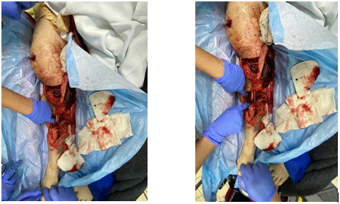
Intraoperatively (12.12.2020 –13H.45): a complete section of the popliteal vascular-nerve bundle is revealed, which is why it is decided to amputate the lower limb segment from the proximal 1/3 of the calf (Figure 1). The fixation of the supracondylar fracture focus was performed with the 3 pins, achieving the bone stabilization. The bony bridge was covered with a muscle-hamstring flap.

Figure 1. Patient pictures (the multiple detached and fractured bone fragments (the tibial fragment 14 cm long and the fibular fragment 17 cm long).
The pathological anatomical examination of the amputated fragment reveals the following:
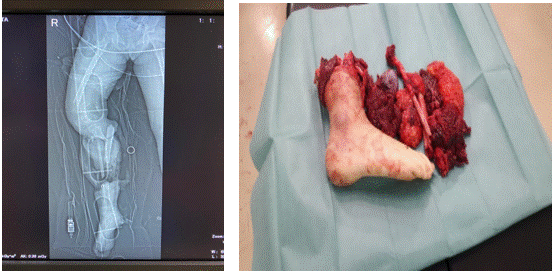
Macroscopically: The surgical resection of the leg and distal 1/3 of the calf, with the multiple detached and fractured bone fragments (the tibial fragment 14 cm long and the fibular fragment 17 cm long), with the 4 detached muscle fragments, showing the haemorrhagic areas and the skin flap with the subcutaneous adipose tissue (Figure 2).

Figure 2. The surgical resection of the leg and distal 1/3 of the calf.
Microscopically: The multiple fragments of the striated muscle tissue with the extensive haemorrhagic areas and the inflammatory foci with the moderate granulocytic infiltrate, the congestive vessels or the leukocytosis were examined. The preloculated fragments including sections from the popliteal artery revealed the small adherent thrombi of the endothelium as well as the neutrophilic inflammatory infiltrate and the periarterial haematous infiltrates. The other large caliber vessels harvested (anterior tibial artery and posterior tibial artery) show no changes of the pathological significance.
The postoperative evolution was remarkable:
- 13th.12.2020: the medium general condition, afebrile, the fit patient, the cardiorespiratory and the digestive balanced.
-14th.12.2020: the general condition affected, afebrile at the time of the consultation, approximately 150 ml of the wound fluid is drained; the wound was the clean appearance, the healing of the amputation wound with the slight areas of the skin damage, with the phlyctenitis and the deep lamellar thickening at the wound edge.
The neurological examination limited by the patient's post-sedation position after the amputation of the right leg surgery; no cranial nerve damage; no swallowing tubes; osteo-tendinous reflexes present bilaterally and symmetrically to the upper limbs; to the left lower limb osteo-tendinous reflexes present and no changes of sensitivity; to the right lower limb: the skin reflexes present; Glasgow score are 15 points. TC scan: the right frontotemporal epicranial hematoma.
The psychiatric examination: the cooperative patient: spontaneous, coherent speech, worrying glimpses of how she will move; what the prosthesis will look like; frequently deflects speech to family members; disappointed that her mother cannot be with her at this time.
- 15th.12.2020: the general condition influenced, the stationary evolution, the neurological investigations and the attached psychiatric examinations are continued underneath.
The psychiatric examination: the anxious mood patient, slightly increased reactivity to auditory stimuli such as: the flinches at the cry of the child; to the ambulance siren; she didn’t want to talk about the accident; although so far, she has been logorrheic about the accident; relatively she improved her sleep and her appetite.
The psychiatric diagnosis: the acute reaction to the life-threatening stressors.
The recommendations were the following: the individual and the family psychological counselling; the treatment with antidepressants; the psychiatric reassessment in case of worsening of the symptoms of psychomotor restlessness, the insomnia. A new psychiatric check-up for reassessment on 18.12.2020. After the discharge she returns periodically for the reassessment medical check-ups.
Subsequently, on 30th.12.2020 under general intravenous anesthesia we have done the clinical toilet with dressing, grasollind application, we changed the dressing under the continuous control with vacuum and 120 bar pressure.
On the 05ft.01.2021 the plastic surgeon he has had surgery again for the performance of degranulation, with the local toileting, use of split liver graft patch, suturing of the areas and dressing with grassolind.
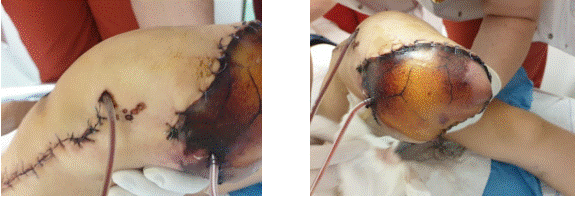
As can be seen (Figure 3) postoperatively, the necrosis of the tegumentary flap was detected, which required the removal and the subsequent coverage of the tegumentary defect with the total skin graft.

Figure 3. The necrosis of the skin flap.
The literature confirms that the wound complication-type phenomena (including the dehiscence, the seroma, the haematoma) may occur in 12% to 34% of below knee amputation of the patients and 6% to 16% of above knee amputation of the patients [8]. The Risk factors for wound complications include the sepsis, the compartment syndrome, the end-stage renal disease, the body mass index above 30 kg/m2 and BKA [9]. A retrospective study showed that to use of the incisional negative pressure wound therapy (NPWT) of the major limb amputation and the revision of the amputation had a demonstrable benefit in decreasing the risk of the wound complications [10].
The amputation of a limb not only reduces the patient's overall mobility but will also promote a decrease to the patient's quality of life, primarily through increased the energy expenditure (directly dependent on the level of amputation) [11]. The extensive research has shown that the patients with the unilateral amputations below the level of the popliteal area have 9% increased the oxygen consumption compared to unaffected subjects. This increase is even more notable to the patients with the amputations above knee level, at about 49% and an overwhelming 280% in those with the bilateral amputations above the knee region [12].
The lower extremity amputations are correlated with the increased incidences of the preoperative morbidity and mortality (ranging from 4%-22%) [13]. According to specialized studies, the following should be mentioned:
- the long-term mortality rates (at 1, 3 and 5 years post-operatively) can reach 15.38% and 68% respectively [14].
- the mortality rates of the patients with the amputation of the lower extremities affected by the diabetic processes can reach 77% within 5 years [15].
For this reason, the risk factors that may influence the adverse outcome of the patients undergoing operative limb amputation have been examined. The results of an analysis of the 2879 amputations showed that the most common post-surgical complications include the respiratory infections such as the pneumonia (22%), the acute respiratory distress syndrome (13%), the acute kidney injury (15%) and the deep vein thrombosis (15%) [10,11].
A particularly important factor to consider to the patients (especially, to the paediatric patients) requiring an important magnitude surgery and the step is to be decided for the psychological therapy and it has been early initiation. The pain of the amputated limb, known as phantom limb pain (PLP), is that a painful phenomenon which persists even after the complete tissue healing. From a medical point of view, it is characterized by the dysesthesia occurring of the absent limb. The patients describe this pain as sharp, burning, stinging, and the sensation that the amputated limb is in an abnormal position [16]. This pain may be present to 67% of the patients at six months and to 50% of the patients at five to seven years [17,18]. There are several risk factors for the development of PLP, which include: the presence of pre-amputation the pain, the female gender, the upper extremity amputations, and the bilateral upper and/or the lower extremity amputations [16].
We decided to conduct the several psychological consultations which provided us the following information:
-Immediately postoperatively, on 14th.12.2020 we concluded that the patient is a member of a legally constituted family with normal family relationships. The patient is actively involved in the community. During the initial assessment, the discrete regressions of the patient's field of the consciousness are noted, with impaired attention, the appearance of the disorientation symptoms associated with the vegetative signs of anxiety. The patient presented a partial amnesia of the traumatic episode. The individual and the family psychotherapy is recommended throughout the hospital stay and after the discharge the outpatient. The therapeutic session at this time is focused on the attempt, to announce the post-operative disability, as well as to detect the psychological impact on the child.
- The therapy on 15.12. 2020 is focused on how to accept the new situation and to overcome the pain associated with the amputation.
- 17.12.2020 – the therapy of adaptation to the changes that have occurred.
Finally, we conclude that, according to the specialized studies, the traumatic injuries caused by the high energy events may lead to the need for the amputation due to the devastating effects that may be present at the time of injury. Alternatively, there are situations where the patients may present to the hospital with an injured limb that cannot be reconstructed.
It is important to note that the victims of the severe traumatic lower extremity injuries who were initially candidates for the limb salvage may become the candidates for the amputation later, due to the infection, the inability to achieve the optimal bone coverage, or even the unwillingness to undergo a series of the reconstructive protocols for the poor functional outcomes.
The particularity of this case consists of the difficulty to explain to a child that an adult's carelessness changed the course of his life, in the difficulty to support her morally to accept this situation.
Sources of Funding: No funding for the research
Ethical approval: This is not a research study. No ethical approval was necessary.
Consent: Consent for publication was obtained from the patient discharged.
MARINA Virginia: Wrote the final form of the article and corresponding author.
E-mail: virginiamarina02@yahoo.com
ANGHELE Mihaela: Collaborating physician
STEFANOPOL Ioana-Anca: Collaborating physician
DRAGOMIR Liliana: Collaborating physician
ANGHELE Aurelian-Dumitrache: Surgeon who took the pictures, processed the pictures; made the surgery and wrote the surgery part of the article.
CIORTEA Diana-Andreea: Collected data and wrote the clinical part of the article
Registration of research studies: This is a case report. Is not a research study.
Guarantor: ANGHELE Aurelian -Dumitrache
Provenance and peer review: Not commissioned externally peer- reviewed.
Declaration of Competing Interest: The author declarate that there is not conflict of interest regarding the publication of this article.
- Dillingham TR, Pezzin LE, Shore AD (2005) Reamputation, mortality, and health care costs among persons with dysvascular lower-limb amputations. Arch Phys Med Rehabil 86: 480-486. [Crossref]
- Beckman JA, Creager MA, Libby P (2002) Diabetes and atherosclerosis: epidemiology, pathophysiology, and management. JAMA 287: 2570-281. [Crossref]
- Moxey PW, Gogalniceanu P, Hinchliffe RJ, Loftus IM, Jones KJ, et al. (2011) Lower extremity amputations--a review of global variability in incidence. Diabet Med 28: 1144-1153. [Crossref]
- Bosse MJ, MacKenzie EJ, Kellam JF, Burgess AR, Webb LX, et al. (2002) An analysis of outcomes of reconstruction or amputation after leg-threatening injuries. N Engl J Med 347: 1924-1231. [Crossref]
- Isaacson BM, Weeks SR, Pasquina PF, Webster JB, Beck JP, et al. (2010) The road to recovery and rehabilitation for injured service members with limb loss: a focus on Iraq and Afghanistan. US Army Med Dep J 31-6. [Crossref]
- Chen SL, Kuo IJ, Kabutey NK, Fujitani RM (2017) Physiologic Cryoamputation in Managing Critically Ill Patients with Septic, Advanced Acute Limb Ischemia. Ann Vasc Surg 42: 50-55. [Crossref]
- MacKenzie EJ, Bosse MJ, Kellam JF, Burgess AR, Webb LX, et al. Factors influencing the decision to amputate or reconstruct after high-energy lower extremity trauma. J Trauma 52: 641-649. [Crossref]
- Morisaki K, Yamaoka T, Iwasa K (2018) Risk factors for wound complications and 30-day mortality after major lower limb amputations in patients with peripheral arterial disease. Vascular 26: 12-17. [Crossref]
- O'Brien PJ, Cox MW, Shortell CK, Scarborough JE (2013) Risk factors for early failure of surgical amputations: an analysis of 8,878 isolated lower extremity amputation procedures. J Am Coll Surg 216: 836-842. [Crossref]
- Zayan NE, West JM, Schulz SA, Jordan SW, Valerio IL (2019) Incisional Negative Pressure Wound Therapy: An Effective Tool for Major Limb Amputation and Amputation Revision Site Closure. Adv Wound Care (New Rochelle) 8: 368-373. [Crossref]
- Poredos P, Rakovec S, Guzic-Salobir B (2005) Determination of amputation level in ischaemic limbs using tcPO2 measurement. Vasa 34: 108-112. [Crossref]
- Moreira CC, Farber A, Kalish JA, Eslami MH, Didato S, et al. (2016) The effect of anesthesia type on major lower extremity amputation in functionally impaired elderly patients. J Vasc Surg 63: 696-701. [Crossref]
- Choksy SA, Lee Chong P, Smith C, Ireland M, Beard J (2006) A randomised controlled trial of the use of a tourniquet to reduce blood loss during transtibial amputation for peripheral arterial disease. Eur J Vasc Endovasc Surg 31: 646-650. [Crossref]
- van Netten JJ, Fortington LV, Hinchliffe RJ, Hijmans JM (2016) Early Post-Operative Mortality After Major Lower Limb Amputation: A Systematic Review of Population and Regional Based Studies. Eur J Vasc Endovasc Surg 51: 248-257. [Crossref]
- Larsson J, Agardh CD, Apelqvist J, Stenström A (1998) Long-term prognosis after healed amputation in patients with diabetes. Clin Orthop Relat Res 350: 149-158. [Crossref]
- Fortington LV, Geertzen JH, van Netten JJ, Postema K, Rommers GM, et al. (2013) Short- and long-term mortality rates after a lower limb amputation. Eur J Vasc Endovasc Surg 46: 124-131. [Crossref]
- López-Valverde ME, Aragón-Sánchez J, López-de-Andrés A, Guerrero-Cedeño V, Tejedor-Méndez R, et al. (2018) Perioperative and long-term all-cause mortality in patients with diabetes who underwent a lower extremity amputation. Diabetes Res Clin Pract 141: 175-180. [Crossref]
- Lim S, Javorski MJ, Halandras PM, Aulivola B, Crisostomo PR (2018) Through-knee amputation is a feasible alternative to above-knee amputation. J Vasc Surg 68: 197-203. [Crossref]


