Abstract
The aim of the study is to determine the effect of the type of hip prosthesis on the oxidative-reduction processes and factors of the inflammatory process, as well as to determine the activity of selected enzymes constituting the oxidative defence of cells against RTF in red blood cells of patients suffering from osteoarthritis of the hip before and after total arthroplasty. Arthroplasty and surgical treatment of fractures mainly concern the elderly, whose oxidative stress reduction abilities are 3 times lower than in the case of a 20-year-old person.
Methods
The study included 200 patients aged 31–81 years old who underwent hip replacement surgery. Depending on the type of implanted prosthesis, patients were divided into two groups. The first group consists of patients who were implanted with a titanium alloy prosthesis in conformity with ISO 5832-1: 1996, and the tribological elements (an acetabulum and a head) were made of porcelain. The second group consists of patients who had a stainless steel prosthesis implanted in conformity with ISO 5832-1: 1997, the tribological elements were a metal head and a polyethylene acetabulum, and the prostheses were mounted on bone cement. Blood samples were taken three times: before the procedure, on the 1st day after the procedure and 6 weeks after the procedure. TAS and the following enzymes were determined: SOD-1, MDA, GSH-Px, Isoprostany 8-isoPGF2α.
The material was taken and secured in a manner typical for biochemical studies. TAS concentration was determined by the immunoenzymatic ELISA method from Immudiagnostic - ImAnOx (TAS / TAC) kit. The activity of SOD-1 superoxide dismutase was determined by the method of Misr and Fridovich. The concentration of MDA malondialdehyde in reaction with thiobarbituric acid was determined by the Placer method. The GSH-Px glutathione peroxidase activity was determined by the method of Pagil and Valentine. The concentration of isoprostane 8-isoPGF2α was determined by the Direct method 8-iso-Prostaglandin F2α (8-iso-PGF2α) Enzyme Immunoassay (EIA) kit. The concentration of IL-6 and CRP was also determined using the immunoenzymatic method (Human IL-6 Elisa, Diallone, Besancon, France) and the concentration of CRP using the immunoenzymatic ELISA Sensitiv method by LDN.
Results
The obtained results were analyzed statistically with the Shapiro-Wilk test. In most studies, the comparison of titanium-porcelain prostheses with those made of stainless steel and polyethylene did not show statistical differences on the first day after the procedure. TAS values before hip replacement surgery in both groups of prostheses are similar (about 200 pg / ml). After 6 weeks after the procedure, the TAS value in relation to the baseline value increases by an average of 25%, and the type of implanted prosthesis affects the body's defenses against ROS.
The results were similar for the following enzymes: SOD-1, GSH-Px, Isoprostany 8-isoPGF2α, MDA. After 6 weeks, MDA levels dropped significantly in patients with implanted titanium-porcelain prostheses, and remained at an elevated level in the second group of prostheses (cement prostheses). SOD-1 activity after 6 weeks was statistically significantly higher in the group with titanium-porcelain prostheses. The activity of glutathione peroxidase GSH-Px was not different for both groups. The concentration of isoprostane 8-iso PGF2α after 6 weeks from the arthroplasty operation was statistically lower after implantation of the titanium-porcelain combination prosthesis, which proves that the oxidation processes are less intensified. The concentration of CRP is significantly lower at the 6th week after the procedure in the group of patients who used porcelain-titanium prostheses. A similar distribution of values was obtained by determining the concentration of interleukin 6.
Conclusions
Chemical compounds from which hip joint prostheses are made affect the values of oxidative stress in the body. The lowest oxidative stress is caused by prostheses, in which the titanium content is 95%, and the tribological elements are made of porcelain (aluminum oxide).
Research confirms the increase in the amount of pro-inflammatory cytokines (CRP and interleukin-6) after implantation of bone cement implants.
Background
Every year, the number of hip prosthesis implantation procedures increases, which forces patients to search for optimal implants that would not cause increased oxidative stress and the hypersensitivity reaction that always arises after the introduction of a foreign body into the body.
In the case of hip prostheses, implant materials implanted intra-tissue, taking over the static-dynamic functions, should be biologically inert and should not cause oxidative stress.
Clinical observations, including the assessment of the survival of joint prostheses, unequivocally prove that prostheses, after implantation in the tissues of a living organism, cause a number of inflammatory processes of various degrees of severity, often leading to loosening of the strain on the implant-connective tissue interface, and the loosening process is preceded by an aseptic inflammatory reaction. It is formed due to the release of biomaterials used to build the prosthesis into the environment of the implant and is typical for the pathophysiology of foreign bodies.
The aim of the study was to determine how the type of hip prosthesis affects the oxidative status and whether it triggers inflammation. The work also determined the activity of selected enzymes constituting the oxidative defense of cells against the effects of RTF in red blood cells of patients suffering from osteoarthritis of the hip joint before and after total arthroplasty. Implantation of a joint prosthesis into the body, fusion of broken bones with metal connectors disturbs homeostasis, which causes a redox reaction. Arthroplasty and surgical treatment of fractures mainly concern the elderly, and the oxidative stress reduction abilities of a 75-year-old person are 3 times lower than that of a 20-year-old person.
The article was written in connection with the lack of literature comprehensively explaining and developing the impact of implants on the antioxidant status, created after implantation of a prosthesis in a diseased hip joint.
Methods
The study included 200 patients aged 31–81 years who underwent hip arthroplasty in 2010–2011. Women constituted 63% of the respondents, and men - 37%.
Blood samples were collected three times: before the procedure, on the 1st day after the procedure and 6 weeks after the procedure. The material was collected from the ulnar vein into heparin tubes and tubes without anticoagulant. Serum was obtained from blood devoid of anticoagulant (approximately 3 ml) by centrifugation for 5 minutes at 5000 × g at + 4˚C. The serum was then separated and stored at -80˚C.
Results
The obtained results were subjected to statistical analysis, in which the statistical mean, maximum and minimum value were calculated for each value. In most studies, the results in the first day after surgery were similar in the case of titanium-porcelain dentures and other dentures (Figure 1).
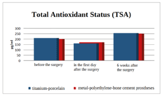
Figure 1. Comparison of the total antioxidant status (TAS) in patients who received titanium-porcelain prostheses with those who received other commonly used prostheses
Total oxidative status (TAS) reflects the body's ability to defend itself against free oxygen radicals. On the basis of the TAS test performed, we conclude that the baseline values before the procedure
hip arthroplasty were similar to each other and oscillated around 200 pg / ml for all types of prostheses. On the first day after surgery, the amount of TAS decreased by ¼ of the baseline values, while 6 weeks after the surgery, the TAS value increased to 255 pg / ml in the case of titanium-porcelain prostheses and to 250 pg / ml in the case of other prostheses (mainly placed on bone cement). The study shows that 6 weeks after the procedure, the TAS value in relation to the baseline value increases by an average of 25% and the type of implanted prosthesis affects the body's defenses against ROS (Figure 2).
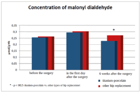
Figure 2. Comparison of the concentration of compounds reactive with thiobarbituric acid in patients who received titanium-porcelain prostheses with patients who were given other commonly used prosthes
The concentration of TBARS before arthroplasty in the studied groups was within the range of ~ 0.25 μmol / g Hb. On the first day after prosthesis implantation, this amount increased to 0.3 μmol / g Hb in both study groups. After 6 weeks, the amount significantly decreased in patients with implanted titanium-porcelain prosthesis and amounted to 0.225 μmol / g Hb, and in the group of other prostheses it remained at an increased level compared to the amount before prosthesis implantation (Figure 3).
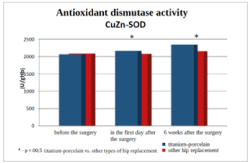
Figure 3. Comparison of SOD-1 superoxide dismutase activity in patients who received titanium-porcelain prostheses with patients who used other commonly used prostheses
The activity of the tested antioxidant enzymes before the procedure did not differ in both groups. On the first day after the procedure, in the group of people who used titanium-porcelain prostheses, statistically significantly higher activity of superoxide dismutase was observed (Figure 3) with significantly lower glutathione peroxidase activity (Figure 4) compared to the group of patients with other commonly used prostheses. Six weeks after the surgery, the dismutase activity was statistically significantly higher in the group with titanium-polymer prostheses. The activity of glutathione peroxidase did not differ between the groups (Figure 5).
Comparison of the concentration of isoprostane 8-iso PGF2α in patients who received titanium-porcelain prostheses with those who received other commonly used prostheses.
Comparing the concentration of isoprostane 8-iso PGF2α in both groups before the procedure and on day 1 after the procedure, we find a higher amount of this compound in the group of patients with prostheses other than titanium ones. Six weeks after the arthroplasty operation, statistically lower amounts of PGF2α were found in the study, which proves that the oxidation processes causing DNA damage are less intensified after the implantation of a titanium-porcelain prosthesis (Figure 6).
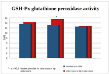
Figure 4. Comparison of glutathione peroxidase activity in patients using titanium-porcelain prostheses with patients using other commonly used prostheses
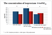
Figure 5. Comparison of the concentration of isoprostane 8-iso PGF2α in patients who received titanium-porcelain prostheses with those who received other commonly used prostheses
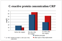
Figure 6. Comparison of CRP C-reactive protein concentration in patients who received titanium-porcelain prostheses with that in patients who used other commonly used prostheses
Graph 6 summarizes the acute phase protein concentration. The concentration of CRP is significantly lower at the 6th week after the procedure in the group of patients who used porcelain-titanium prostheses. It should be noted that within the studied groups the differences regarding the period from surgery were also statistically significantly different. This proves that the healing of postoperative wounds is always associated with inflammation. This is confirmed by the concentrations of the pro-inflammatory cytokine, the concentrations of which were analogous to those of CRP, and were summarized in Graph 7. It is clearly visible that the inflammatory reaction decreases much faster when using titanium-porcelain prostheses.
The amount of CRP before the procedure was higher in patients qualified for non-titanium-porcelain arthroplasty, after the procedure, both on the 1st day and after 6 weeks, the amount of CRP decreased in patients with titanium-porcelain prostheses, which proves a statistically lower the ability to generate an inflammatory reaction in the area of arthroplasty tissues.
A similar distribution of values was obtained by determining the concentration of interleukin 6 (Graph 7). Also in this study, statistically higher median values of the inflammatory process, i.e., interleukin 6, were found (Figure 7).
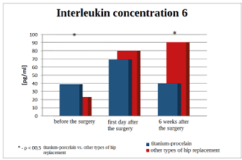
Figure 7. Comparison of interleukin 6 concentration in patients using titanium-porcelain prostheses with that in patients using other commonly used prostheses
Discussion
The introduction of foreign bodies into the body, such as elements of hip prostheses, causes a disturbance of the pro-oxidative and antioxidant balance through the increased generation of reactive oxygen species. This is accompanied by an excessive accumulation of H + protons, which activate such enzymes: parathyroid hormone, Vit D3, corticosteroids, interleukin 1 and 6, TNFα, and these enzymes lead to the activation of osteoclasts, which in turn causes bone resorption. This is an unfavorable phenomenon in the place of implantation of the prosthesis.
The analysis of the results of oxidative stress showed variable values of antioxidant enzymes depending on the implanted elements of the prosthesis.
Joint alloplasty enables the replacement of pathological structures and the restoration of normal function, however, it is associated with the introduction of foreign bodies into the body, which in the long run may be rejected by anatomical tissues. When interpreting the obtained results of oxidative stress tests - apart from the influence of post-traumatic stress, short-term hypokinesia experienced by the patient during the procedure, and the patient's age - the inflammatory process should be taken into account. For this reason, apart from the determination of TAS and the amount of selected antioxidant enzymes, the amount of IL-6 and CRP were also determined as markers of the inflammatory process.
Until recently, the prevailing view was that the course of osteoarthritis was not accompanied by inflammation due to the lack of vascularization of the cartilage. Thanks to the new possibilities in the field of immunohistopathological diagnostics, it has been established that the processes causing the degradation of the articular cartilage are inflammatory lesions. In recent years it has been proven that osteoarthritis is accompanied by synovitis. As a result, the pro-oxidative-antioxidant balance is disturbed by the increased generation of ROS, which are formed, among others, by in the presence of NADH and NADPH during the conversion of arachidonic acid along the cyclooxygenase and lipoxygenase pathway to prostaglandins and leukotrienes. Histopathological examinations clearly show that there are numerous macrophages in the junction of the tissue with the prosthesis during phagocytosis. Phagocytic cells in response to inflammation can generate ROS, because the phagocytosis process is associated with increased oxygen consumption in the so-called oxygen explosion. Of course, the phenomena of hypoxia and reperfusion in the affected area are not without significance. This phenomenon is related to the conversion of dehydrogenase under hypoxic conditions into xanthine oxidase, which catalyzes the oxidation of hypoxanthine to uric acid with the formation of ROS compounds. The research conducted by Gallo J. and Co. [1] and Ding H. and Co. [2] showed that the size, shape, amount and, above all, the material of the prosthesis have a great influence on the biological response of the organism.
The main problem in arthroplasty procedures is aseptic loosening of the endoprosthesis elements, the main cause of which is a phagocytic reaction, as a response to abrasion of its movable joints. To this day, the mechanism of this phenomenon has not been clearly explained. In recent years, more and more attention has been paid to the role of undiagnosed and oligosymptomatic infections in the pathogenesis of joint endoprostheses loosening. Aseptic loosening of the endoprosthesis occurs in the form of chronic inflammation in the tissues surrounding the implant, and its intensity depends both on the material of the prosthesis and the reactivity of the tissues surrounding the implant, as evidenced by the studies by Ding Y. and Co [3].
Unlike most organic compounds, metals cannot be eliminated from tissues by metabolic degradation. The only way of their elimination from the tissues is excretion in the urine via the kidneys or via the digestive system. It has been proven in animal studies that most metals are accumulated in the liver for many years. There are also studies such as [4], which showed that in the perspective of only 16.3% of the examined patients, the level of chromium increased or remained at an elevated level. The results of the research conducted by [5] prove that in the perspective of 4 years, the increased results of the cobalt level after hip arthroplasty do not affect the functioning of the kidneys or neurological or cardiological dysfunctions. Nevertheless, many authors emphasize the need to investigate the risk of clinical diseases caused by elevated concentrations of metal ions. On the basis of 8-year studies of patients after hip arthroplasty, [6] state that the concentration of metal ions may have a negative systemic effect and affect the skeletal system and cardiological functions. The studies conducted by [7] showed an increased content of cobalt and chromium ions in the blood of patients undergoing capoplasty, where the tribological elements are formed by metal-metal. Clinically unjustified increased pain was found in 69% of respondents. Most likely, the pain inducing factor is increased oxidative stress caused by excess metal ions.
The type of prosthesis fixation is important from the point of view of oxidative stress, and thus the speed of wound healing. The comparison of the examined parameters in the case of the application of the bonding of the prosthesis to the bone with the use of bone cement and without the use of an additional binder showed a differentiated effect on the induction of SOD, MDA and GSHPx. In the case of SOD-1 activity before the procedure and in the first day after the procedure, no significant differences in the activity of the tested enzyme were observed. 6 weeks after surgery.
In the case of patients with cementless bonding, the SOD-1 activity was statistically significantly higher than in patients with bone cement prostheses. Ryan et al. [8] found on the basis of 10-year observations that - contrary to the opinion of the Scandinavian Register of Prostheses - the survival of cementless dentures is much longer, while those fixed on bone cement are less durable, which is confirmed by the results of the level of oxidative stress. TAS decreases with cemented dentures, and with cementless dentures it has a higher value.
Conclusions
- The type of chemical compounds from which hip joint prostheses are made affects the values of oxidative stress in the body.
- The research shows that the lowest oxidative stress is caused by prostheses made of titanium alloys, in which titanium is 95% of the content, and the tribological elements are made of porcelain (aluminum oxide).
- The study of the value of pro-inflammatory cytokines (CRP and interleukin-6) allows to determine the differentiation of the biotolerance of the implants depending on the materials used for the production of prostheses and confirm the increase in the amount of pro-inflammatory cytokines after implantation of bone cement implants.
References
- Gallo J, Slouf M, Goodman SB (2010) The relationship of polyethylene wear to particle size, distribution, and number: A possible factor explaining the risk of osteolysis after hip arthroplasty. J Biomed Mater Res B Appl Biomater 94: 171-177. [Crossref]
- Ding H, Zhu Z, Tang T, Yu D, Yu B, et al. (2012) Comparison of the cytotoxic and inflammatory responses of titanium particles with different methods for endotoxin removal in RAW264.7 macrophages. J Mater Sci Mater Med 23: 1055-1062. [Crossref]
- Ding Y, Qin CQ, Fu YR, Xu J, Huang DS (2012) In vitro comparison of the biological activity of alumina ceramic and titanium particles associated with aseptic loosening. Biomed Mater 7: 045019. [Crossref]
- Maezawa K, Nozawa M, Yuasa T, Aritomi K, Matsuda K, et al. (2010) Seven years of chronological changes of serum chromium levels after Metasul metal-on-metal total hip arthroplasty. J Arthroplasty 25: 1196-200. [Crossref]
- Van Lingen CP, Ettema HB, Timmer JR, de Jong G, Verheyen CC (2013) Clinical manifestations in ten patients with asymptomatic metal-on-metal hip arthroplasty with very high cobalt levels. Hip Int 23: 441-444. [Crossref]
- Prentice JR, Clark MJ, Hoggard N, Morton AC, Tooth C, et al. (2013) Metal-on-metal hip prostheses and systemic health: a cross-sectional association study 8 years after implantation. PLoS One 8: e66186. [Crossref]
- Hart AJ, Matthies A, Henckel J, Ilo K, Skinner J, et al. (2012) Understanding Why Metal-on-Metal Hip Arthroplasties Fail: A Comparison Between Patients with Well-Functioning and Revised Birmingham Hip Resurfacing Arthroplasties AAOS Exhibit Selection. J Bone Joint Surg Am 94: e22 1-10. [Crossref]
- Takenaga RK, Callaghan JJ, Bedard NA, Liu SS, Klaassen AL, et al. (2012) Cementless Total Hip Arthroplasty in Patients Fifty Years of Age or Younger: A Minimum Ten-Year Follow-up. J Bone Joint Surg Am 94: 2153-2159.