The Lyme disease produced by Borrelia burgdorferi generates a multiorgan affectation. Mainly dermatological, rheumatic, neurological and cardiac lesions. The detection and treatment in the first stages are basic to prevent its progression.
This presentation is based on a clinical case of the patient with Lime disease, late diagnosis, not diagnosed in the acute phase.
Patient currently presents skin lesion compatible with chronic erythema migrans, arthralgias. Current Serology: Borrelia IgG Immunoblot Positive pattern; Ac. Anti-Borrelia burgdorferi (IgG) Positive; Ac. Anti-Borrelia burgdorferi (IgM) Negative.
In addition, the patient suffers from depression as side effects of treatment used before the definitive diagnosis.
lyme disease, borrelia burgdorferi, late diagnosis
The Lyme disease produced by Borrelia burgdorferi, is transmitted by ticks, generates a multiorganic affectation. Mainly causes dermatological, rheumatic, neurological and cardiac lesions. The detection and treatment in the first stages are basic to prevent its progression.
In localized infection, erythema migrans can be detected early. It is a plaque, erythematous, non-painful skin lesion at the site of the tick bite. Erythema migrans is the most common manifestation of Lime Borreliosis, but it is not always present. The rash occurs from the second day of the tick bite until one month later [1,2]. Erythema migrans may be accompanied by a clinical picture similar to influenza that includes fever, chills, myalgia, arthralgia and stiff neck. If not treated, infection at an early stage of the disease can cause more severe manifestations, of which Lyme neuroborreliosis and Lyme arthritis are the most frequent1. There may also be other less frequent manifestations, such as borrelial lymphocytoma, Lyme carditis, ocular affectations and chronic atrophic acrodermatitis. Neurological manifestations may include headache, pain or tingling in the limbs, Bell's palsy, mood disorders, memory deficits, sleep disturbances and so on [2].
The prevention of Lyme borreliosis consists mainly of avoiding tick bites through personal protection measures.
We present the case of a patient with Lime disease who has been treated for 3 years by several other diagnoses, not successful. Subsequently, after long suffering and symptomatic treatments, the disease has been diagnosed.
Male patient of 59 years, has gone to the emergency department of the hospital for diffuse abdominal pain, low-grade fever and malaise and myalgia, several days of evolution. The examination, imaging tests, blood and urine tests have not shown alterations. The patient has been prescribed a soft diet for 48 hours and symptomatic treatment and dopamine antagonists. Several months later the patient returns to the hospital with the presence of a bulge in the right abdominal region.
Exploration shows a painless swelling in the right iliac fossa that can be appreciated only in bipedistacion. Additional tests have been carried out. The radiological study has not shown alterations. Analysis of bleeding and urine have been normal. The patient was diagnosed with probable Spiegel hernia, painkilling analgesics in case of pain. At the next consultation, a month later, the bulge is no longer detected. Only abdominal distention is appreciated.
The following month the patient goes to the hospital for pain and paresthesias in both lower limbs and paresthesias in glove in both hands. It also refers to painful discomfort throughout the body, arthralgias and generalized myalgias. The neurological examination is normal. In the blood analysis the alteration of Lipase 375 U / L, ALT 83 U / L and PCR 8.2 mg / L, Rheumatoid factor <10 UI / ml, CK 406 U / L, with the rest of the parameters was detected within normality. The study of autoimmunity with Indirect Immunofluorescence technique has resulted in negative antinuclear antibodies. The abdominal ultrasound did not detect alterations. Magnetic resonance imaging showed signs of disc dehydration in the area of L3-L4-L5-S1. The CT scan presents normality. The patient has been prescribed treatment with analgesics associated with corticosteroids.
After one month the patient continued with sick leave, painful discomfort throughout the body, malaise and worried about his physical condition, he also presented the anxious-depressive symptoms. He is prescribed a treatment with antidepressant drugs. With the treatment of analgesics and antidepressants, the patient has been for more than a year and has only achieved the occasional slight improvement.
In a review the patient came to the hospital due to discomfort from a leg injury, after doing the usual hiking on a mountain route. The lesion was erythematous, indurated, 20 mm in diameter. Due to a suspicion of Erythema Migrans, the treatment with Proderma 100 mg is prescribed every 12 hours and the serology of Borrelia is requested in the microbiology laboratory. In addition, a biopsy of the skin lesion is performed to analyze it in the Anatomia Patologica laboratory.
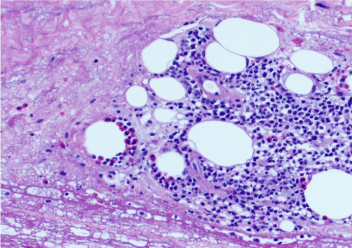
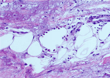
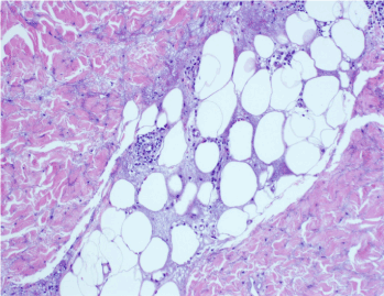
The results of Pathological Anatomy: Inflammatory changes compatible with insect bite (Figures 1-3).

Figure 1. Interstitial infiltrate predominantly eosinophilic (H&Ex20)

Figure 2. Degradation of collagen with necrobiotic appearance (H&Ex40)

Figure 3. The alterations affect the entire thickness of the dermis and reach subcutaneous fat (H&Ex40)
The results of microbiological study (repeated twice): Borrelia IgG Immunoblot Positive pattern; Ac. Anti-Borrelia burgdorferi IgG Positive; Ac. Anti-Borrelia burgdorferi IgM Negative. The last Lyme disease is diagnosed.
Currently the patient is better, but with musculoskeletal pain of a periodic nature. It also continues with antidepressant treatment. The long period of uncertainty in its diagnosis and the consumption of analgesic and antidepressant medications (Gabapentin, Lorazepam, Lyrica, etc.) could have been reasons for the depressive state.
Lyme disease is often not easily diagnosed, especially when the bite is not visible and is not detected in the beginning. Specific IgM antibodies usually reach their peak between the third and sixth week of the disease, while IgG antibodies begin to increase slowly after weeks or months of infection initiation [3]. A percentage of patients have been described who, after receiving an adequate treatment with resolution of the objective signs of the disease, have fatigue, musculoskeletal pain, difficulty in concentration and other symptoms that last more than 6 months and that have been denominated "Post-Lyme syndrome" [4]. The development of this post-Lyme syndrome is rare after adequate antibiotic treatment. In the case of our paceinte the amtibiotic treatment has not been adequate at the time of transmission of the disease. In addition, the treatments with the drugs used during a long period of time could have induced a stage of depression in the patient.
Usually, up to 50% of those affected also do not have the characteristic skin lesion, erythema migrans. The later phases of the disease are characterized by the non-specificity of the signs and symptoms [5]. Therefore, it is very important to perform microbiological laboratory tests at the appropriate time, to the appearance of symptoms and suspicion of disease, to confirm the existence of infection.
- Steere AC, Strle F, Wormser GP, Hu LT, Branda JA, et al (2016) Lyme borreliosis. Nat Rev Dis Primers 2: 16090.
- Müllegger RR, Glatz M (2008) Skin manifestations of lyme borreliosis: diagnosis and management. Am J Clin Dermatol 9: 355-368. [Crossref]
- Evison J, Aebi C, Francioli P, Péter O, Bassetti S, et al. (2006) [Lyme disease Part I: epidemiology and diagnosis]. Rev Med Suisse 2: 919-924. [Crossref]
- Wormser GP, Dattwyler RJ, Shapiro ED, Halperin JJ, Steere AC, et al. (2006) The clinical assessment, treatment, and prevention of Lyme disease, human granulocytic anaplasmosis, and babesiosis: clinical practice guidelines by the Infectious Diseases Society of America. Clin Infect Dis 43: 1089-1134.
- Portillo A, Santibáñez S, Oteo JA (2014) Lyme disease. Enferm Infecc Microbiol Clin 32 Suppl 1: 37-42. [Crossref]


