Abstract
The COVID-19 pandemic posed challenges in patient management and resource allocation. To date, there have been no documented cases of COVID-19 patient survival with extracorporeal cardiopulmonary resuscitation (eCPR). We present a case demonstrating the role of eCPR in COVID-19 infection and the coordination necessary for its utilization during a pandemic.
Keywords
COVID-19, acute myocardial injury, veno-arterial extracorporeal membrane oxygenation
Introduction
The emergence of the novel coronavirus (SARS-CoV-2) global pandemic has created new challenges in healthcare as COVID-19 patients present with unique constellations of symptoms. In a subset of patients, the virus manifests as acute COVID-19 cardiovascular syndrome (ACovCS), a myocarditis-like syndrome causing acute myocardial injury and reduced left ventricular ejection fraction [1]. ACovCS can be further complicated by arrhythmias, heart failure, and shock [1]. The application of veno-arterial extracorporeal membrane oxygenation (VA-ECMO) has expanded to include use for resuscitation during CPR or immediately post-CPR to prevent re-arrest, also known as extracorporeal cardiopulmonary resuscitation (eCPR). A randomized control trial showed significant improvement in survival to discharge, 43% with ECMO versus 7% with standard CPR [2]. However, as the pandemic progressed, many established eCPR programs were shuttered out of concern for healthcare worker safety [3]. This case demonstrates the successful use of eCPR in a patient suffering cardiac arrest and cardiogenic shock from sequelae of COVID-19.
Case report
The patient is a 29-year-old male smoker with a history of obesity (body mass index 41.9), Type-2 diabetes mellitus and depressive disorder who developed a dry cough. After three days of worsening symptoms and the development of a fever, he was found to be COVID-19 positive via RT-PCR. Chest imaging revealed opacities throughout both lungs concerning for infiltrate including COVID-19. The cardiac silhouette and pulmonary vasculature were normal at this time, and no pleural effusion or pneumothorax was noted (Figure 1). The patient’s dyspnea progressed for three days, prompting him to present to an outside emergency department for evaluation. Upon admission, his oxygen saturation was 94% and he received oxygen but did not require intubation. He was discharged home after an uneventful five-day hospital course. Twenty-one days after testing positive for COVID-19, he developed progressively worsening chest pain. At an outside facility, an EKG showed changes consistent with an anterolateral STEMI. Coronary angiography demonstrated complete thrombosis of the left proximal anterior descending artery. He underwent thrombectomy, which was unsuccessful, followed by intracoronary tPA and placement of a 4.0 x 32 mm everolimus-eluting stent in the left anterior descending artery. Left descending artery blood flow after stent placement was evaluated as TIMI 2 likely due to slow flow phenomenon. The right coronary artery did not have significant stenosis.
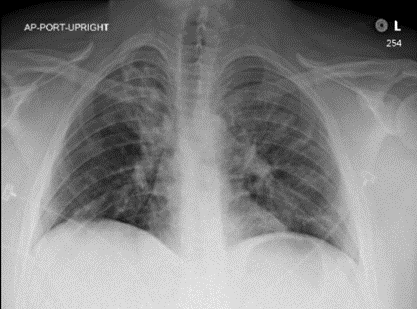
Figure 1. Patient’s chest X-Ray at the time of COVID-19 diagnosis revealed irregular opacities throughout both lungs concerning for infiltrate including COVID-19. The cardiac silhouette and pulmonary vasculature were normal at this time and no pleural effusion or pneumothorax was noted.
The patient was transferred to the ICU but became increasingly tachypneic four hours after percutaneous intervention. A transthoracic echocardiogram showed a large area of akinesis in the LAD distribution involving the anterior, septal and apical segments, ventricular ejection fraction of 5-10%, protruding LV apical thrombus measuring 1.4 x 1.1 cm and mildly reduced global right ventricular function. His condition deteriorated further with worsening cardiogenic shock and hypoxia, warranting intubation. He developed recurrent ventricular fibrillation and asystolic arrests requiring intermittent cardiopulmonary resuscitation (CPR) for approximately three hours, resulting in anoxic brain injury, subarachnoid hemorrhage, pericardial effusion, acute kidney injury and systolic heart failure with an LVEF of 35%.
Repeat angiogram showed thrombosis of the newly placed stent (Figure 2). Due to the incomplete revascularization during the first intervention, he remained in severe cardiogenic shock with repeat echocardiogram demonstrating an EF of 25% and was transferred to our facilities for ECMO. Given his ongoing shock and intermittent cardiac arrest, eCPR was initiated to stabilize the patient. His blood gas values prior to the initiation of ECMO were pH 7.41, CO2 46 mmHg, O2 98 mmHg, HCO3 28.8, lactic acid of 13.4 and O2 saturation of 97.7%. A 17 Fr arterial ECMO cannula was inserted into the right femoral artery and a 25 Fr venous cannula was inserted into the right femoral vein. Therapeutic anticoagulation was achieved using unfractionated heparin. Of note, he had not received any therapeutic interventions for COVID-19 specifically.

Figure 2. Coronary angiography demonstrating thrombosis of everolimus-eluting type of stent placed after initial intervention (Panels 1, 2). We were able to reestablish TIMI 2 flow (Panel 3).
The patient was decannulated from ECMO-on-ECMO day 5 following an uncomplicated run and successful turn-down study demonstrating an ejection fraction of 15% at x 4.7 L of flow. He was extubated after 13 days of mechanical ventilation and transferred to the floor after 16 days in the ICU. After a hospitalization of 23 days, he went to inpatient rehabilitation for seven days followed by subsequent discharge home.
Discussion and conclusion
Emerging evidence supports the novel coronavirus as a respiratory virus that can have effects on the cardiovascular system. A study by Puntmann et al. [4] demonstrated that of 100 unselected participants who had recently recovered from COVID-19, 60% continued to have myocardial inflammation on cardiac MRI, independent of pre-existing conditions [4]. Additionally, similar studies have found that in a sample of 416 patients who were hospitalized for the coronavirus, nearly 20% sustained cardiac injury during their hospitalization [5]. These sequelae, called Acute COVID-19 Cardiovascular Syndrome (ACovCS), has a diverse symptomatic manifestation, including myocardial infarctions and arrhythmias, but diagnosis and management of these patients remains unclear [4].
Extracorporeal cardiopulmonary resuscitation (eCPR) has been utilized in the rescue of patients experiencing cardiac arrest and serious cardiopulmonary compromise. However, the use of eCPR is complicated by issues of patient selection and optimization of resource utilization [6]. The global COVID-19 pandemic has amplified the complexity of these issues, posing additional challenges including staff safety and scarcity of resources [3]. Our facility designated one ICU for COVID-19 patients who require ECMO. All ECMO patients who are positive or are under investigation are located on the designated COVID-19 ICU and placed on COVID-19 precautions. If the patient is positive, they remain in the designated COVID-19 ICU regardless of the primary indication for ECMO. This process includes our eCPR patients whose COVID-19 status is typically unknown at the time of ECMO initiation, especially in out-of-hospital eCPR cases. In addition to the internal COVID-19 ECMO process, our facility partners with other ECMO centers in the state. Together, we created ECMO selection criteria during times of high ECMO utilization and scarce resources [7]. When two of the centers close to ECMO for any reason, this triggers the agreed upon resource allocation process where the state determines who is placed on ECMO based on the highest likelihood of survival. Even though eCPR outcomes are improving [2,8], it comparatively remains one of the lowest probabilities of survival.
COVID-19 is often characterized as more severe in the elderly population, however, increasing evidence demonstrates the vulnerability of young patients to some of the virus’ most severe complications [9]. The timing of our patient’s clinical presentation is notable. He developed ACovCS symptoms 21 days after the initial diagnosis of COVID-19, after his respiratory symptoms had resolved. This clinical presentation underlies the importance of continued monitoring of patients who have recovered from the virus, even those considered low risk. Additional resources will be required to care for patients with complications from the disease, and must be considered by policy makers and physicians, as well as the potential need for resource intensive therapy like eCPR in symptomatic and recovered COVID-19 patients.
There remain many unknowns about the long-term effects of COVID-19 and who is most vulnerable. Our case demonstrates that patients suffering from cardiac arrest from COVID-19 can benefit from eCPR. Patient selection for eCPR should consider scarcity of resources and complex constellations of symptoms in COVID-19 patients. This case demonstrates one successful outcome, but it should be noted that more data for this patient population is crucial. ECMO programs should collaborate with public health and hospital officials to devise a plan for this therapy so it may be offered to patients who can benefit from it.
References
- Hendren NS, Drazner MH, Bozkurt B, Cooper LT (2020) Description and Proposed Management of the Acute COVID-19 Cardiovascular Syndrome. Circulation 141: 1903-1914. [Crossref]
- Yannopoulos D, Bartos J, Raveendran G, Walser E, Connett J, et al. (2020) Advanced reperfusion strategies for patients with out-of-hospital cardiac arrest and refractory ventricular fibrillation (ARREST): A phase 2, single centre, open-label, randomised controlled trial. The Lancet 396: 1807-1816. [Crossref]
- Worku E, Gill D, Brodie D, Lorusso R, Combes A et al. (2020) Provision of ECPR during COVID-19: evidence, equity and ethical dilemmas. Crit Care 24: 462. [Crossref]
- Puntmann VO, Carerj ML, Wieters I, Fahim M, Arendt C et al. (2020) Outcomes of Cardiovascular Magnetic Resonance Imaging in Patients Recently Recovered From Coronavirus Disease 2019 (COVID-19). JAMA Cardiol 5: 1265-1273. [Crossref]
- Shi S, Qin M, Shen B, Cai Y, Liu T, et al. (2020) Association of Cardiac Injury With Mortality in Hospitalized Patients With COVID-19 in Wuhan, China. JAMA Cardiol 5: 802-810. [Crossref]
- Pappalardo F, Montisci A (2017) What is extracorporeal cardiopulmonary resuscitation? Journal Thorac Dis 9: 1415-1419.
- Patient Care Strategies for Scarce Resource Situations. Minnesota Health Care Preparedness Program, Minnesota Department of Health (2020) www.health.state.mn.us/communities/ep/surge/crisis/standards.pdf.
- Bartos J, Frascone R, Conterato M, Wesley K, Lick C, et al. (2020) The Minnesota mobile extracorporeal cardiopulmonary resuscitation consortium for treatment of out-of-hospital refractory ventricular fibrillation: Program description, performance and outcomes. E Clinical Medicine 29-30: 100632. [Crossref]
- Oxley T, Mocco J, Majidi S, Kellner C, Shoirah H, et al. (2020) Large-vessel stroke as a presenting feature of Covid-19 in the young. N Engl J Med 382: e60. [Crossref]