Objective: Extramedullary plasmacytoma (EMP) is a rare variant of plasma cell myeloma that affects soft tissues. The most affected sites of EMP are primarily the head and neck regions. In this study, we report a case of this rare neoplasm in the larynx and its management using an advanced radiation therapy protocol.
Methods: A 42-year-old Hispanic female presented with a history of progressive hoarseness for several weeks. Initially the patient underwent endoscopic evaluation of her throat to find any tumour lesion in the region of her false vocal cord. The patient was diagnosed with the presence of EMP of the larynx using microscopic laryngoscopy followed by computed tomography (CT)/positron emission tomography (PET)/magnetic resonance imaging (MRI) and pathological evaluations.
Results and discussion: The findings of the endoscopic evaluation and microscopic laryngoscopy of the patient’s throat indicated a submucosal mass in the region of her false vocal cord that was keeping the true vocal cords from adequately apposing. The CT/PET/MRI and pathology evaluations revealed the presence of a stage I EMP of the larynx. Following the diagnosis of her disease, the patient was administered a course of conformal 3-Dimentional intensity modulated radiation therapy (IMRT)/image-guided radiation therapy (IGRT) to a total dose of 5,040 cGy in 28 fractions over 44 calendar days. A post-operative treatment evaluation after more than 7 years indicated no evidence of a recurrent neoplasm or distant metastasis.
Conclusion: The findings of this case study demonstrated that conformal 3-D IMRT/IGRT is clinically effective in managing the EMP of the larynx successfully.
head and neck cancer, intensity modulated radiation therapy, image guided radiation therapy, laryngeal cancer
Plasmacytoma is a rare neoplastic disorder of a monoclonal plasma cell proliferation of B lymphocyte lineage that originates in bone or soft tissues [1,2]. This rare neoplasm can occur in the form of either a solitary plasmacytoma or progresses to multiple myeloma. A solitary plasmacytoma is a single localized mass of neoplastic plasma cells occurring in either bone (medullary) or soft tissue predominantly in the head and neck area (extramedullary). Extramedullary plasmacytoma (EMP) is uncommon, accounting for only 3% of all plasma cell neoplasms [3]. Most lesions occur in the head and neck [4], primarily in the upper aerodigestive tract including the nasal cavity, paranasal sinuses, or nasopharynx; only a minority occurs in the larynx [5]. EMP of the larynx represents from 0.04% to 0.45% of the malignant tumours of the larynx, with an incidence of less than 1% of all head and neck malignancies [5,6].
EMP occurs approximately three times more often in men than in women. It is predominantly found in males between 50 and 70 years, but there are cases reported in the second decade of life [7]. The most common sites for laryngeal EMP are, in order of decreasing frequency, the epiglottis, true vocal cords, false vocal cords, ventricles, arytenoids, and subglottic space [3]. The clinical presentation of EMP varies according to its location, which includes hoarseness, cough, dyspnoea, and stridor. These symptoms can last from months to years before a diagnosis is made [8]. The diagnosis of EMP is based primarily on the histopathological evaluations that demonstrate the presence of neoplastic monoclonal plasma cells [9,10]. In addition, the diagnosis of EMP is made by the exclusion of multiple myeloma. A computed tomography (CT) or magnetic resonance imaging (MRI) examination is necessary to define the margins of the homogeneous laryngeal mass [11].
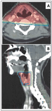
Due to the rarity of this neoplasm, there are no generally accepted guidelines for the treatment of patients with EMP and most recommendations remain a consensus of expert opinion. In general, these neoplasms are radiosensitive; therefore, radiation therapy is considered the treatment of choice for patients with EMP [9,10]. However, the optimal dosage regimen for radiotherapy is a matter of debate. Innovations in radiation therapy techniques have led to the introduction of intensity modulated radiation therapy (IMRT) and image-guided radiation therapy (IGRT) for the treatment of various cancers [12-15]. IMRT is an advanced form of high-precision radiotherapy that uses computer-controlled multiple small radiation beams of varying intensities to deliver precise radiation doses to the target tissues while sparing adjacent healthy tissues [16]. By incorporating 3-dimentional (3-D) CT or positron emission tomography (PET) imaging technology, IMRT allows the radiation dose to conform more precisely to the three-dimensional shape of the tumour while modulating the intensity of the radiation beam and minimizing its dose to those sensitive and unaffected organs. IGRT uses a variety of 2-D, 3-D and 4-D imaging techniques that improve the precision and accuracy of the delivery of the radiation dose to the targeted tumour tissue while minimizing the dose to the surrounding normal tissue during the course of radiation therapy (Figure 1).

Figure 1. These figures represent the axial (A) and sagittal (B) view of isodose coverage of the primary tumour site. This targeted three-dimensional intensity modulated radiation therapy and image-guided radiation therapy (IMRT/IGRT) plan depicts the high dose areas of radiation treatment to the primary tumour. Colour changes from bright red to blue indicate reduction of the radiation dose from highest to the lowest
At the University of Cancer Centres, IMRT/IGRT techniques have used to successfully manage a variety of tumours including this malignant tumour [17,18]. In this report, we present a challenging case of EPM and describe our experience in managing this disease using an IMRT/IGRT based 3-D conformal radiation therapy protocol.
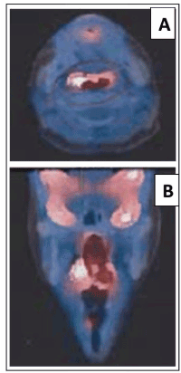
In October 2012, a 42-year-old Hispanic woman presented with a history of progressive hoarseness for several weeks. At presentation, the patient reported that she had not been exposed to hazardous materials and denies ever using controlled substances. Prior to the diagnosis, the patient had history of occasional cigarette smoking at social gatherings. However, currently she did not smoke or use tobacco products. The patient complained of seasonal sinusitis. She reported no problems with her muscles, bones, or joints. A bone survey was performed and was negative for bone disease. Both serum and urine protein electrophoresis were performed and had no laboratory abnormalities suggestive of multiple myeloma. Further endoscopic evaluation and PET imaging of the patient’s throat revealed the presence of a submucosal mass in the region of her false vocal cord that was keeping the true vocal cords from adequately apposing (Figure 2).

Figure 2. Positron emission tomography (PET) Imaging of the patient’s primary tumour site reveal slight uptake in the area of the larynx but negative for metastasis. A. Axial view of the PET imaging. B. Coronial view of PET imaging
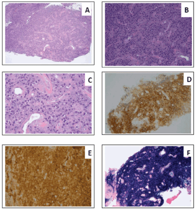
During surgery, microscopic suspension laryngoscopy was performed under anaesthesia, and a submucosal mass was found in the area of the false vocal cord. During this procedure, the mass was gently moved away from the right anterior true vocal cord, it was apparent at that time that this mass extended down to the true vocal cord. Post-operative CT/MRI of the neck showed no residual disease or metastasis. Pathology revealed the presence of EMP of the larynx. Immunohistochemical evaluation and Hematoxylin-Eosin (H and E) staining indicated cells were positive for CD138, Kappa (Figure 3) and an atypical plasma cell infiltrate was seen consistent with plasmacytoma. The patient underwent a course of post-operative IMRT/IGRT delivered in 28 fractions over 44 calendar days to a total dose of 5,040 cGy. Post-treatment evaluation after more than 7 years indicated no evidence of a recurrent neoplasm or development of multiple myeloma.

Figure 3. Histopathologic evaluation and Hematoxylin-Eosin staining demonstrated an atypical plasma cell infiltrate consistent with plasmacytoma. A. Magnification of 10X, B. Magnification of 20X, C. Magnification of 40X, D. CD138 antibody staining, E. Kappa staining, and F. In situ hybridization with kappa restricted
EMP is a rare neoplasm of plasma cells that occurs in the soft tissue outside of the bone marrow [19]. The first EMP report was made in 1905 by Schridde [20]. The most frequently affected sites are the submucosal lymphoid tissue of the nose and sinuses [19]. The larynx is rarely reported but it appears that the incidence of involvement appears to be increasing with 4.5% to 18% of the reported cases of the head and neck occurring in the larynx [10]. Currently, information is limited regarding its diagnosis, staging and natural history [21]. In general, the diagnosis of EMP of larynx is often delayed. Presenting symptoms are usually limited and non-specific, slowly progressive hoarseness over the period of months to years appears to be its most common feature.
Although the clinical presentation varies depending on the organ involved [3], EMP of the larynx often presents with hoarseness and/or dysphagia [8]. Gross appearance may vary from a yellow grey to dark brown polypoid or sessile mass or diffuse thickening of the involved organ. The surface is usually smooth and consistency semi-firm and rubbery. Common sites of laryngeal involvement in order of frequency are the epiglottis, ventricles, the vocal folds, false cords, the aryepiglottic folds, arytenoids, and subglottis. EMPs of the larynx are generally submucosal but may be polypoid and may involve single or multiple contiguous sites [22]. It is believed that these neoplasms are non-disseminating in nature but appears to be locally aggressive [4] and about 10 to 20% of these tumours metastasize to cervical lymph nodes [23]. Approximately one-third of patients who have EMPs will eventually progress to multiple myeloma within 2 years of presentation [24]. Although it is yet to be established, time to presentation, the size of the tumour at diagnosis, total serum protein level, and a monoclonal spike observed on serum protein electrophoresis seem to predict the progression to multiple myeloma [25] in 10%-40% of cases. This progression occurs within the first two years from disease onset [26].
Although radiation therapy is considered the standard treatment for patients with EMP of the larynx [9,10], the optimal dosage regimen has not been firmly defined. Innovations in radiation therapy have led to the development of novel techniques such as IMRT/IGRT for the treatment of cancers. The clinicians at the University Cancer Centres, Houston, Texas have used these innovative therapy techniques for the treatment of a variety of malignant tumours including the EMP of the larynx [17,18]. These innovative techniques offer the potential to deliver highly conformal dose-intense radiation to targeted regions of disease, while sparing adjacent critical non-malignant tissue. The improved shaping of high dose distributions with IMRT/IGRT potentially mitigates treatment-related toxicities. For example, the use of advanced radiation therapy techniques has been associated with increased preservation of parotid salivary flow [27-29].
In this study, we have demonstrated that by using conformal 3-D IMRT/IGRT we were able to treat the EMP of the larynx while sparing adjacent critical structures that were not involved by the disease, and reduce many of the detrimental side effects associated with the radiation therapy. Therefore, an IMRT/IGRT should be considered as the standard of care for the management of the EMP of the larynx. However, long-term follow-up is also important, because of the high long-term risk for the development of malignant myeloma. In our patient’s case after more than 7 years from her treatment, the patient has or laboratory evidence of systemic disease.
- Rutherford K, Parsons S, Cordes S (2009) Extramedullary plasmacytoma of the larynx in an adolescent: a case report and review of the literature. Ear Nose Throat J 88: E1-E7. [Crossref]
- Soni NK, Trivedi KA, Kumar A, Prajapati JA, Goswami JV, et al. (2002) Solitary extramedullary plasmacytoma - larynx. Indian J Otolaryngol Head Neck Surg 54: 309-310.
- Maniglia AJ, Xue JW (1983) Plasmacytoma of the larynx. Laryngoscope 93: 741-744. [Crossref]
- Wax MK, Yun KJ, Omar RA (1993) Extramedullary plasmacytomas of the head and neck. Otolaryngol Head Neck Surg 109: 877-885. [Crossref]
- Ramirez-Anguiano J, Lara-Sanchez H, Martinez-Banos D, Martinez-Benitez B (2012) Extramedullary plasmacytoma of the larynx: a case report of subglottic localization. Case Rep Otolaryngol 2012: 437264. [Crossref]
- Strojan P (2002) Extramedullary plasmacytoma of the larynx: a report of three cases. Radiol Oncol 36: 225-229. [Crossref]
- Gorenstein A, Neel HB, Devine KD, Weiland LH (1977) Solitary extramedullary plasmacytoma of the larynx. Arch Otolaryngol 103:159-161.
- Zbaren P, Lang H, Beer K, Becker M (1995) Plasma cell granuloma of the supraglottic larynx. J Laryngol Otol 109: 895-898. [Crossref]
- Velez D, Hinojar-Gutierrez A, Nam-Cha S, Acevedo-Barbera A (2007) Laryngeal plasmacytoma presenting as amyloid tumour: a case report. Eur Arch Otorhinolaryngol 264: 959-961. [Crossref]
- Pino M, Farri F, Garofalo P, Taranto F, Toso A, et al. (2015) Extramedullary Plasmacytoma of the Larynx Treated by a Surgical Endoscopic Approach and Radiotherapy. Case Rep Otolaryngol 2015: 951583.
- Saad R, Raab S, Liu Y, Pollice P, Silverman JF (2001) Plasmacytoma of the larynx diagnosed by fine-needle aspiration cytology: a case report. Diagn Cytopathol 24: 408-411. [Crossref]
- D'Andrea MA, Reddy GK (2019) The systemic immunostimulatory effects of radiation therapy producing overall tumor control through the abscopal effect. J Radiation Oncol 8: 143-156.
- Hajj C, Goodman KA (2015) Role of Radiotherapy and Newer Techniques in the Treatment of GI Cancers. J Clin Oncol 33: 1737-1744. [Crossref]
- O'Sullivan B, Rumble RB, Warde P (2012) Intensity-modulated radiotherapy in the treatment of head and neck cancer. Clin Oncol (R Coll Radiol) 24: 474-487. [Crossref]
- Allison RR, Sibata C, Patel R (2013) Future radiation therapy: photons, protons and particles. Future Oncol 9: 493-504. [Crossref]
- Yao M, Dornfeld KJ, Buatti JM, Skwarchuk M, Tan H, et al. (2005) Intensity-modulated radiation treatment for head-and-neck squamous cell carcinoma--the University of Iowa experience. Int J Radiat Oncol Biol Phys 63: 410-421. [Crossref]
- D'Andrea MA, Reddy GK (2015) Management of metastatic malignant thymoma with advanced radiation and chemotherapy techniques: report of a rare case. World J Surg Oncol 13: 77. [Crossref]
- D'Andrea MA, Reddy GK (2017) Management of tonsillar carcinoma with advanced radiation therapy and chemotherapy techniques. J Commu Support Oncol 15: e268-e273. [Crossref]
- Lewis K, Thomas R, Grace R, Moffat C, Manjaly G, et al. (2007) Extramedullary plasmacytomas of the larynx and parapharyngeal space: imaging and pathologic features. Ear Nose Throat J 86: 567-569. [Crossref]
- Schridde H (1905) Weitere Untersuchungen uber die Kornelungen der Plasmazellen. Centralbl Allg Pathol Anat 16: 433-435.
- Liebross RH, Ha CS, Cox JD, Weber D, Delasalle K, et al. (1999) Clinical course of solitary extramedullary plasmacytoma. Radiother Oncol 52: 245-249. [Crossref]
- Nofsinger YC, Mirza N, Rowan PT, Lanza D, Weinstein G (1997) Head and neck manifestations of plasma cell neoplasms. Laryngoscope 107: 741-746. [Crossref]
- Kapadia SB, Desai U, Cheng VS (1982) Extramedullary plasmacytoma of the head and neck. A clinicopathologic study of 20 cases. Medicine (Baltimore) 61: 317-329. [Crossref]
- Husain M, Nguyen GK (1996) Primary pulmonary plasmacytoma diagnosed by transthoracic needle aspiration cytology and immunocytochemistry. Acta Cytol 40: 622-624. [Crossref]
- Holland J, Trenkner DA, Wasserman TH, Fineberg B (1992) Plasmacytoma. Treatment results and conversion to myeloma. Cancer 69: 1513-157. [Crossref]
- Corwin J, Lindberg RD (1979) Solitary plasmacytoma of bone vs. extramedullary plasmacytoma and their relationship to multiple myeloma. Cancer 43: 1007-1013. [Crossref]
- Little M, Schipper M, Feng FY, Vineberg K, Cornwall C, et al. (2012) Reducing xerostomia after chemo-IMRT for head-and-neck cancer: beyond sparing the parotid glands. Int J Radiat Oncol Biol Phys 83: 1007-1014. [Crossref]
- Eisbruch A (2007) Reducing xerostomia by IMRT: what may, and may not, be achieved. J Clin Oncol 25: 4863-4864. [Crossref]
- Pow EH, Kwong DL, McMillan AS, Wong MC, Sham JS, et al. (2006) Xerostomia and quality of life after intensity-modulated radiotherapy vs. conventional radiotherapy for early-stage nasopharyngeal carcinoma: initial report on a randomized controlled clinical trial. Int J Radiat Oncol Biol Phys 66: 981-991. [Crossref]


