Abstract
Abomasal issues such as bloat and ulcers commonly occur in milk-fed calves worldwide. Mild cases could be effectively treated using drugs to optimize abomasum motility. However, severe cases especially those with perforated ulcers and peritonitis can lead to death. The aim of this study was to present a case of abomasal bleeding in Holstein calves which was accompanied by abnormalities in other splanchnic organs (e.g., liver and lung). In the current case, necropsy findings on-farm indicated that the problem was more complicated than a simple abomasal bleeding. A 20-d old male Holstein calf was primarily diagnosed with abomasal bloat by observing severe distention on the right side of the calf. Veterinary intervention was immediately performed to empty abomasum contents (i.e., gas and digesta). Nonetheless, the calf died approximately one week after the initial diagnosis. In the necropsy, anemia was clearly observed with definitive failure in liver and lungs’ function. These findings demonstrated that abomasal bleeding might affect other organs’ functionality, and that calf death can be related to a set of failures in splanchnic organs. Whether the organs’ failure observed in the current case was a direct consequence of bleeding or occurred independently remains unknown and needs to be further investigated.
Key words
Splanchnic failure, anemia, liver, abomasum, calf
Case Presentation
This case was diagnosed in a large commercial dairy farm (> 1000 milking dairy cows) in Tehran, Iran. A 20-d-age calf (fed whole milk and commercial starter) was primarily diagnosed with abomasal bloat by observing a distinct distention on the right side of the abdomen. Splashing sound was heard when the abdomen was tested due to the accumulation of fluids in the abomasum. According to the herd protocol, the following steps were taken to treat the calf: injection of metoclopramide, tylosin, and indigest (i.e., a drug triggering digestive enzymes) as well as penicillin to protect the calf from clostridium perfringens. The treatment worked well and the calf recovered following the treatment; however, distention of the right side of calf occurred again and the calf’s general health situation became worse. The feces had a color change to black, followed by diarrhea two days before dying. The calf was depressed and became weaker gradually and died about 1 week after the initial diagnosis. Necropsy was performed on-farm. The abomasum was dilated following knife collision. Watery black-colored blood poured out because of the higher pressure inside the abomasum. The liver, lungs and the entire carcass was pale and the signs of anemia were clear. Gallbladder’ color and its content was abnormal. The heart was normal in shape. The small intestine was empty of digesta indicating that the abomasal obstruction or torsion may have occurred. In summary, abomasal bleeding and abnormalities in splanchnic function as reflected in the liver and lungs’ color was confirmed as main causes of calf death. The Figure 1 presents the post-necropsy findings.
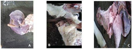
Figure 1. A) Liver and gallbladder with pale color, B) watery black-colored blood pouring out from the abomasum, C) lungs with pale color
The abomasal damages are amongst fatal diseases threatening calves’ survival [1]. There are many types of abomasal issues from mild to severe cases. Abomasal ulcers of non- perforated types mostly do not show clinical signs but sometimes can cause some blood loss, leading to anemia [2]. In agreement with our case, pale mucous membrane and pale udders have been reported in adult cows [2]. Environmental and nutritional stresses may be associated with abomasal ulcers. In veal calves, ingesting large volumes of milk replacer can be associated with higher rates of pyloric contractions, causing local ischemia and focal erosions [2,3]. The abomasal ulcers are typically classified into 4 types: type 1 includes non-perforating ulcers without excessive bleeding, type 2 consists of non-perforating ulcers with excessive bleeding, type 3 ulcers are perforating with local peritonitis, and type 4 are those with perforating ulcers and diffused peritonitis [3]. According to these abomasal damages, the case reported in the current article is categorized as type 2. In addition, the watery black-colored fluids pouring out of the abomasum further indicates that severe bleeding had occurred on the abomasal wall.
As noted, pale liver and lungs were observed in the present case which have not been reported in similar studies previously, to our knowledge. It was uncertain if the abomasal bleeding caused the hepatic pale and failure or the hepatic failure triggered anemia. Theoretically, hepatic failure and bile ducts problem would lead to anemia because of greater plasma bilirubin concentrations [4]. It has been reported that greater plasma bilirubin levels induce erythrocytes death and cause anemia [4]. Consequently, the severe anemia observed in the present case may be related to a failure of hepatic, gallbladder and bile duct functions. Further pathological and biochemical investigations are needed to confirm such a hypothesis.
Conclusion
A case of abomasal bleeding in a young male Holstein calf was reported that was accompanied by abnormalities in other splanchnic organs (e.g., liver and lung). Necropsy findings on-farm indicated that the problem was more complicated than a simple abomasal bleeding. Abomasal ulcers may lead to severe bleeding and calf death in dairy farms. According to our findings, this event can become complicated with the failure of other splanchnic organs such as liver and lungs. However, the anemia observed in the current case can be attributed to the failure of the liver. The hepatic failure is associated with higher plasma bilirubin concentrations which can induce erythrocytes death. Future pathological and biochemical examinations of similar cases are warranted to better understand the etiology of such problems.
References
- Bus JD, Stockhofe N, Well LE (2019) Invited review: Abomasal damage in veal calves. J Dairy Sci 102: 943-960. [Crossref]
- Kuiper R (1991) Abomasal diseases. Am Asso Bovine Pract: 26.
- Bus JD, Stockhofe N, Webb LE (2019) Invited review: Abomasal damage in veal calves. J Dairy Sci 102: 943-960.
- Lang E, Gatidis S, Freise NF, Bock H, Kubitz R, et al. (2015) Conjucated bilirubin triggers anaemia by inducing erythrocyte death. Hepatol 61: 275-284.