Extrahepatic manifestations occur in 10% to 20% of patients infected with hepatitis B virus. The most described include arthritis, arthralgia, glomerulonephritis, polyarteritis nodosa, popular acrodermatitis, skin rash, etc. Digestive and biliary involvement are rarely described. We report in this paper an original observation of an 87-year-old man who presented sphincter of Oddi dysfunction (SOD) during acute hepatitis B.
viral hepatitis b, extrahepatic manifestations, sphincter of oddi dysfunction
Viral hepatitis B (VHB) is a public health problem, with more than 300 million people infected worldwide. In the common acute form, clinical presentation is made of a preicteric phase (including nausea, vomiting, decreased appetite, weight loss, fever, tiredness, headache and joint pain, urticarial, etc.) followed by the onset of jaundice which decreases on average in 2 to 6 weeks. In adults, acute VHB can progress to chronicity in 5 to 10% with risk of cirrhosis and hepatocellular carcinoma [1].
Extrahepatic manifestations occur in 10% to 20% of patients infected with hepatitis B virus (HBV); their pathogenesis is unclear in most cases; it is believed to be mediated by virus-specific immune complex injury. These disorders include essentially acute necrotizing vasculitis (polyarteritis nodosa), membranous glomerulonephritis, skin rash, arthritis, arthralgia, and popular acrodermatitis. Essential mixed cryoglobulinemia is possible during VHB but less frequently than viral hepatitis C (VHC). Other disorders have been less reported such as Encephalitis, Guillain-Barré syndrome, Myelitis, myasthenia gravis, Myocarditis, thrombocytopenic purpura, etc. [2].
Digestive and biliary involvement, as cholecystitis and pancreatitis, are rarely described during VHB. We report in this paper an original observation of a patient who presented sphincter of Oddi dysfunction (SOD) during acute VHB.
K.R, 87 years old consulted for frank jaundice. He is an old self-sufficient man, with no pathology that hinders his daily life. The only history reported is a surgery for hiatus hernia 30 years ago. He had used tobacco before the age of 60, had never taken alcohol, and had not taken drugs or other hepatotoxic products. However, we note repeated practice of hijama (traditional scarification).
In May 2019, the patient presented a moderate fever, asthenia, and vomiting followed by the onset of frank jaundice and right upper quadrant pain. The general condition gradually deteriorated with anorexia, weight loss, intense pruritus, dark urine, and pale stools. He had no neurological disorders or bleeding syndrome; the BMI was at 21 Kg/m2.
Liver function tests showed serum aspartate aminotransferase: 1200 IU/L, alanine aminotransferase: 1100 IU/L, alkaline phosphatase: 500 IU/L, total bilirubin: 110 mg/L, direct bilirubin 64 mg/L, y-glutamyltransferase 282 IU/L, amylasemia: 88 IU/L, lipasemia: 35UI/L, and prothrombin ratio: 76.9%. Blood count indicated white blood cell count: 7000 /mm3, hemoglobin: 11.9 g/dl, platelet: 239000 /mm3. The creatinine clearance was 51 ml/min.
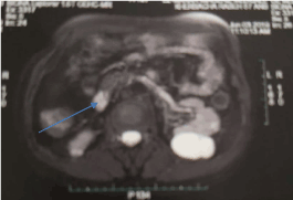
Abdominal ultrasound revealed dilation of the main bile duct (10 mm) without visible obstacle. At MRI, intra-hepatic bile ducts are also dilated; these dilations were upstream of an inflammatory thickening of the duodenal papilla (Figure 1).

Figure 1. Dilatation of Oddi sphincter without visible obstacle at MRI
We noted in virological tests a positive HBsAg, positive HBeAg, and positive IgM-HBc antibodies; the HBV viral load was 223 copies/ml. The hepatitis A and C serologies were negative as was the HIV serology.
SOD was evoked as extra hepatic manifestation during acute VHB, and the patient was treated with tenofovir on 06/19/2019. He recovered quickly and, after a month of treatment, the liver function tests became normal, the follow-up ultrasound found no bile duct dilatation, and there was loss of HBsAg and HBeAg followed by the appearance of HBsAb. Antiviral treatment was stopped after 3 months.
The sphincter of Oddi, described in 1887 by the Italian anatomist, Ruggero Oddi, is a smooth muscle valve regulating the flow of biliary and pancreatic secretions into the duodenum. SOD is a clinical syndrome secondary to functional impairment (dyskinesia: a pure motor disorder) or mechanical (stenosis: anatomical obstruction). Synonyms for SOD include biliary spasm, biliary dyskinesia, ampullary stenosis, papillitis, oddities, biliary dys-synergia and post-cholecystectomy syndrome. It occurs mainly during the follow-up after cholecystectomy but also before surgery. Its prevalence in the general population is estimated at 1.5% and it is most often found in women aged 20 to 50 years.
Mechanistically, SOD causes increased resistance to the passage of bile and pancreatic juice into the duodenum, resulting in raised choledochal pressure. This backpressure on the biliary tree is thought to be the primary mechanism of pain, causing distension, spasm, and inflammation. In about half of the cases, the stenosis or hyper pressure of the sphincter of Oddi is related to fibrosis of the sphincter of Oddi and the remaining cases are due to motor disorders [3].
Biliary pain and biliary colics are the more frequent symptoms (characterized as disabling epigastric or right upper quadrant pain lasting 30 min to several hours) although pancreatitis may also be present. The Milwaukee classification forms the basis of the diagnostic and therapeutic strategy for Oddian dysfunction; it classifies SOD patients into three types (I, II, and III) based on clinical presentation as well as laboratory and/or imaging abnormalities. The predictability of true SOD using this classification is category I: 65–95%, category II: 50-63% and category III: 12-28% [4].
Evaluation of patients with suspected sphincter of Oddi dysfunction includes biological (standard serum liver chemistries, serum amylase and lipase levels) and morphological tests (abdominal ultrasound examination or CT scan or even Cholangiopancreatography). Biliary manometry may be practiced but it is associated with a high risk of pancreatitis, not acceptable for a diagnostic procedure. Biliary scintigraphy offers an alternative without any risk although the sensitivity is not so high [5].
Besides biliary pathologies (post-cholecystectomy, preoperative cholelithiasis, agenesis of the gallbladder, gallstone lithotripsy), SDO has also been linked to liver transplantation, alcoholism, hypothyroidism (Delayed emptying of the biliary tract), and irritable bowel. Some medications such as opiates (morphine, tramadol, buprenorphine, pentazocine, etc.) are known to alter flow through the sphincter of Oddi [3,6].
HBV has not been described as a risk factor for SOD in clinical series; however, a study, reported in 2005, checked whether there is a pathogenic correlation between the affection of the biliary system and viral hepatitis B and C in 183 patients using fractionated duodenal tubing with biochemical and bacteriological bile testing and dynamic ultrasonic investigation. This study found that 69.9% of patients have hypertonicity of the sphincter of Oddi and 4.4% have hypotonicity. In addition, SOD was a risk factor for chronic acalculous cholecystitis [7].
All the clinical, biological, morphological, and evolutionary data from our observation are in favor of SOD being an extrahepatic manifestation in hepatitis B.
This is an original observation of SOD during acute hepatitis B. SOD can be considered as an extrahepatic manifestation, certainly underestimated, which requires prospective studies to better establish its pathogenesis and its frequency.
- WHO (2017) Global hepatitis report 2017. Available from: https://www.who.int/publications/i/item/global-hepatitis-report-2017
- Shim M, Han SH (2006) Extrahepatic manifestations of chronic hepatitis B. Hepatitis B Annual 3: 1. [Crossref]
- Afghani E, Lo SK, Covington PS, Cash BD, Pandol SJ (2017) Sphincter of Oddi function and risk factors for dysfunction. Front Nutr 4: 1. [Crossref]
- Silverman WB, Slivka A, Rabinovitz M, Wilson J (2001) Hybrid classification of sphincter of Oddi dysfunction based on simplified Milwaukee criteria: effect of marginal serum liver and pancreas test elevations. Dig Dis Sci 46: 278-281. [Crossref]
- Bhandari M, Toouli J (2007) Motility of the biliary tree. in TextBook of Hepatology from Basic Science to Clinical Practice. (3rd edn), Blackwell Publishing Ltd.
- Barthet M, Gonzalez JM (2015) Dysfonction du sphincter d’Oddi. HepatoGastro 22: 708-716.
- Pal'tsev AI, Osipenko MF, NB Voloshina NB (2005) Biliary tract in patients with chronic viral hepatitides. Ter Arkh 77: 72-76. [Crossref]

