Rhomboencephalosinapsi is an extremely rare malformation of the posterior cranial fossa, characterized by the fusion of the cerebellar hemispheres, the medial peduncles, the dentate nuclei and the hypogenesis of the cerebellar vermis. It is considered a sporadic malformation due to an autosomal dominant mutation that arises "de novo"; The incidence at birth is estimated at around 0.13% [1,2]. In this article we present the case of a male patient of 6 years of age with global delay of the diagnosis diagnosed with rhombencephalosinopsis and the MRI findings made in the Central Military Hospital.
anomalies, agenesis encephalopathies, rhomboencephalosinapsis
A 6-year-old male patient with a global developmental delay diagnosed with rhombencephalosinopsis [1,2]. Magnetic resonance imaging (MRI) practiced the following findings:
Rhombencephalosinapsi (RES) is an extremely rare malformation of the posterior cranial fossa; first described in 1914 by Obersteiner during an autopsy [1,3]. According to the most established theory, the fusion of the cerebellar hemispheres is due to a primitive defect in the differentiation of the cerebellar vermis. The literature describes RES cases in which chromosomal alterations have been identified (chromosome 2q deletion and unbalanced translocation 2p, 10q); This would explain the cases of family recurrence. Environmental factors have also been considered in the genesis of RES, in particular insulin dependent diabetes mellitus, the intake of phenylcyclidine at the beginning of pregnancy and prenatal contact with cytomegalovirus or ionizing radiation [3,4].
The diagnosis can be assigned in the course of the second-trimester ultrasound examination when there is no visualization of the cerebellar vermis. In this case, the transverse diameter of the cerebellum is smaller, but the cerebellum is not hypoplastic. In the cases reported in the literature, RES is suspected after the diagnosis of ventriculomegaly [4]. The diagnosis is confirmed exclusively by MRI in which it is observed: fused cerebellar hemispheres, hypoplastic or absent vermis, IV small ventricle and fused nuclei [4,5]. RES may present as an isolated malformation of the posterior cranial fossa or in association with other intracranial anomalies. Frequent dilation of the supratentorial ventricular system is observed. Hydrocephalus, secondary to the obstruction of the Silvio aqueduct, is often the first sign found during an ultrasound examination. The intracranial anomalies described in the literature are: oloprosencephaly, absence of the septum septum, absence / hypoplasia of the corpus callosum, olive hypoplasia, anomalies of the limbic system, fusion of the thalamus, septo-optic dysplasia, anomalies of the optic chiasm, cortical malformations and multiple cranial sutures [1,3,4]. It also describes the association with craniofacial dysmorphism (hypertelorism, prominent forehead, low or later rotated ears, microretrognathia, etc.) and extracranial anomalies that affect the cardiovascular, respiratory, urinary and skeletal systems (segmentation and fusion of the vertebrae; radio, polydactyly, syndactyly and phalanges of hypoplasia) [3]. The RES can also be included in different syndromic images, such as: Secuencia di Gomez-Lopez-Hernandez, in which turricephaly, temporoparietal alopecia, trigeminal anesthesia is observed; VACTREL sequence; VACTREL-H sequence [5].
Based on the participation of the supratentorial structures, the RES manifests clinically with ataxia, muscular hypotonia, spasticity, strabismus, abnormal eye movements, dysarthria and developmental delay [1,4,5] (Figures 1-4).
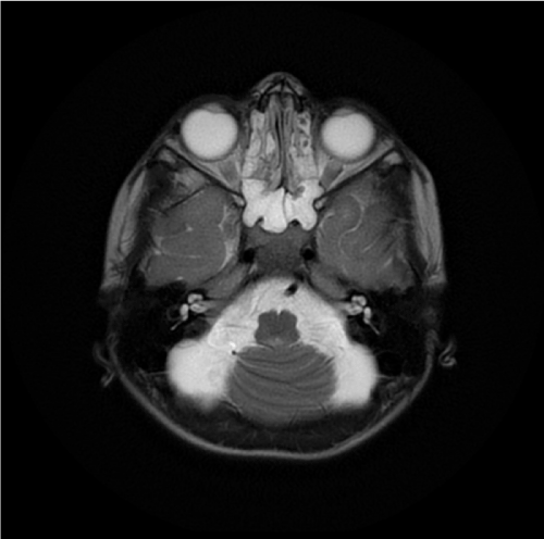
Figure 1. MRI shows the fusion of cerebellar hemispheres with absent vermis. The 4th ventricle is small and has diamond shaped
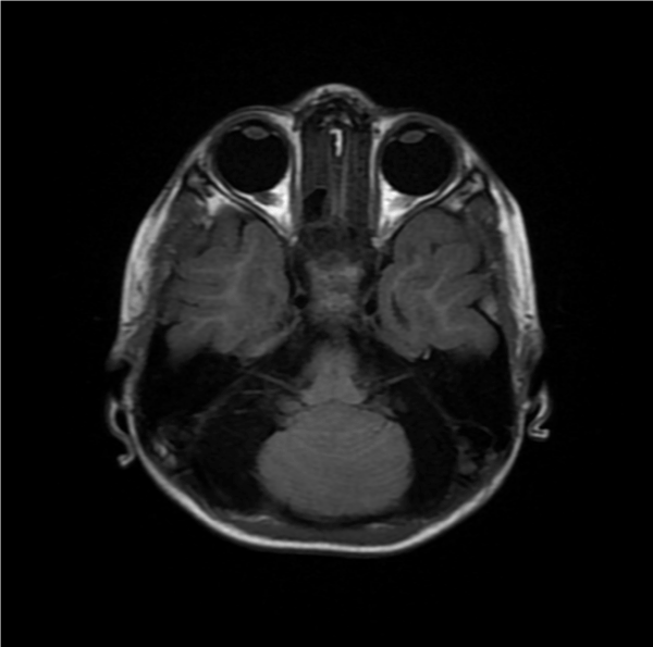
Figure 2. MRI of the posterior fossa demonstrates the absence of the cerebellar vermis with complete fusion of the cerebellar hemispheres Also, notice the absent septum pellucidum giving a mono-ventricle appearance
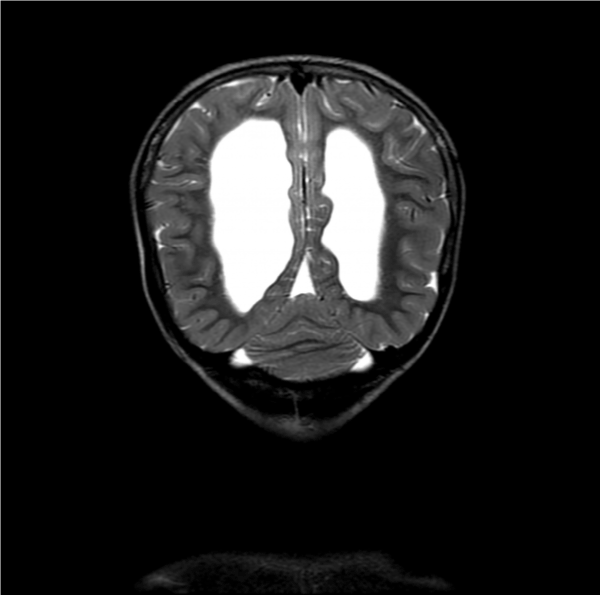
Figure 3. Coronal MRI show an absence of a vermis with continuous cerebellar white matter tracts and cortex across the midline
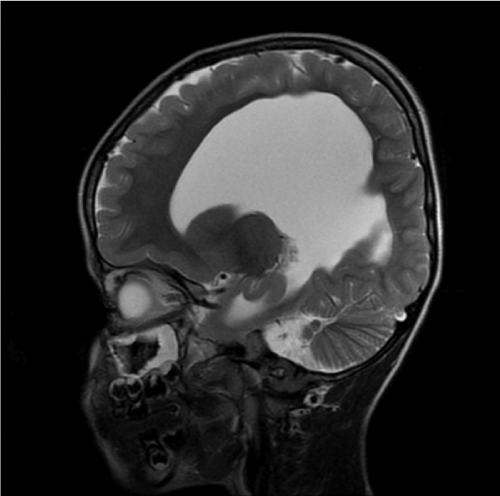
Figure 4. There is a fusion of cerebellar hemispheres with absent vermis, ventricular prominence without aqueductal stenosis and the absent septum pellucidum giving a mono-ventricle appearance
- Arango G, Muñoz E, Narváez J, Garcia E (2008) Romboencefalosinapsis: reporte de un caso. Rev Colomb Radiol 3: 2483-2484.
- Sener RN et al., 2003 Diffusion magnetic resonance imaging in infantile neuroaxonal dystrophy. J Comput Assist Tomogr 27: 34-37. [Crossref]
- Cabaj A, Bekiesinska M, Mądzik J (2012) MRI patterns of hypoxic-ischemic brain injury in preterm and full term infants - classical and less common MR findings. Pol J Radiol 77: 71-76. [Crossref]
- Mendonça JLF (2004) Rhombencephalosynapsis: CT and MRI findings. Neurol India 52: 118-120. [Crossref]
- Savolaine ER, Fadell RJ, Patel YP (1991) Isolated rhombencephalosynapsis diagnosed by magnetic resonance imaging. Clin Imaging 15: 125-129. [Crossref]