MRI, RARS1, leukodystrophy, hypomyelination, brain atrophy
A 3-year-old boy presented in the neonatal period with drug-resistant epileptic seizures, followed by severe developmental delay, microcephaly, and extrapyramidal movements. Brain MRI showed diffuse absence of myelination (Figure A-D). Examination at last follow-up demonstrated tetraparesis and nystagmus. Brain MRI revealed severe brain atrophy with marked white matter volume loss and hypomyelination (Figure E-H). Next-generation exome sequencing showed compound heterozygous variants ((c.173T>C(p.L58P); c.1790T>C(p.L597P)) in RARS1, encoding the cytoplasmic arginyl-tRNA synthetase (ArgRS), linked to Leukodystrophy, Hypomyelinating 9 (MIM 616140) [1]. A wide clinical spectrum has been described, with severe forms showing hypomyelination and early-onset brain atrophy, suggesting a primarily neuronal phenotype [2].
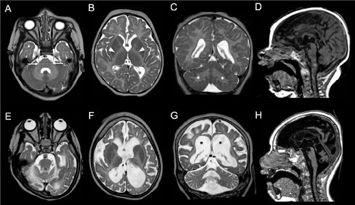
Figure 1: (A-C) T2-weighted images at 5 months of age reveal homogeneous white matter hyperintensity, with no myelination in the internal capsules (arrows), pons (arrowheads) and cerebellum (asterisks). D) T1-weighted image demonstrates small corpus callosum and pons. E-H) Corresponding T2 and T1-weighted images at 3 years of age show marked white matter thinning with ventricular dilatation (asterisks), subarachnoid spaces enlargement, callosal atrophy (empty arrow), and small midbrain and pons.
AA and MS had the idea of this work. AA drafted the manuscript, MS reviewed brain MRI, assembled the figure, revised and edited the manuscript. LP and MDR clinically evaluated the patient and revised the manuscript.
We are grateful to the parents for their willingness to contribute to this study.
- Wolf NI, Salomons GS, Rodenburg RJ, Pouwels PJ, Schieving JH, et al. (2014) Mutations in RARS cause hypomyelination. Ann Neurol 76: 134-139. [Crossref]
- Mendes MI, Green LMC, Bertini E, Tonduti D, Aiello C, et al. (2020) RARS1-related hypomyelinating leukodystrophy: Expanding the spectrum. Ann Clin Transl Neurol 7: 83-93. [Crossref]
Editorial Information
Editor-in-Chief
Article Type
Case Report
Publication history
Received: July 14, 2020
Accepted: July 28, 2020
Published: July 31, 2020
Copyright
©2020 Accogli A. This is an open-access article distributed under the terms of the Creative Commons Attribution License, which permits unrestricted use, distribution, and reproduction in any medium, provided the original author and source are credited.
Citation
Accogli A, Pisciotta L, Figà-Talamanca L, Di Rocco M, Severino M (2020) RARS1-related leukodystrophy: When hypomyelination meets brain atrophy. Med Res Innov 4: DOI: 10.15761/MRI.1000178.
Corresponding author
Dr. Mariasavina Severino
Neuroradiology Unit, IRCCS Istituto Giannina Gaslini, Genoa, Italy.
E-mail : bhuvaneswari.bibleraaj@uhsm.nhs.uk