Introduction
Oncocytoma is a rare benign neoplasm of oncocytes characterized by granular eosinophilic cytoplasm with abundance of mitochondria originally found associated with Hurthle cell tumor of the thyroid gland [1]. It usually occurs in the kidney, thyroid and salivary gland [2]. Oncocytoma involving the lungs is extremely rare with less than 10 reported cases in the past three decades. We present a case of an oncocytoma that presented as a distal tracheal mass.
Case description
A 65-year- old male with chronic obstructive pulmonary disease presented to his primary care physician with symptoms of dysphagia and odynophagia. He was an active smoker with 96 pack-year smoking history. He was thought to have gastroesophageal reflux for which omeprazole was given with some clinical improvement but given his symptoms along with a heavy smoking history, a chest CT to screen for lung malignancy was ordered. The CT showed a distal tracheal abnormality on the right (Figure 1A). Secretions or a tracheal wall abnormality were thought to be possible causes of the abnormality. A repeat CT scan was planned and performed 4 months later. During this interval, the patient complained of development of cough with yellowish phlegm production. A repeat CT showed a persistent presence of a distal right tracheal wall lesion (Figure 1B).

Figure 1. A. CT chest showing a right distal trachea lesion. B. A repeat CT chest showing a persistent right distal trachea lesion.
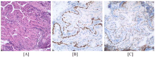
Given the persistent imaging abnormality, it was decided that a bronchoscopy evaluation with possible biopsy should be performed as a diagnostic procedure. A fiberoptic bronchoscopy was performed and the presence of a raised smooth walled lesion was found in the distal trachea. Four needle biopsies using a 22G Wang™ needle was performed. Cytology and immunohistochemistry showed oncocytic glandular proliferation in bronchial wall, consistent with oncocytoma arising from bronchial glands, with no evidence of malignancy. AE1-AE3. was positive. P 63, CK5/6 and TTF-1 were focally positive (Figure 2). P53 was positive for few cells. Ki 67 showed the result of 1-4%. SYN was positive for rare cell. Calponin and S100 were negative. The patient’s case was discussed at the hospital’s tumor board meeting. Given the location of the neoplasm and its reportedly benign course, it was decided that surveillance at regular intervals through imaging studies would be the best plan for the patient.

Figure 2. Histopathological and immunohistochemical image of the patient.
Discussion
Pulmonary oncocytoma is a very rare benign tumor. It has been reported to occur in the trachea, bronchus and the lung parenchyma itself. Pulmonary oncocytoma involving trachea has only been reported one other time and this would be the first reported case in English. Distal tracheal lesions could be due to mucoid impaction, primary malignant tumors, secondary malignant tumors, or benign tumor. Proper evaluation, especially in smokers, should be undertaken given the high probability of malignancy. Oncocytoma should be considered as part of the differential diagnosis of a distal tracheal lesion and biopsy should be obtained. Although most cases in the literature were surgically resected, patients with high risk of complications from an operation due to the location of tumor or comorbidities may require conservative treatment [3]. The prognosis of oncocytoma is generally good and usually does not impact lifespan of the patient [4]. Given its reported benign nature, surveillance at regular intervals could be a viable option.
References
- Hamperl H (1950) Oncocytes and the so-called Hürthle cell tumor. Arch Pathol 49: 563-570.
- Roy AA, Jameson C, Christmas TJ, Aslam Sohaib S (2011) Retroperitoneal oncocytoma: case report and review of the imaging features. Br J Radiol 84: e161-163. [Crossref]
- Vogel Y, Wolff I, Zienkiewicz T, Buttner R, Schulte (2010) A very rare cause of haemoptysis- coexistence of primary oncocytic adenoma of trachea with bronchial carcinoma. Dtsch Med Wochenschr 139: 1295-1298.
- Kuroda N, Toi M, Hiroi M, Shuin T, Enzan H (2003) Review of renal oncocytoma with focus on clinical and pathobiological aspects. Histol Histopathol 18: 935-942. [Crossref]
2021 Copyright OAT. All rights reserv


