Multiple pulmonary opacities on chest radiographs, often construed as cannon balls, are the classical presentation of haematogenous dissemination of malignancy to lungs. In contrast, such presentation may be associated with a non-malignant, extra-pulmonary aetiology. Thus, such radiographs require meticulous interpretation.
We present a case report of a young patient who suffered from spontaneous rib fractures, mimicking cannon balls appearance with a review of literature.
Conclusion: Attention to past medical history coupled with recognising the cannonball like pattern on chest radiographs is paramount in order to reach the most likely diagnosis.
cannon ball lung opacities; multiple pulmonary lesions; spontaneous rib fractures
Cannonball-like pulmonary opacities on chest radiographs are often attributed to a number of diagnoses such as sarcomas, seminomas, choriocarinomas, colon, prostate, thyroid, breast, head and neck, renal cell and squamous cell carcinomas. In addition, they may arise from fungal, parasitic infections, hydatid disease, rheumatoid nodules, wegener's granulomatosis and pulmonary tuberculosis [1,2]. Moreover, lesions external to lungs may mimic the cannonball-like appearance.
In order to avoid pitfalls on plain films, pattern-recognition is key in differentiating pulmonary from non-pulmonary opacities. On chest radiographs, pulmonary metastases typically appear bilaterally as peripheral rounded nodules of variable sizes [3]. Not only are small or medium-sized pleural effusions often misidentified as lung opacities, [4] thoracic cage lesions such as rib fractures or osteochondroma may also mimic lung lesions. [5] Additionally, a vascular thoracic lesion may well be confused for a pulmonary neoplasm [6]. Furthermore, Skin lesions namely nipple shadow, moles, neurofibromas, lipomas may resemble pulmonary nodules [7].
Computed tomography (CT) is usually invaluable in visualising pulmonary nodules and similar to chest radiographs, metastatatic lesions appear rounded, well-circumscribed and of soft tissue attenuation peripherally. [8] MRI, albeit not routinely used, may potentially be as sensitive as CT in spotting pulmonary metastases [9].
A young patient, with no history of trauma, attended the emergency department with sharp chest pain associated with mild shortness of breath. Past medical history included cushing’s syndrome. A chest radiograph, which was performed to exclude pneumothorax, showed a right fifth posterior rib fracture and well-defined opacities overlying the ribs in the right middle and left upper zones (Figure 1). All relevant laboratory data including white cell count, erythrocyte sedimentation rate, C-reactive protein, liver enzymes, calcium and phosphate levels, thyroid and parathyroid hormones were within normal ranges.
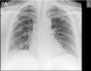
Figure 1. Chest X-ray showed well-defined opacities overlying the ribs in the right middle and left upper zones and right fifth posterior rib fracture
A subsequent contrast enhanced computer tomogram (Figure 2, 3, 6) of the thorax, abdomen and pelvis revealed multiple sclerotic ribs with periosteal bone formation. No destructive bony lesion or fractures were found elsewhere within the visualised skeleton (Figure 4) and pituitary gland appeared normal (Figure 5).
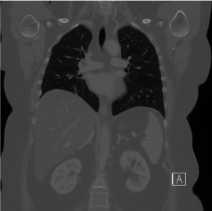
Figure 2. Initial Coronal CT image demonstrating multiple sclerotic ribs with periosteal bone formation
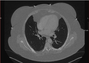
Figure 3. Initial axial CT image demonstrating multiple sclerotic ribs with periosteal bone formation
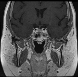
Figure 4. MRI T1 coronal image of pituitary fossa demonstrating normal appearances of the pituitary gland
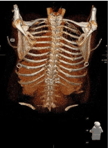
Figure 5. 3D reconstruction demonstrating multiple bilateral posterior rib fractures
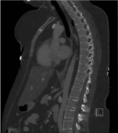
Figure 6. Sagittal CT image of chest and abdomen demonstrating the normal vertebral height and alignment of vertebral spine with no evidence of osteoporotic fractures
The most likely diagnosis was Cushing’s syndrome-associated spontaneous rib fractures. The patient was managed conservatively then discharged. Pituitary adenomectomy was performed later. Unfortunately, the patient passed away a few days following the surgery.
Cannonball-like lung opacities are often the classic presentation of haematogenous dissemination of malignancy to the lungs; therefore, indicating advanced stage of the disease with a very grim prospective cure or survival [10]. Thus, thorough assessment may reduce avoidable patient’s anxiety and potentially needless examinations.
In this case, the primarily diagnosed cushing’s syndrome may have possibly led to secondary osteoporosis, which may have given the described findings. Cushing’s syndrome-related osteopenia and osteoporosis ranges between 60-80% and 30-65% respectively. Around one-third to half the patients who suffer from Cushing’s syndrome experience non-traumatic fractures, which may be the presenting manifestation of this syndrome. Spontaneous fractures may occur in the ribs or pelvis and less commonly in long bones. Approximately one-third of patient would develop vertebral fractures especially at the thoracic and lumbar levels [11].
Several terms are used interchangeably for spontaneous rib fractures including pathological, fragility, compression and insufficiency fractures. Aetiology includes osteoporosis (calcium deficiency and corticosteroid –induced), malignancy, Vitamin A hypervitaminosis, cerebral palsy (in children) and osteodystrophy in chronic renal failure. Bisphosphonates-associated adverse outcomes are a rare cause [12].
To evaluate the degree of osteoporosis, bone density scan should be performed. Not only does it act as a comparative study following treatment, bone scan may also reveal multifocal uptake in the ribs and reflect the extent of bony involvement; an appearance that may potentially resolve following treatment. Skeletal metastases commonly manifests with osteolytic rib lesions on plain chest radiographs and hot spots on bone scan [13]. Furthermore, multiple spontaneously-distributed foci of increased radionuclide uptake in ribs, mimicking osseous metastases, have been reported in post-traumatic conditions, extreme altitudes, tuberculosis, brucellosis, coccidiomycosis and Cushing's syndromes [14].
The treatment for acutely symptomatic spontaneous rib fractures is analgesia, orally administered bisphosphonate, with supplemental calcium and vitamin D. This regimen is well-tolerated with unremarkable gastrointestinal side effects [14]. Patients with Cushing’s syndrome often require definitive intervention for hypercortisolism by either adrenalectomy or pituitary adenomectomy, with subsequent disappearance of the multiple hot spots found on bone scan [11].
Spontaneous rib fractures are likely to be misinterpreted as metastatic lung lesions, particularly in young patients with Cushing’s syndrome. Awareness of such presentation and linking it to past medical history, which proved to have a significant weight in diagnosis, is essential in order to guide treatment and avoid unwarranted further investigations.
- Seaton A (2000) Other pulmonary neoplasm and related conditions. Crofton and Douglas's Respiratory Diseases. In: Seaton A, Seaton D, Leitch AG, editors. [5th Edn]. Oxford: Blackwell science ltd.
- The WHO manual of diagnostic imaging (2006) Radiographic anatomy and interpretation of the chest and the pulmonary system. In: Ostensen H, Petterson H, Editors. Geneva: World Health Organization.
- Seo JB, Im JG, Goo JM, Chung MJ, Kim MY (2001) Atypical pulmonary metastases: spectrum of radiologic findings. Radiographics 21: 403-417. [Crossref]
- Kitazono MT, Lau CT, Parada AN, Renjen P, Miller WT Jr. (2010) Differentiation of pleural effusions from parenchymal opacities: accuracy of bedside chest radiography. AJR Am J Roentgenol 194: 407-412. [Crossref]
- Lass RB, Norton KI, Mitre SA, Kang E (2002) Pediatric ribs: a spectrum of abnormalities. Radiographics 22: 87-104.
- Raymond GS, Miller RM, Müller NL, Logan PM (1997) Congenital thoracic lesions that mimic neoplastic disease on chest radiographs of adults. AJR Am J Roentgenol 168: 763-769.
- Wolfgang Dahnert (2011) Radiology Review Manual, [7th Edn] ISBN-10: 1609139437.
- Collins J, Stern EJ (2007) Chest radiology, the essentials. Lippincott Williams & Wilkins. ISBN: 0781763142.
- Naidich DP, Srichai MB, Krinsky GA (2007) Computed tomography and magnetic resonance of the thorax. Lippincott Williams & Wilkins.
- C Greenfield LJ, Mulholland MW (1997) Essentials of surgery, scientific principles and practice. Lippincott Williams & Wilkins. ISBN: 0397515324.
- Lee HJ, Je JH, Seo JH, Na YJ, Yoo HJ (2014) Mutiple Spontaneous Rib Fractures in Patient with Cushing's Syndrome. J Bone Metab 21: 277-282. [Crossref]
- Wick JY (2009) Spontaneous fracture: multiple causes. Consult Pharm 24: 100-2, 105-8, 110-2.
- Wang K, Allen L, Fung E, Chan CC, Chan JC, et al. (2005) Bone scintigraphy in common tumors with osteolytic components. Clin Nucl Med 30: 655-671.
- Vrbanić TS, Novak S, Sestan B, Tudor A, Gulan G (2008) A case of pathological rib fractures: focal osteolysis or osteoporosis? West Indian Med J 57: 178-181.