Congenital high airway obstruction syndrome (CHAOS) is an extremely rare life-threatening condition. The sonographic findings are very characteristic. Generally the diagnosis is made with the 4-chamber view of the fetal heart. Typically, both lungs appear severely enlarged and highly echogenic. The heart points towards the midline of the thorax. Fetal ascites is determined commonly on ultrasound examination. A case of CHAOS, diagnosed antenatally via ultrasound is reported here.
congenital high airway obstruction syndrome, tracheal atresia, ultrasonography
Congenital high airway obstruction syndrome (CHAOS) is defined as complete or partial obstruction of the fetal upper airways. This clinical condition was brought into notice firstly by Hedrick in the late 1900s [1]. The true incidence of CHAOS is unknown. Laryngeal atresia seems to be the most frequent cause [2]. CHAOS has been considered an almost invariably fatal condition [3] which often goes incorrectly diagnosed until autopsy. Fortunately, more cases can be recognized in utero nowadays, as there are significant technical improvements in prenatal imaging. Bilaterally enlarged hyperechoic lungs, dilated airways, and flattened or inverted diaphragm are the typical prenatal sonographic findings. Fetal ascites and non-immune hydrops may also be associated with the clinical condition [4]. We report here a case of CHAOS due to tracheal atresia diagnosed by antenatal ultrasonography.
A 25-year-old woman, gravida 1, parity 0, was referred to our perinatal unit at 24 weeks of gestation due to fetal ascites. There was no history of consanguinity. She and her family had both unremarkable medical histories. Ultrasound examination showed that the fetus had bilateral large hyperechoic lungs. The diaphragm was inverted. The heart seemed small and was centrally displaced (Figure 1). Main bronchi and trachea were full of fluid (Figure 2).There was massive ascites (Figure 3). Based on ultrasound findings, diagnosis of CHAOS was made. The possible unfavorable outcome of the pregnancy was discussed with the parents. The couple was referred to a genetics specialist and cordocentesis procedure is offered. The parents accepted an cordocentesis and the pregnancy was terminated due to request of the parents. Autopsy was declined. There were no other gross abnormalities. The diagnosis of congenital high airway obstruction syndrome was confirmed. Fetal karyotype was found to be normal.
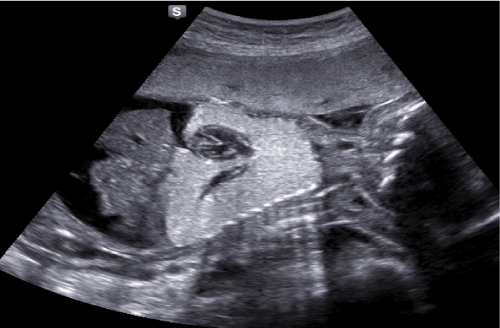
Figure 1. Echogenic lungs, inverted diaphragm and fetal ascites are shown on sagittal plane.
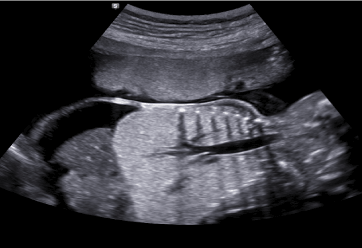
Figure 2. Dilated airways including trachea and main bronchus are demonstrated on coronal plane.
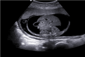
Figure 3. Fetal ascites on transvers plane.
Tracheal atresia is a very rare congenital malformation which takes place by deficient recanalization of the upper airways around the 10th week of gestation resulting in a clinical spectrum defined as congenital high airway obstruction syndrome (CHAOS) [1]. CHAOS is a rare abnormality, usually with a lethal outcome. The primary abnormality is an intrinsic obstruction of the upper airway. Causes of obstruction include laryngeal atresia, laryngeal web, laryngeal cyst, and tracheal atresia or stenosis. The obstructed airway causes decreased clearance of fluid produced by the fetal lungs as well as an increase in intratracheal pressure resulting in lung hyperexpansion and abnormal development [3,5]. The hyperexpanded lungs cause compression of the heart and inferior vena cava, decreasing venous return and leading to nonimmune hydrops [3,5,6]. These series of events are responsible for the prenatal imaging characteristics of CHAOS. Our case shows that fetuses with CHAOS have typical ultrasound findings, which include markedly increased lung volumes with flattened or inverted hemidiaphragms, a dilated airway below the level of obstruction, massive ascites, centrally positioned. Regarding the amniotic fluid index, compression of the esophagus by dilated airways may lead to polyhydramnios, as the fetal swallowing of the fluid is disrupted. On the hand, impaired swallowing of the fetus may also cause oligohydramnios [7]. In our case, amniotic fluid index was normal. The identification of airway obstruction is key to establishing the diagnosis of CHAOS and to differentiating it from bilateral lung masses such as a bilateral congenital pulmonary airway malformation (CPAM) or other causes of extrinsic airway obstruction such as a double aortic arch [8]. In utero death of the affected cases; being a part of some genetic syndromes. The most common associated genetic disorder with CHAOS is Fraser’s syndrome which is inherited by autosomal recessive form and characterized by urogenital defects, laryngeal atresia, syndactyly, and cryptophthalmos [7]. CHAOS may be also a part of Cri-du-Chat syndrome, short-rib polydactyly syndrome, and velocardiofacial syndrome [5,7]. In our case, karyotype was normal and there was no other suggestion findings of these genetic disorders. Live born neonates with CHAOS often have several postnatal symptoms/abnormalities including respiratory distress caused by the abnormal development of the lungs, diaphragmatic dysfunction likely caused by stretching of the diaphragm, tracheomalacia and capillary leak syndrome [5]. There are few cases with spontaneous antenatal improvements. This progress may be due to spontaneous perforation or tracheoesophageal fistula with the refining of the obstructed fluid leading to decrease in the pressure of airways and reversal of the process [9].
In conclusion, Postnatal management of newborn babies with CHAOS is difficult and the prognoses of the affected infants are often poor. The perinatal mortality of CHAOS is 100% without intervention. Antenatal sonographic imaging shows typical findings which can lead to an early diagnosis. This is important especially if any fetal intervention is considered.
- Roybal JL, Liechty KW, Hedrick HL, Bebbington MW, Johnson MP (2010) Predicting the severity of congenital high airway obstruction syndrome. J Pediatr Surg 45: 1633-1639. [Crossref]
- Sanford E, Saadai P, Lee H, Slavotinek A (2012) Congenital high airway obstruction sequence (CHAOS): a new case and a review of phenotypic features. Am J Med Genet A 158A: 3126-3136. [Crossref]
- Hedrick MH, Ferro MM, Filly RA, Flake AW, Harrison MR, et al. (1994) Congenital high airway obstruction syndrome (CHAOS): a potential for perinatal intervention. J Pediatr Surg 29: 271-274. [Crossref]
- Garg MK (2008) Case report: antenatal diagnosis of congenital high airway obstruction syndrome. Laryngeal atresia. Indian J Radiol Imaging 18: 350-351. [Crossref]
- Lim FY, Crombleholme TM, Hedrick HL, Flake AW, Johnson MP, et al. (2003) Congenital high airway obstruction syndrome: natural history and management. J Pediatr Surg 38: 940-945. [Crossref]
- Onderoglu L, Saygan Karamursel B, Bulun A, Kale G, Tuncbilek E (2003) Prenatal diagnosis of laryngeal atresia. Prenat Diagn 23: 277-280. [Crossref]
- Joshi P, Satija L, George RA, Chatterjee S, D’Souza J (2012) Congenital high airway obstruction syndrome-antenatal diagnosis of a rare case of airway obstruction usingmultimodality imaging. Med J Armed Forces India 68: 78-80.
- Shum DJ, Clifton MS, Coakley FV, Hornberger LK, Joe BN, et al. (2007) Prenatal tracheal obstruction due to double aortic arch: a potential mimic of congenital high airway obstruction syndrome. AJR Am J Roentgenol 188: W82-W85. [Crossref]
- Kuwashima S, Kitajima K, Kaji Y, Watanabe H, Watabe Y (2008) MR imaging appearance of laryngeal atresia (congenital high airway obstruction syndrome): unique course in a fetus. Pediatr Radiol 38: 344-347. [Crossref]
2021 Copyright OAT. All rights reserv