Background: Myocarditis is a possible complication of COVID vaccinations that has been observed in previously published case reports (1). In the following case report, we discuss the presentation, diagnosis, and management of a case of myocarditis that occurred after the Oxford-AstraZeneca COVID-19 vaccination. Almost all of the reported cases of myocarditis were post mRNA COVID vaccinations (Pfizer and Moderna).
Case Report: A 47-year-old male presented to our cardiac center with dyspnea. The patient is an ex-smoker with no known prior medical history. Echocardiography followed by cardiac MRI showed severe reduction in the left ventricular ejection fraction (LVEF) and late gadolinium enhancement (LGE), consistent with myocarditis.
Conclusion: We report a case of classic myocarditis on cardiac MRI secondary to COVID vaccination. Exact prevalence, mechanism, and prognosis of COVID-19 vaccination- related myocarditis is unknown.
myocarditis, myocardium, covid-19, dyspnea, vaccination, astrazeneca covid-19 vaccination
A 47 year-old male presented to our cardiac center with dyspnea. He had no prior medical history. His symptoms started a day after receiving his first dose of Oxford-Astrazeneca Covid 19 vaccination. Initial symptoms were generalized symptoms like body aches, fatigue, fever and dyspnea, all of which resolved except for the dyspnea. His shortness of breath progressed a week later and he developed lower extremity edema. He was seen at a periphery urgent care facility with limited resources, where he was given a diuretic and his symptoms improved. He was referred to our outpatient center. The patient was evaluated in our cardiology clinic. His main symptoms at this point were orthopnea, exertional dyspnea, NYHA class II paroxysmal nocturnal dyspnea.
On examination, his vital signs were unremarkable, and rest of his examination is significant for classic findings of heart failure: jugular venous distension, lung crackles bilaterally, apical pansystolic murmur and bilateral lower extremity edema. ECG showed sinus rhythm with multiple premature ventricular complexes (PVCs) and lateral T wave inversions (Figure 1). Echocardiography was performed and showed a severely reduced left ventricular ejection fraction (LVEF) of ~30.8% estimated by Simpson’s biplane with global hypokinesis and moderate to severe mitral regurgitation and moderate left atrial dilation (Figure 2). Additional work-up included a 24-hour holter, which showed frequent PVC burden (36.1%) with no evidence of sustained ventricular tachycardia.
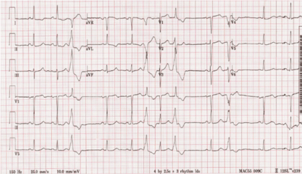
Figure 1. ECG showed sinus rhythm with multiple premature ventricular complexes and lateral T wave inversions
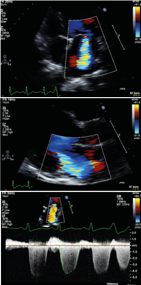
Figure 2. Echocardiography with Color-Doppler showing moderate-severe mitral regurgitation
The patient was electively admitted and coronary angiography was done, which showed a mid focal left anterior descending (LAD) 80% eccentric lesion at the bifurcation with the diagonal artery and a proximal 40% lesion in diagonal branch with no significant lesions in other coronaries (Figure 3). He underwent successful percutaneous coronary intervention (PCI) to mid LAD using DES with provisional approach to the diagonal.
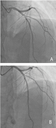
Figure 3. Coronary angiography in cranial views. A. Pre-intervention angiogram showing a mid focal LAD eccentric lesion at the take-off of first diagonal. B. Post-intervention angiogram after provisional stenting of LAD
Post PCI, he continued to have a significant PVCs burden. A repeat 24-hour holter showed more frequent PVC burden (37.9%), indicating that the PVCs were not ischemic. Additionally, the LV dysfunction is out of proportion to his coronary artery disease with global dysfunction. Therefore, additional work-up was needed to look for the etiology of his cardiomyopathy. He underwent a cardiac MRI, which showed a severely reduced LVEF with severe global hypokinesia, subepicardial late gadolinium enhancement (LGE) in the basal lateral wall, and a significant increase in T2 signal intensity in the basal to mid lateral wall, suggestive of acute or subacute myocarditis (Figure 4).
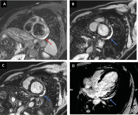
Figure 4. Cardiac MRI A) Short Axis view T2 with fat suppression showed high T2 signal intensity in the subepicardial part of the basal lateral wall (red arrow) indicate myocardial edema. B,C,D) Short Axis view and 4-Chamber view showing subepicardial late gadolinium enhancement (LGE) in the basal and part of mid lateral wall (blue arrow) along with the high T2 signal intensity are consistent with myocarditis
The patient was discharged on dual antiplatelet therapy, beta blocker, sacubitril-valsartan and oral amiodarone. The patient was seen in follow-up and remains stable with no recurrent heart failure admission.
Myocarditis is an inflammation of the myocardium, which could be secondary to infection, autoimmune conditions and many other etiologies. One of the recently recognized causes of myocarditis is COVID-19 vaccinations [1-4]. There are several reported cases, the majority of which are related to mRNA vaccinations [2]. The incidence is overall rare and reported to be 0.8 per 1 million first doses and 5.8 per 1 million second doses of BNT162b2 (Pfizer/BioNTech) and mRNA-1273 (Moderna) vaccines [2]. There have been reports of Oxford- AstraZeneca COVID-19 vaccination myocarditis mostly from the UK. As of July 28th, 2021, myocarditis was seen in 149 cases after the Pfizer/BioNTech vaccine, 82 cases after Oxford- AstraZeneca vaccine and 25 cases after the Moderna vaccine [5]. The UK data suggests 1.7 cases of myocarditis per million doses of Oxford- AstraZeneca vaccine.
While most cases of myocarditis are asymptomatic, some of the possible symptoms include chest pain, fever, dyspnea, and syncope [6]. Prodromal manifestations such as respiratory and gastrointestinal symptoms are also reported in some cases [6]. In one study, 26.6% of patients were reported to have systolic dysfunction (defined as a LVEF of <50% at the initial in-hospital echocardiography) [7].
The diagnosis of myocarditis can be established with endomyocardial biopsy or with the less invasive strategy of cardiac MRI (7). The use of cardiac MRI has allowed for the diagnosis of milder cases of myocarditis, as endomyocardial biopsy was typically reserved for critically ill patients [7]. The implementation of the new Lake Louise Criteria for cardiac MRI has a sensitivity of 87.5% and a specificity of 96.2% for the detection of acute myocarditis in patients with clinical suspicion [8].
The management of myocarditis includes supportive therapy for systolic dysfunction. Heart failure guideline-directed medical therapy is recommended in patients with depressed EF.
Arrhythmias are generally managed conservatively, as they tend to resolve on their own. However, in patients with sustained bradycardia or complete heart block, a pacemaker may be indicated. In addition, in patients with ventricular tachycardia, the antiarrhythmic amiodarone can be used [9]. Many patients with COVID-19 vaccination myocarditis respond well with medical treatment [5].
Myocarditis has been described post mRNA COVID-19 vaccinations but rarely after Oxford- AstraZeneca COVID-19 vaccinations. Patients typically do well and the risks of COVID-19 infections remain higher than the risk of myocarditis.
- Mei R, Raschi E, Forcesi E, Diemberger I, De Ponti F, et al. (2021) Myocarditis and pericarditis after 1immunization: Gaining insights through the Vaccine Adverse Event Reporting System.
- Simone A, Herald J, Chen A, Gulati N, Shen AY, et al. (2021) Acute Myocarditis Following COVID-19 mRNA Vaccination in Adults Aged 18 Years or Older. JAMA Intern Med : e215511. [Crossref]
- Engler RJ, Nelson MR, Collins Jr LC, Spooner C, Hemann BA, et al. (2015) A prospective study of the incidence of myocarditis/pericarditis and new onset cardiac symptoms following smallpox and influenza vaccination. PLoS One 10: e0118283. [Crossref]
- Salah HM, Mehta JL (2021) COVID-19 Vaccine and Myocarditis. Am J Cardiol 157: 146-148. [Crossref]
- Singer ME, Taub IB, Kaelber DC (2021) Risk of Myocarditis from COVID-19 Infection in People Under Age 20: A Population-Based Analysis. medRxiv 1: 21260998. [Crossref]
- Ammirati E, Frigerio M, Adler ED, Basso C, Birnie DH, et al. (2020) Management of acute myocarditis and chronic inflammatory cardiomyopathy: an expert consensus document. Circ Heart Fail 13: e007405. [Crossref]
- Ammirati E, Cipriani M, Moro C, Raineri C, Pini D, et al. (2018) Clinical presentation and outcome in a contemporary cohort of patients with acute myocarditis: multicenter Lombardy registry. Circulation 138: 1088-1099 [Crossref]
- Luetkens JA, Faron A, Isaak A, Dabir D, Kuetting D, et al. (2019) Comparison of original and 2018 Lake Louise criteria for diagnosis of acute myocarditis: results of a validation cohort. Radiol Cardiothorac Imaging 1: e190010.
- Cooper Jr LT (2009) Myocarditis. New Eng J Med 360: 1526-1538.