Background: Acute renal failure associated to renal cortical necrosis is a rare condition that can appear in pregnancy, during or after the delivery. It can be caused by various factors which must all be excluded in order to reach the correct diagnosis. Due to its low incidence in developed country, it presents a challenging diagnosis, cause management and treatment.
Case report: Here we report a rare case of acute renal failure with renal cortical necrosis probably caused by post-partum hemorrhage. A 36-years-old primiparous woman, after a post-partum hemorrhage, presented low platelets count, anuria and increase serum creatinine level that necessitate daily dialysis and Continuous Veno-Venous Hemofiltration. After the discharge she presented persisting stage IV chronic renal insufficiency, currently being treated. After excluding all possible causes of pregnancy-related acute renal failure, the only reasonable explanation was renal cortical necrosis due to post-partum hemorrhage.
Conclusion: Due to a loss of 3000 cc of blood, the patient suffered of a life threating disease. The acute renal failure is a serious condition that must be taken into consideration in case of post-partum bleeding and promptly.
acute renal failure, post-partum hemorrhage, renal cortical necrosis, delivery, dialysis
Acute renal failure (ARF) is a rare complication of pregnancy and delivery. In the Unites States of America the estimated incidence varied from 2 to 4.5 per 10,000 deliveries [1]. The long-term outcome of pregnancy related ARF is usually favorable unless renal cortical necrosis (RCN) occurs. RCN accounts for only 2% of all cases of ARF and can lead to chronic renal failure (CRF) due to the ischemic destruction of the renal cortex. The incidence of pregnancy-related RCN is declining in developing countries with only few cases reported [2] and Pakistan [3]. The most frequent causes of obstetric RCN are placenta previa, abruption placentae and postpartum hemorrhage (PPH). Other less frequent conditions associated to pregnancy-related RCN are amniotic fluid embolism, septic abortions, atypical hemolytic uremic syndrome (SEU) and thrombotic thrombocytopenic purpura (TTP) [4].
Postpartum hemorrhage (PPH) is defined as a blood loss of 500 ml or greater after vaginal birth, or 1000 ml or greater after cesarean delivery, or a blood loss with associated signs or symptoms of hypovolemia, that occurs within 24 h of delivery [5]. PPH is a relatively common complication of delivery (4-6% of pregnancy) and the most common etiology is uterine atony [6]. Recognition of PPH is challenging and the estimation of blood loss is frequently not accurate.
We present a rare case of postpartum acute renal failure with renal cortical necrosis due to postpartum hemorrhage without other possible causes of this condition.
A 36-year-old primiparous woman presented to the labor ward at 39 weeks of gestation. She had no significant medical history and was not on any medications. She presented polyamnios and a reciprocal translocation (3;11) at the chorionic villus sampling (CVS). The only abnormality seen on the admission blood tests was a platelet count of 106,000. After an operative delivery there was an umbilical cord rupture during the active management and a manual placenta removal was necessary. A persistent uterine atony occurred with a post-partum hemorrhage despite uterine massage, oxytocin infusion, Tranexamic Acid (TXA) administration, 2 unit of red blood cells and the placement of a Bakri balloon. After about 3000 ml of estimated blood loss the patient needed uterine artery embolization performed by an interventional radiologist. On the first day after the delivery the patient presented anuria, elevated body temperature, nausea and vomit. The blood tests showed hemoglobin level of 6.7 gr/dL and a platelet count of 30,000 after 3 unit of packed red blood cells and 1 unit of whole blood platelets. On the second day after delivery, the patient’s clinical picture persisted and she underwent thorax and abdomen computed tomography. The contrast enhanced CT scan revealed ischemic hepatic areas, avascularized renal cortical, abdominal and pleural effusions (Figure 1). Due to persistence of anuria and increased creatinine levels, the patient required daily dialysis. All autoantibodies tests turned out negative and ADAMTS-13 activity was normal. Due to the lack of improvement after 11 days of daily dialysis, she performed two treatment cycles of plasma exchange. Another CT scan shown the persistence of the hepatic and renal conditions and the appearance of a paravaginal hematoma. It required a drainage in the operating theater and a subsequent Intensive Care Unit (ICU) hospitalization. During the ICU stay, Continuous Veno-Venous Hemofiltration (CVVH) was performed until resumption of diuresis. Upon reaching a stable hemodynamic condition, the patient was hospitalized in nephrology unit for the treatment of the stage IV renal insufficiency with a persistent serum creatinine between 3.5 and 4.0 gr/dL. She was discharge at 90 days after the delivery with improvement of the renal insufficiency and oral anticoagulant therapy. After 3 months, the serum creatinine level was 2.58 gr/dL with persisting stage IV chronic renal insufficiency.
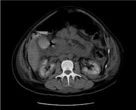
Figure 1. CT-scan showing avascularized renal cortical
This case appears to be an example of Acute Renal Failure with Renal Cortical Necrosis probably caused by Post-Partum Hemorrhage. The differential diagnosis to be excluded are amniotic fluid embolism, HELLP syndrome and thrombotic microangiopathies like atypical hemolytic uremic syndrome (SEU) and thrombotic thrombocytopenic purpura (TTP) [4]. Thrombotic microangiopathy (TMA) is a heterogeneous group of disorders features by thrombocytopenia, hemolytic anemia and organ failure. Severe post-partum hemorrhage can mimic thrombotic microangiopathy with thrombocytopenia, elevated lactate dehydrogenase levels, anemia with detectable schistocytes and acute renal injury [7]. Like in uremic hemolytic syndrome, the PPH can lead to renal cortical necrosis due to disseminated intravascular coagulation (DIC) [8].
In our case, normal ADAMTS-13 activity excluded TTP and normal value of complement factors with absence of mutation of complement genes excluded atypical SEU.
In Frimat, et al. [8] case series of renal cortical necrosis, the initial presentation features were major blood loss (average 2.6 L), oliguria and frequent DIC and/or hemolysis. The range of volume of blood loss was 1.3 L – 3.5 L and the mean volume of packed red blood cells was 1.45 ± 0.9 L. None of the patients had recovered normal kidney function at last follow-up (12-55 months). In our case the volume of blood loss was 3 L, in range with Frimat’s case series, and, as well as his patients, she didn’t recover normal kidney function at last follow-up.
The management of PPH consists of pharmacological agents that target uterine atony (like oxytocin), antifibrinolytics (like TXA) and massive transfusion protocols [5]. In our case, the hospital protocol of PPH treatment was followed and all therapeutic measures recommended have been performed. Nonetheless, Acute Renal Failure occurs and daily dialysis and CVVH was necessary. To our knowledge, no guideline exists for the treatment of post-partum acute renal failure.
Acute renal failure is a rare but serious post-partum complication and, if associated with cortical renal necrosis, can lead to important morbidity for the patient and poor outcome. One of the most common cause is post-partum hemorrhage. It’s important exclude other less frequent causes and to deal promptly any worsening of the patient’s symptoms without underestimating the possible consequences of PPH. Dialysis and Continuous Veno-Venous Hemofiltration are essential for treat renal insufficiency.
- Callaghan WM, Creanga AA, Kuklina EV (2012) Severe maternal morbidity among delivery and postpartum hospitalizations in the United States. Obstet Gynecol 120: 1029-1036. [Crossref]
- Prakash, Vohra R, Wani IA, Murthy AS, Srivastva PK, et al. (2007) Decreasing incidence of renal cortical necrosis in patients with acute renal failure in developing countries: a single-centre experience of 22 years from Eastern India. Nephrol Dial Transplant 22: 1213-1217. [Crossref]
- Ali A, Ali MA, Ali MU, Mohammad S (2011) Hospital outcomes of obstetrical-related acute renal failure in a tertiary care teaching hospital. Ren Fail 33: 285-290. [Crossref]
- Meibody F, Jamme M, Tsatsaris V, Provot F, Lambert J, et al. (2020) Post-partum acute kidney injury: sorting placental and non-placental thrombotic microangiopathies using the trajectory of biomarkers. Nephrol Dial Transplant 35: 1538-1546. [Crossref]
- Higgins N, Patel SK, Toledo P (2019) Postpartum hemorrhage revisited: new challenges and solutions. Curr Opin Anaesthesiol 32: 278-284. [Crossref]
- Bateman BT, Berman MF, Riley LE, Leffert LR (2010) The epidemiology of postpartum hemorrhage in a large, nationwide sample of deliveries. Anesth Analg 110: 1368-1373. [Crossref]
- Fakhouri F (2016) Pregnancy-related thrombotic microangiopathies: Clues from complement biology. Transfus Apher Sci 54: 199-202. [Crossref]
- Frimat M, Decambron M, Lebas C, Moktefi A, Lemaitre L, et al. (2016) Renal cortical necrosis in postpartum hemorrhage: A case series. Am J Kidney Dis 68: 50-57. [Crossref]