Introduction: The pelvic floor dysfunction leading to muscle hypertonia is often idiopathic, but sometimes recognisable disorders may explain the steadily increased pelvic muscle tone in women and men. If tissue temperatures remain below 42°C, radiofrequency technologies induce pain relief and muscle relaxation, probably through neuromodulation, without heat-induced tissue damage. The Dynamic Quadripolar Radiofrequency™ (DQRF™) technology has the further benefit of being non-invasive. The paper reports on the outcomes of the first real-life exploratory study with the DQRF™ technology, combined with radioporation of lenitive and pro-trophic agents (UPR™), in subjects with pelvic floor muscle hypertonia.
Methods: Office-based, prospective pilot cohort study in 31 subjects with painful posterior pelvic floor muscle hypertonia, 20 women and 11 men 23 to 60 years old, without discriminating between idiopathic or secondary pelvic floor hypertonia. The DQRF™ device incorporates a novel flat probe ergonomically designed for the radiofrequency treatment of the posterior pelvic floor and anorectal areas. Assessments, at baseline (first treatment session) and end of treatment (last treatment session): carried out according to the validated criteria for pelvic floor dysfunction (IUGA/ICS Joint Report on the Terminology for Female Pelvic Floor Dysfunction); 10-cm VAS for pain; yes/no for evidence of constipation.
Results: Normal voluntarily and involuntarily muscle contraction and relaxation at the end of the planned DQRF™ treatment cycle; no loss of benefits over the following weeks and months thanks to the satisfactory compliance to the prescribed at-home exercise and massage program. Pain at the end of the treatment cycle: –83.9% vs baseline.
Conclusions: A short cycle of weekly, non-invasive DQRF™ + UPR™ sessions controls pain and the obstructed defecation syndrome effectively in subjects with idiopathic and non-idiopathic pelvic floor muscle hypertonia. The preliminary DQRF™ + UPR™ relaxation of pelvic muscles also facilitates all following physiokinetic therapies.
pelvic floor muscle hypertonia, dynamic quadripolar radiofrequency™, DQRF™, ultra-pulsed radioporation™, UPR™
ATF3: Activating Transcription Factor 3; DQRF™: Dynamic Quadripolar Radiofrequency™; MDa: x106 dalton; MHz: Megahertz; PRF: Pulsed RadioFrequency; SEM: Standard error of the mean; UPR™: Ultra-Pulsed Radioporation™; VAS: Visual Analogue Scale
The factory production line has its times: has the long habit of self-imposed prolonged holding of urine or stool and the stress to avoid involuntary bowel or bladder incontinence led to Mary’s current nonrelaxing pain dysfunction? Alternatively, the fault may have been Mary’s longstanding atrophic vaginitis: she never wants to displease her partner, but more and more frequently, she has felt dyspareunia to trigger the involuntary muscle contraction of the pelvic floor. Perhaps, those painful moments of intimacy have perfidiously led to her current persistent pelvic hypertonia and chronic pain [1,2].
Jane’s and Elizabeth’s cases are seemingly less ambiguous: a transvaginal mesh kit placement a few years ago to correct Jane’s urinary incontinence and endometriosis for Elizabeth [3]. Similarly, Helen’s pelvic floor hypertonicity seems related to her interstitial cystitis/bladder pain syndrome [4]. Somehow all those conditions lower nociceptive thresholds and led to neuropathic up-regulation, hypersensitivity, and allodynia [5].
What about Richard’s similar syndrome? The insidiously developing non relaxing pelvic floor dysfunction, with variable pain and voiding associated with anorectal and sexual dysfunctions, is not a female monopoly. The legs, the hips, the pelvis, and the spine are a single kinetic unit, and possibly Richard’s car accident and lumbar spine injury some months ago triggered muscle overcompensation despite his strengthening physiotherapy sessions and his current, chronic and painful non relaxing pelvic floor dysfunction [6].
Often the inciting cause or event remains foggy, and even more so because those factors that often perpetuate chronic pain-depression, anxiety, sleep disorders-often settle in and contribute to the fog [2].
Radiofrequency techniques like mini-invasive Pulsed RadioFrequency (PRF) and non-invasive Dynamic Quadripolar Radiofrequency™ (DQRF™) avoid temperatures in the targeted tissues to rise beyond 42°C, the critical threshold for neuronal damage, and induce pain relief without thermal tissue damage [7]. A neuromodulatory effect might be the underlying analgesic mechanism. Revealing are the increased expression of the early gene c-Fos in pain-transmitting small-diameter axons (C and A-delta fibres) in the dorsal spinal horn, a marker of enhanced neuronal activity, and the Activating Transcription Factor 3 (ATF3) gene, a marker of cellular stress [8]. Electromagnetic waves in the frequency range 3 to 6 MHz, generating electric fields that oscillate at frequencies of two to three thousand per second, also have a relaxant effect on somatic muscles like the masseter and so, presumably, also on the anal sphincter complex and pelvic floor muscles [9].
However useful, PRF requires mini-invasive needles. The new non-invasive anatomical tip of the EVA™ device, based on the Dynamic Quadripolar RadioFrequency™ (DQRF™) technology, could obviate such limitation, possibly in synergy with the in-depth tissue penetration of active principles via Ultra-Pulsed Radioporation™ (UPR™). The paper illustrates the outcomes of the first study that explored the real-life effectiveness of the novel non-invasive DQRF™ technology in relieving the pain and muscle spasm associated with pelvic floor hypertonia.
Real-life study design, exploratory rationale, and cohort profile
In the authors’ opinion, a small exploratory cohort of subjects with painful posterior pelvic floor muscle hypertonia, prospectively selected among those attending the authors’ office, could give a first idea of how effective the non-invasive DQRF™ technology may be in real-world clinical practice. Even if preliminary, the outcomes in such a small uncontrolled cohort could also usefully help to dimension the future well-controlled studies.
Attenuating the muscle hypertonia and related pain was the DQRF™ treatment goal: in fact, a pre-condition to make the following individualised physiotherapy sessions tolerable. Due to the study’s preliminary and exploratory nature, the investigators did not discriminate whether the idiopathic forms of pelvic floor hypertonia and the variants associated with recognisable causes answered differently to the radiofrequency treatment. The study remained at a purely symptomatic level.
All study materials were peer-reviewed for ethical problems, and all enrolled subjects gave full written informed consent.
The DQRF™ device
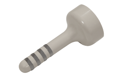
The DQRF™ device used in the study is an adaptation of the DQRF™-based EVA™ device (Novavision Group S.p.A., Misinto, Monza-Brianza, Italy) extensively used to deliver radiofrequency energy non-invasively to gynaecological tissues [10-12]. The DQRF™ device incorporates a flat probe of novel design, ergonomically suited to the posterior pelvic floor and anorectal anatomical areas (Figure 1).

Figure 1. Evidence of the four-ring quadripolar electrode system and the flat-tipped DQRF™ probe. The system continuously generates variable and dynamically controlled electromagnetic fields within the four electrodes that allows the associated diathermic effect to concentrate in the posterior pelvic floor areas without the need for invasive needles penetrating tissues.
The DQRF™ technology, centred on four stainless steel ring electrodes on the probe, operates at radio wave frequencies between 1.0 to 1.3 MHz and has a maximum emitting power of 55 watts. The DQRF™ device’s electrodes steadily cycle between radio wave receiver and transmitter status. The cycling electromagnetic fields confined within the four electrodes and the electric current flows thus generated in pelvic floor tissues eliminate the need for an external grounding pad on thighs or buttocks as required by other radiofrequency technologies.
In the ideal configuration controlled by self-regulating automatisms, the confined electric fields so generated concentrate the pain-relieving and muscle-relaxant thermal effect with high topographical precision in the hypertonic posterior pelvic floor muscles and the anal tube while minimising the total energy burden delivered to target tissues and eliminating burn risks. Electronic movement and temperature sensors (RSS™, Radiofrequency Safety System technology) rigidly control temperatures in the targeted muscles and eliminates the need for systemic or local anaesthesia.
The proprietary Ultra-Pulsed Radioporation™ (UPR™) technology [12], used in combination with DQRF™ in the same session, deliver a lenitive and pro-trophic mixture of two-third glucogel and one-third hyaluronic acid (molecular weight, 1.5 to 2.2 MDa, concentration 0.2%), previously spread on the rectal flat tip, to the target pelvic skin and mucosal areas below and above the dentate line. UPR™ acts by opening aqueous channels in cell membranes through modulation of the DQRF™ effects [12].
DQRF™ and pelvic floor massage sessions
The data already available from preclinical investigations with the DQRF™ device in laboratory animals and clinical experiences with other pelvic DQRF™ devices helped identify the DQRF™ program fixed points.
Before each DQRF™ session, the device power was set at 18-20% of its maximum emitting power (about 10 watts) to reach tissue temperatures of 38-39°C in the target pelvic floor muscles. Each DQRF™ weekly session lasted 10 minutes, with the subject in the left lateral decubitus position and the probe steadily inserted in the rectum. The “Tenesmus” proprietary software controlled the 10-min radiofrequency energy delivery to the target hypertonic muscles. The treatment cycles were individually planned between five and ten weekly DQRF™ sessions, always performed according to the ethical standards laid down in the Declaration of Helsinki as revised in Brazil 2013.
To further relieve muscle tension and pain, a massage session by the anal route of the hypertonic pelvic floor muscles followed each DQRF™ sessions. A regular program of at-home respiration and relaxing exercises complemented the office DQRF™ and massage sessions; the regular at-home use of a footstool helped relieve the tension in the puborectalis muscle sling and pubococcygeus muscle during defecation.
Muscle tone, trophism, and pelvic floor muscle function-baseline (first treatment session) and end of treatment (last treatment session)
Procedure: digitally in the vaginal introitus to assess the right and left aspects of the levator ani muscle and intra-anally to assess the external anal sphincter and then the puborectalis muscle—the latter distinguishable from other pelvic floor muscles thanks to its insertion on the inferior aspect of the os pubis. According to the International Urogynecological Association (IUGA)/International Continence Society (ICS) Joint Report on the Terminology for Female Pelvic Floor Dysfunction, the following four validated statements define the pelvic floor muscle function (tone at rest and strength of a voluntary contraction) [13]:
“Normal pelvic floor muscles” — Pelvic floor muscles which can voluntarily and involuntarily contract and relax.
“Overactive pelvic floor muscles” — Pelvic floor muscles which do not relax or may even contract when relaxation is functionally needed, for example, during micturition or defecation.
“Underactive pelvic floor muscles” — Pelvic floor muscles, which cannot voluntarily contract when this is appropriate.
“Non-functioning pelvic floor muscles” — Pelvic floor muscles where there is no action palpable.
Always according to IUGA/ICS Joint Report on the Terminology for Female Pelvic Floor Dysfunction, the strength of a voluntary contraction was rated as “strong”, “normal”, “weak” or “absent”, and voluntary muscle relaxation as “absent”, “partial” or “complete” [13].
Pain baseline and end of treatment
Standard 10-cm Visual Analogue Scale (VAS) - 0, no pain; 1 to 3, mild pain; 4 to 6, moderate pain; 7 to 10, severe pain.
Constipation associated with the pelvic floor hypertonia baseline and end of treatment
Binary assessment-Yes / No.
The exploratory cohort of ambulatory subjects with painful posterior pelvic floor muscle hypertonia, 20 women and 11 men 23 to 60 years old (mean, 42.3 ± 9.92; median 44.0), was prospectively selected from September to December 2020. At baseline, all subjects showed a picture of “overactive pelvic floor muscles” and “weak” strength of voluntary contraction. The posterior pelvic floor muscle hypertonia appeared purely dysfunctional in most subjects (29 out of 31 or 93.6%) with no evidence of anatomical disorders in the pelvic floor region like anal lesions, rectocoele or enterocoele and rectal intussusception or prolapse. Conversely, there was evidence of haemorrhoids and linear fissures in the remaining two subjects.
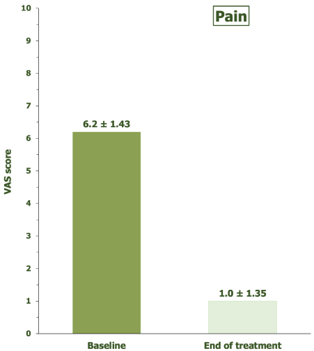
Voluntary muscle relaxation was “absent” at baseline in 20 subjects (64.5%) and “partial” in 11 (35.5%). An overall 22 out of 31 cohort subjects reported constipation, or a more correctly defined general obstructed defecation syndrome with specific reference to the two subjects with anal lesions. The mean VAS pain score at baseline was 6.2 ± 1.43 (median, 6.0).
The cohort subjects underwent a mean of 7.4 ± 1.91 DQRF™ plus massage sessions (median, 7.0; range, 5 to 10).
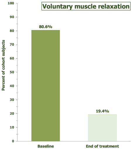
At the end of the individual treatment program, all cohort subjects showed a picture of regular voluntarily and involuntarily muscle contraction and relaxation (“normal pelvic floor muscles”); the strength of voluntary contraction was “normal” in all subjects but one, who showed a “strong” voluntary contraction. The end-of-treatment voluntary pelvic muscle relaxation was “complete” in most subjects (Figure 2), whilst no cohort subject reported an obstructed defecation syndrome, including the two subjects with anal lesions at baseline. No subject reported any clinically troublesome worsening of defecation control over the following weeks and months, provided they continued to comply with their at-home plan of self-administered massages and exercises and acceptable defecation practices in daily life. Figure 3 compares the mean pain VAS scores at baseline and end of treatment; 18 cohort subjects (58.1%) reached full pain control (zero pain VAS score) at the last treatment session. No subjects reported discomfort during and after the endocavitary DQRF™ rectal treatment.

Figure 2. Voluntary pelvic floor muscle relaxation, end of treatment cycle vs baseline, **p <0.01 vs baseline.

Figure 3. Pelvic pain, end of treatment cycle vs baseline, 10-cm VAS, ** p <0.01 vs baseline.
Subjects with pelvic floor muscle hypertonia frequently show a lost, or at least reduced, control of the levator ani muscle, with uncertain or impossible voluntary contraction and muscle relaxation. Pain is likely to contribute to poor levator ani control. All treatments, including kinesitherapy, aim to eliminate the pelvic pain that impedes daily activities and restore the normal tone and voluntary control of pelvic floor muscles [1,2].
As a subtype of obstructed defecation syndrome, constipation is also frequent-for instance, in 20 out of the 29 cohort subjects with purely dysfunctional posterior pelvic floor muscle hypertonia. In subjects with dysfunctional constipation, barium defecography could quickly reveal the anorectal angle’s failure to increase beyond the resting 90 degrees during defecation straining; standard manometry and electromyography at rest and during squeezing could confirm the functional nature of the pelvic floor musculature dysfunction [14]. In everyday clinical practice, procedures are more straightforward, as happened in the prospective cohort subjects.
The baseline at-rest muscle tone was “overactive”, according to the IUGA/ICS Joint Report on the Terminology for Female Pelvic Floor Dysfunction classification, in all cohort subjects. Voluntary pelvic muscle relaxation was also “absent” in more than one-third of subjects; in no case, relaxation was more than “partial”, meaning difficulties and shame whenever prompt muscle relaxation is crucial, like during micturition or defecation [13]. The end of the DQRF™ + UPR™ individualised treatment cycle saw a picture largely reversed, with only 24% of subjects still reporting a “partial” voluntary muscle relaxation and no report of constipation or, in general, obstructed defecation syndrome. The pain had also disappeared in more than half cohort subjects.
Quite impressive results indeed, that raise the placebo effect issue. The lack of a (sham-treated?) control group is acceptable in a probing study like this one, conceived as introductory to well-designed and adequately controlled clinical trials, but any outcome is only provisional and aimed at giving an idea of what to expect.
At present, we can definitively say the DQRF™ + UPR™ idea is non-invasive and innovative. We might also say that will it likely benefit subjects with a miserable life because of pelvic floor muscle hypertonia, be it idiopathic and purely functional or secondary to haemorrhoids or similar conditions. Only future well-designed studies compared with the therapies currently accepted as gold standard will define how much useful. Although possibly overestimated by the placebo effect bias, this exploratory cohort study’s outcomes will be useful to dimension the future trials that will give the definitive answer about how effectively can DQRF™ + UPR™ counteract pelvic floor muscle hypertonia.
A short cycle of weekly DQRF™ + UPR™ sessions is likely to control pain and the obstructed defecation syndrome very effectively in subjects with idiopathic and non-idiopathic pelvic floor muscle hypertonia, with the benefit of non-invasiveness already firmly established. The preliminary DQRF™ + UPR™ relaxation of pelvic muscles also facilitates all following physiokinetic therapies.
- Whitehead WE, Bharucha AE (2010) Diagnosis and treatment of pelvic floor disorders: what is new and what to do. Gastroenterology 138: 1231-1235, 1235.e1-e4. [Crossref]
- Butrick CW (2009) Pathophysiology of pelvic floor hypertonic disorders. Obstet Gynecol Clin North Am 36: 699-705. [Crossref]
- Hurtado EA, Appell RA (2009) Management of complications arising from transvaginal mesh kit procedures: a tertiary referral center’s experience. Int Urogynecol J Pelvic Floor Dysfunct 20: 11-17. [Crossref]
- Dias N, Zhang C, Smith CP, Lai HH, Zhang Y (2020) High-density surface electromyographic assessment of pelvic floor hypertonicity in IC/BPS patients: a pilot study. Int Urogynecol J: 10.1007/s00192-020-04467-2. [Crossref]
- Faubion SS, Shuster LT, Bharucha AE (2012) Recognition and management of nonrelaxing pelvic floor dysfunction. Mayo Clin Proc 87: 187-93.
- Akuthota V, Nadler SF (2004) Core strengthening. Arch Phys Med Rehabil 85: S86-S92.
- Byrd D, Mackey S (2020) Pulsed radiofrequency for low-back pain and sciatica. Expert Review of Medical Devices 17: 83-86. PMID: 31973587.
- Hamann W, Abou‐Sherif S, Thompson S, Hall S (2006) Pulsed radiofrequency applied to dorsal root ganglia causes a selective increase in ATF3 in small neurons. Eur J Pain 10: 171-176. [Crossref]
- Pihut M, Górnicki M, Orczykowska M, Zarzecka E, Ryniewicz W, et al. (2020) The application of radiofrequency waves in supportive treatment of temporomandibular disorders. Pain Res Manag: 6195601. [Crossref]
- Vicariotto F, Raichi M (2016) Technological evolution in the radiofrequency treatment of vaginal laxity and menopausal vulvovaginal atrophy and other genitourinary symptoms: first experiences with a novel dynamic quadripolar device. Minerva Ginecol 68: 225-236. [Crossref]
- Vicariotto F, DE Seta F, Faoro V, Raichi M (2017) Dynamic quadripolar radiofrequency treatment of vaginal laxity/menopausal vulvovaginal atrophy: 12-month efficacy and safety. Minerva Ginecol 69: 342-349. [Crossref]
- Tranchini R, Raichi M (2018) Ultra-Pulsed Radioporation further enhances the efficacy of Dynamic Quadripolar RadioFrequency in women with post-menopausal vulvovaginal atrophy. Clin Obstet Gynecol Reprod Med 4: 1-5.
- Haylen BT, de Ridder D, Freeman RM, Swift SE, Berghmans B, et al. (2010) An International Urogynecological Association (IUGA)/International Continence Society (ICS) joint report on the terminology for female pelvic floor dysfunction. Int Urogynecol J 21: 5-26. [Crossref]
- Kuijpers HC, Bleijenberg G (1985) The spastic pelvic floor syndrome. A cause of constipation. Dis Colon Rectum 28: 669-672. [Crossref]


