FDG PET-CT imaging is a standard method in diagnosis, staging and follow-up in lymphoma patients. In this case report, leukemic-lymphomatous infiltration findings on PET/CT imaging in a NHL patient was demonstrated and differential diagnosis were presented in patients with diffuse large B-cell lymphoma diagnosed by PET-CT for initial staging.
Leukemic-Lymphomatous Infiltration; PET/CT; Lymphoma

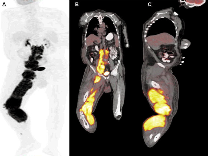
Figure 1. A 63-year-old man with a diffuse large B-cell lymphoma diagnosed after excisional biopsy of the lymph node located in the right inguinal region was directed to our department for performing PET-CT imaging for initial staging. Following 6 hour of fasting, while the patient had a blood glucose level of 122 ml/ dL, whole-body PET-CT imaging with low-dose non- diagnostic CT in 3D mode was performed 60 min later after injection of 8.5 mCi (314.5 MBq) i.v. 18-F-FDG. On PET/ CT imaging (GE Discovery PET-CT 610), hypermetabolic (SUVmax: 14.21) conglomerated lymphadenopathies were observed in bilateral paraaortic, aortocaval, prevertebral, retroperitoneal, right paralic areas, right pelvic chain and right inguinofemoral region. In addition; Edematous appearance in the lower right extremity, hypermetabolic nodular soft tissue densities and diffuse FDG uptake in the right lower extremity (SUVmax: 15.14) in the posterior muscle structures extending from the right crural region to the right pelvic level. Maximum intensity Projection (MIP) (A), coronal (B) and sagittal (C) PET-CT fusion images demonstrate diffuse FDG uptake in the right lower extremity with widespread lymphadenopathies.

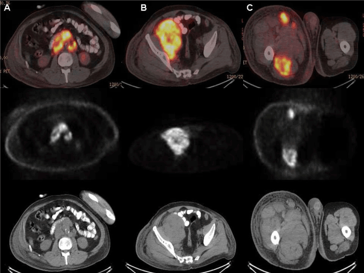
Figure 2. Bilateral paraaortic, aortocaval, retroperitoneal lymphadenopathies in axial PET-CT fusion, axial PET and CT images (A), right pelvic lymphadenopathies in axial PET-CT fusion, axial PET and CT (B) images and right lower extremity muscle uptake in axial PET-CT fusion, axial PET and CT (C) images. According to PET-CT imaging findings, significant increase in metabolic activity observed in muscle structures in the lower right extremity was interpreted as compatible with leukemic-lymphomatous infiltration.

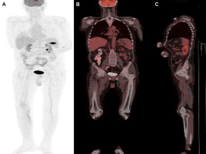
Figure 3. Complete improvement of the findings after chemotherapy in MIP (A), coronal fusion (B) and sagittal fusion (C) PET-CT images. Primary skeletal lymphoma is rare. It occurs in 0.5% of all extranidal lymphomas. Muscle involvement of the lymphoma is rare but most commonly seen in the diffuse B-cell lymphoma subtype as seen in this case. Most moderate to high grade have aggressive histology [1]. A systematic study of 82 autopsy cases showed that contrary to common assumption and scant literature reports, skeletal muscle involvement in leukemia-lymphoma is not a rare phenomenon [2]. HTLV-I infected synovial cells in conjunction with leukemic-lymphomatous infiltration of synovial tissue is reported in a case by Dennis G et al [3]. There are some similar case reports in the literature with muscle involvement [4-7]. Muscular disorders mimicing NHL involving skeletal muscles are primary soft tissue sarcoma, muscle sarcoidosis, intramuscular metastases, myositis, inflammatory pseudo-tumors, and skeletal muscle lymphoma. Extremity muscles; especially the lower extremity muscles are the most frequently affected areas, as in this case. Systemic lymphoma spreads to the skeletal muscles by hematogenous and lymphatic pathways. In this case report, PET-CT imaging findings were presented at the time of leukemic-lymphomatous infiltration in rare muscle structures.
- Keung YK, Liang R (1996) Report of a case of primary skeletal muscle lymphoma and review of the literature. Acta Haematol 96: 184-186. [Crossref]
- Buerger LF, Monteleone PN (1966) Leukemic-lymphomatous infiltration of skeletal muscle. Systematic study of 82 autopsy cases. Cancer 19: 1416-1422. [Crossref]
- Dennis G, Chitkara P (2007) A case of human T lymphotropic virus type I-associated synovial swelling. Nature Reviews Rheumatology 3: 675-680.
- Dai Y, Sowjanya M, You J, Xu K (2015) Non-Hodgkin's Lymphoma of Multiple Skeletal Muscles Involvement Seen on FDG PET/CT Scans. Medicine (Baltimore) 94:833. [Crossref]
- Bennett S, Slocombe R, Holloway S, et al. (2005) Lymphoma(s) showing epitheliotropism and diffuse skeletal muscle involvement presenting as a polymyopathy in a young dog. Aust Vet J 83: 612-615. [Crossref]
- Lee VS, Martinez S, Coleman RE. (1997) Primary muscle lymphoma: clinical and imaging findings. Radiology 203: 237-244. [Crossref]


