Penetrating neck injury is often associated with the destruction of airway structures and large vessels. In cases with spinal cord injury, hemodynamic instability may present secondary to neurogenic shock. We report a case of penetrating neck injury with complete spinal cord transection. Extracorporeal membrane oxygenation (ECMO) was applied to provide respiratory and cardiopulmonary support during the perioperative period. After surgery, neurogenic shock leading to hemodynamic instability was treated with sufficient hydration and vasopressors. ECMO may be a sound choice to provide respiratory support and even cardiopulmonary support when managing patients with potential upper airway injury resulting from penetrating neck injury.
wounds, penetration; neurogenic shock, spinal cord injuries, extracorporeal membrane oxygenation, airway management
Penetrating neck injury (PNI) is relatively uncommon, but all clinicians should be aware of its significance and the potential hazards. The anatomy of the neck is quite complex because it contains major vascular structures like the carotid and vertebral arteries, important nerves like the vagus, phrenic, and recurrent laryngeal nerves, and the esophagus and trachea, which are vital structures for a patient with trauma. The mortality of penetrating neck injury can be as high as 10% [1], and mortality is associated with massive blood loss from accompanying vascular injuries and/or failure of airway securement. To the best of our knowledge, arterial injury and aerodigestive injury may occur in approximately 25% and 23–30% of penetrating neck injuries, respectively [2,3].
Despite the importance of prompt and precise management of patients with penetrating neck injury, consensus guidelines do not exist. Most clinicians must manage such cases based on their own experiences and limited case reports and reviews. Here, we report a case of penetrating neck injury in a patient with a knife stuck in the neck. The patient underwent surgical exploration under general anesthesia along with extracorporeal support.
An 85-year-old female patient was found with a knife stuck in her neck. The knife measured approximately 15 centimeters in length and was stuck in zone II of the neck, from the anterior to the posterior portion. Active bleeding was noted at the site of the injury. The patient’s Glasgow Coma Scale was 3 with response to painful stimulus. She had no other medical history. At arrival, her vital signs were relatively stable, with blood pressure (BP) 106/46 mm Hg, heart rate (HR) 57 beats per min, respiratory rate 20 breaths per min, and oxygen saturation (SpO2) 100%. The initial complete blood count revealed hemoglobin (Hb) 11.6 g/dL and hematocrit (Hct) 33.5%. However, after 25 minutes, the Hb and Hct dropped to 7.2 g/dL and 20.8%, respectively. The patient received 3 unit of red blood cell (RBC) and 5 units each of platelets and fresh frozen plasma (FFP) in the emergency department. A neck computed tomography (CT) scan revealed that the stab wound involved her esophagus, thyroid gland, first thoracic vertebral body, and the spinal cord, without injuries to the trachea (Figures 1A and 1B).
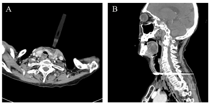
Figure 1. Neck Computed Tomographic Images of a Patient with Penetrating Neck Injury. (A) The knife, stuck in the patient's neck, penetrated her left thyroid gland and esophagus, without injuring the trachea or major vessels, such as the carotid arteries (axial view). (B) The knife penetrated the patient's entire neck, including two vertebral bodies (sagittal view)
When the patient arrived at the operating room, her BP, HR and SpO2 were 105/65 mm Hg, 65 beats per min and 100%, respectively. An electrocardiogram (ECG) showed non-sustained atrial premature complexes. Considering the securement of a patent airway, we carefully evaluated any potential injury to the trachea during endotracheal intubation. In addition, because of the possibility of cardiac events from neurogenic shock, we decided to conduct veno-arterial extracorporeal membrane oxygenation (VA-ECMO) via right femoral cannulation. The cannulas were connected to the circuit, and the circuit was turned on with initial pump flow of 3,000 mL per min and 2,200 revolutions per minute. After application of extracorporeal membrane oxygenation (ECMO), the patient’s vital signs remained stable during the surgical procedure. As the result of exploring the injured site, the knife was placed between the left carotid artery and the trachea (Figure 2A). We prepared for intubation with a video-assisted laryngoscope and an endotracheal tube with a stylet. Because the knife was still stuck in the neck, we proceeded with caution to not cause additional injury to the trachea by the knife during intubation. Therefore, we placed an aseptic surgical plate under the knife along its margin to prevent the knife piercing more deeply. After successful intubation, mechanical ventilation through the endotracheal tube was started. During surgical exploration, the lacerations of the thyroid gland and esophagus were sutured, and bleeding from two stab wounds in the seventh cervical and first thoracic vertebra was controlled by applying bone wax (Figure 2B). At the end of the operation, the patient’s vital signs were stable without any positive inotropic agent or vasopressor, so ECMO was weaned. The patient received 7 units of RBC and 3 units of FFP, and the last blood test in the operating room showed Hb 10.6 g/dL and Hct 32%. The patient was transferred to the intensive care unit (ICU) without extubation.
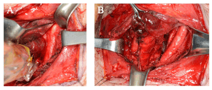
Figure 2. Surgical Manipulation of the Injured Site. (A) Before removal, the knife was placed between the trachea and the left common carotid artery. (B) After removal of the knife, the major vessels were intact and no active bleeding was seen at the site of the injury
Arriving in the ICU, the patient’s vital signs remained stable, with BP 100/60 mm Hg, HR 70 beats per min, and SpO2 98%. About 10 hours later, the patient’s systolic blood pressure dropped and was first measured at below 80 mm Hg. An infusion of norepinephrine was started, and the proper fluid supply was archived. Although the patient’s consciousness had returned, her hemodynamic status deteriorated. On postoperative day 4, widening of the QRS complex on ECG and severe bradycardia of 26 beats per min developed. The patient was resuscitated after chest compressions for 3 minutes and a bolus injection of 1 mg epinephrine. Despite sufficient intensive care and fluid treatment, cardiovascular collapse due to neurogenic shock resulted in death on postoperative day 17.
When assessing and managing patients with penetrating neck injury, we must be aware of the potential hazards of the injury, especially the destruction of airway structures and large vessels. In addition, we should not overlook the possibility of neurogenic shock if there is an accompanying spinal cord injury.
ECMO provides temporary respiratory support. Currently, ECMO is an effective option not only for adequate oxygenation and ventilation during tracheobronchial surgery, but it is also an effective treatment for patients with acute respiratory distress syndrome to “rest the lung” [4]. There are two kinds of ECMO: veno-venous ECMO (VV-ECMO) and veno-arterial ECMO (VA-ECMO). Compared with VV-ECMO, VA-ECMO provides both simple respiratory support and cardiopulmonary support. During VA-ECMO, blood is drained from a large vein, moved through the circuit of ECMO, and oxygenated, while carbon dioxide is simultaneously removed. Eventually, “fresh” blood returns to the patient via the circuit to an arterial system and contributes to hemodynamic support. Since the patient in the present case was elderly and cardiopulmonary bypass might have been needed for the possible development of neurogenic shock, we thought VA-ECMO a better choice for surgery. Despite such advantages of the VA-ECMO, it should be noted that vascular complications are more common in VA-ECMO. In general, because of the large-bore cannulas used for femoral arterial cannulation and the subsequent hemodynamic instability, the risk of thromboembolic events is higher in patients under VA-ECMO. Therefore, vascular complications, such as limb ischemia and vascular injury leading to dissection or pseudoaneurysm, are relatively common in the VA-type of ECMO [4,5].
We evaluated the cause of the patient’s cardiac event in the ICU. The possibility of cardiogenic or hypovolemic shock was very low because the ejection fraction of the left ventricle was normal and there was no regional wall motion abnormality seen in the echocardiogram. In addition, there was no evidence of volume depletion in the echocardiogram. We concluded that the event was most likely due to neurogenic shock secondary to impairment of the sympathetic pathways induced by spinal cord injury.
Neurogenic shock, also known as vasogenic shock, is a disruptive consequence after spinal cord injury (SCI). SCI leads to sudden dysfunction of sympathetic outflow and autonomic instability and is manifested by significant hypotension, bradycardia, and even temperature dysregulation [6,7]. These hemodynamic changes directly result from the impairment of the sympathogenic excitatory input to sympathetic preganglionic neurons. The incidence of neurogenic shock is higher when SCI is above the first thoracic vertebra [8]. In recent years, the reported incidence of neurogenic shock after SCI has varied. It is approximately 29% in the cervical SCI population and 19% in the thoracic SCI population [6,7].
The treatment of neurogenic shock is administration of IV fluid and vasopressors, with the goal of mean arterial pressure measuring 85–90 mm Hg [9,10]. Severe hypotension from neurogenic shock may decrease the perfusion of microvessels within the spinal cord, leading to profound ischemia, and may deteriorate neurologic function [11-13]. A vicious cycle could develop. Therefore, treating neurogenic shock with only vasopressors is not sufficient and may even exacerbate the patient’s condition. The more important treatment is the administration of IV fluids to maintain an adequate blood volume status. Therefore, in this case, we tried to sufficiently hydrate the patient, and we used vasopressors to maintain her systolic blood pressure above 90 mm Hg. Unfortunately, she eventually expired due to irremediable disruption of the autonomic system.
This case demonstrated that ECMO may be a sound choice to provide respiratory support and even cardiopulmonary support (depending on the type of ECMO) when managing patients with potential upper airway injury resulting from penetrating neck injury. Also, physicians must be aware of hemodynamic changes that suggest the probability of neurogenic shock in patients with spinal cord injury. If neurogenic shock is suspected, sufficient hydration along with the proper vasopressors are the mainstays of treatment.
No potential conflict of interest was reported by the authors.
- Nowicki JL, Stew B, Ooi E (2018) Penetrating neck injuries: a guide to evaluation and management. Ann R Coll Surg Engl 100: 6-11. [Crossref]
- Sperry JL, Moore EE, Coimbra R, Croce M, James W Davis JW, et al. (2013) Western Trauma Association critical decisions in trauma: penetrating neck trauma. J Trauma Acute Care Surg 75: 936-940. [Crossref]
- Saito N, Hito R, Burke PA, Sakai O (2014) Imaging of penetrating injuries of the head and neck:current practice at a level I trauma center in the United States. Keio J Med 63: 23-33. [Crossref]
- Ali J, Vuylsteke A (2019) Extracorporeal membrane oxygenation: indications, technique and contemporary outcomes. Heart 105: 1437-1443. [Crossref]
- Lorusso R, Barili F, Mauro MD, Gelsomino S, Pariseet O (2016) In-Hospital neurologic complications in adult patients undergoing venoarterial extracorporeal membrane oxygenation: Results from the extracorporeal life support organization registry. Heart (British Cardiac Society) 44: e964-e972. [Crossref]
- Partida E, Mironets E, Hou S, Iacobini MA, Stoian R, et al. (2016) Cardiovascular dysfunction following spinal cord injury. Neural Regen Res 11: 189-194. [Crossref]
- Ruiz IA, Squair JW, Phillips AA, Lukac CD, Huang D, et al. (2018) Incidence and natural progression of neurogenic shock after traumatic spinal cord injury. J Neurotrauma 35: 461-466. [Crossref]
- Matthews S, Shenvi CL (2017) Airway obstruction and neurogenic shock due to severe cervical spine injury. Am J Emerg Med 35: 196.e1-196.e2. [Crossref]
- Yue JK, Tsolinas RE, Burke JF, Deng H, Upadhyayula PS, et al. (2019) Vasopressor support in managing acute spinal cord injury: current knowledge. J Neurosurg Sci 63: 308-317. [Crossref]
- Ball PA (2001) Critical care of spinal cord injury. Spine (Phila Pa 1976) 26: S27-S30.
- Furlan JC, Fehlings MG, Shannon P, Norenberg MD, Krassioukov AV, et al. (2003) Descending vasomotor pathways in humans: correlation between axonal preservation and cardiovascular dysfunction after spinal cord injury. J Neurotrauma 20: 1351-1363. [Crossref]
- Al Dera H, Brock JA (2018) Changes in sympathetic neurovascular function following spinal cord injury. Auton Neurosci 209: 25-36. [Crossref]
- Witiw CD, Fehlings MG (2015) Acute spinal cord injury. J Spinal Disord Tech 28: 202-210. [Crossref]