Abstract
In patients suffering from paradoxical embolism and cryptogenic stroke, percutaneous transcatheter closure of a patent foramen ovale (PFO) has been utilized as a means of therapy. The most frequently used approach to this procedure has been via entrance from the right or left femoral vein. However, this approach is not feasible in instances where there is an occlusion of the inferior vena cava (IVC), and thus a right internal jugular approach was used as an alternative in this case. Herein, we discuss the percutaneous transcatheter PFO closure using a GORE® CARDIOFORM Septal Occluder device via the right internal jugular approach.
Key words
patent foramen ovale, GORE® CARDIOFORM Septal Occluder, structural heart disease intervention, stroke, inferior vena cava filter, thrombosis
Introduction
Patent foramen ovale (PFO) has been increasingly diagnosed in patients suffering from paradoxical embolism and cryptogenic stroke [1-3]. As PFO has become more recognized as a source for these conditions, percutaneous transcatheter closure has been increasingly used as a therapeutic solution. The typical technique utilizes percutaneous entrance from the right or left femoral veins. However, in the case of concomitant occlusion of the inferior vena cava (IVC), alternative venous access is required [4]. Our case represents this challenge and illustrates the technique for deployment of a GORE® CARDIOFORM Septal Occluder device (W. L. Gore & Associates, Inc., Newark, DE) for PFO closure from the internal jugular venous approach.
PFO closure via the internal jugular venous approach has been utilized in various previously published reports, but to our knowledge, this is the first such report using the GORE® CARDIOFORM Septal Occluder device.
Case report
A 50-year-old female with a history of multifocal cerebrovascular accident, recurrent deep vein thrombosis, and an occluded IVC filter presented with recurrent neurological symptoms. She was referred for PFO closure after she was diagnosed with a large PFO and a transient right to left shunt.
In 2007, this patient presented with acute transient aphasia, left arm weakness, memory deficiency, and headache. This patient had a history of PFO as a child however a transthoracic echocardiogram failed to identify residual PFO at this time. Her workup included an elevated serum homocysteine level (17 μmol/L, normal range of 5-13.9 μmol/L), while the rest of the thrombophilia panel was negative and within normal limits. In August 2014, the patient was admitted to the emergency room with right-sided weakness of arms and legs and expressive aphasia. She received intravenous tissue plasminogen activator (0.9 mg/kg) but did not show any improvement in symptoms and therefore underwent diagnostic cerebral angiography, which showed resolution of the thrombus and thus no intervention was necessary. A transesophageal echocardiogram was performed at this time and showed a positive PFO by bubble study (Figure 1). In September of 2014, the patient was admitted to the emergency room with a painful left hand found to be due to an occlusion of the left subclavian artery, and at the same time she had a documented right popliteal vein thrombus, left basilic vein thrombus, left popliteal artery thrombus, and an embolism to the renal, hepatic, pulmonary, and left vertebral arteries. The patient subsequently underwent a right common femoral vein and popliteal vein thrombectomy and a left brachial/subclavian artery thrombectomy. She was heparinized, however she subsequently developed an intracranial hemorrhage, and thus anticoagulation was held. An IVC filter was placed during this admission. Following her hospitalization, the patient was successfully maintained on Xarelto 20 mg daily for anticoagulation. Subsequently, in December of 2014 during an attempt to close the PFO via the femoral approach, it was found that the IVC was thrombosed (Figure 2). Given her recurrent paradoxical embolic events and documented PFO she was referred for percutaneous PFO closure via the internal jugular vein.
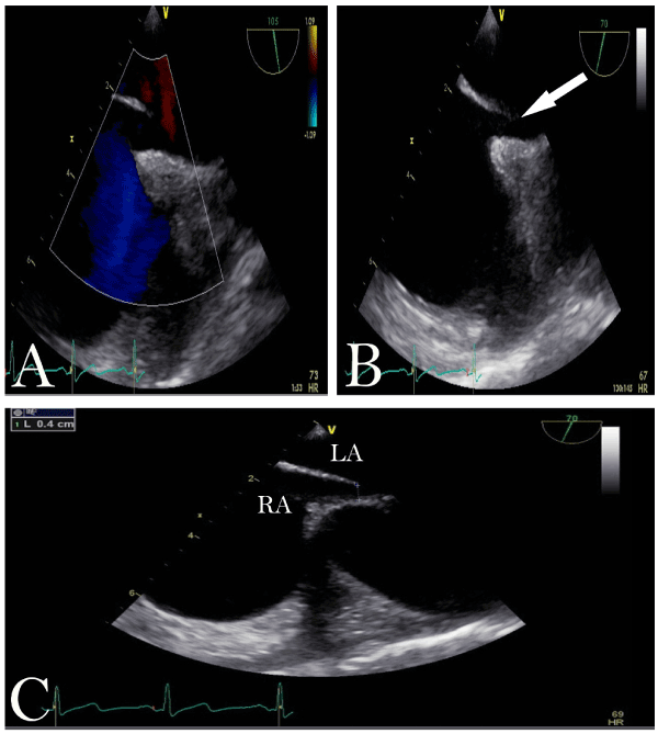
Figure 1. Transesophageal echocardiography images depicting the Patent foramen ovale (PFO). A: PFO with color doppler. B: Arrow: detailed image of PFO tunnel. C: RA: right atrium and LA: left atrium, demonstration of PFO tunnel diameter.
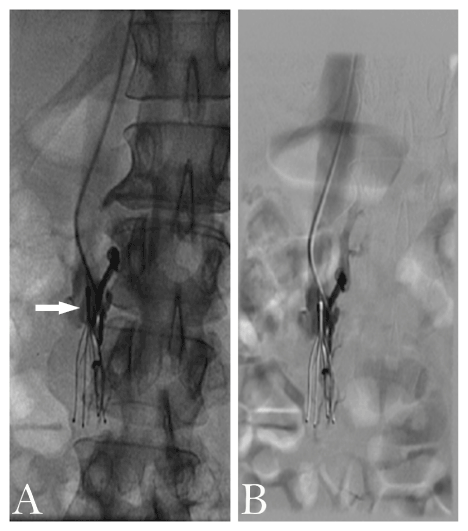
Figure 2. Occluded IVC filter. A and B depict the inferior vena cava filter thrombosed using digital subtraction angiography. Arrow: site of thrombosis.
The procedure was performed in the Cardiac Catheterization laboratory with general anesthesia and transesophageal echocardiographic guidance given lack of access for intracardiac echocardiography. After the right internal jugular vein was accessed using standard technique, the patient was heparinized. A GLIDECATH® Simmons 1 catheter (Terumo Medical Corporation, Somerset, NJ) was utilized to cross the interatrial septum. An Amplatz Extra-Stiffä J-Tip guidewire (Boston Scientific, Marlborough, MA) was positioned distally in the left ventricle, and then the 10-French sheath was positioned across the septum into the left ventricle. The sheath and a 25 mm GORE® CARDIOFORM Septal Occluder were carefully deployed using Echocardiographic guidance to confirm the mitral valve was not entrapped by the left atrial disc. Ultimately, the device was deployed successfully with no residual shunt (Figures 3-4).
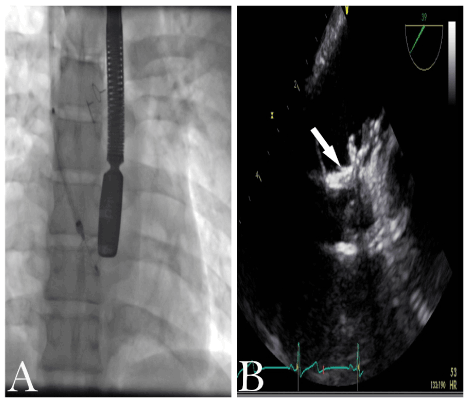
Figure 3. Depictions of the left atrial disc deployed visualized by fluoroscopy (A) and transesophageal echocardiogram (B) to avoid entrapment of mitral valve leaflets/apparatus.
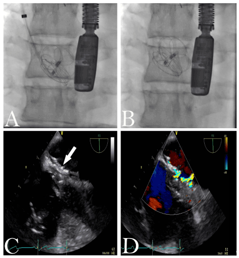
Figure 4. Depictions of the GORE CARDIOFORMâ Septal Occluder during full deployment and release evaluated by fluoroscopy (A and B respectively) and transesophageal echocardiography (C and D). Arrow: fully sealed PFO without residual shunt.
A follow-up bubble study with agitated normal saline contrast showed no significant residual shunting. Hemostasis was obtained in the right internal jugular vein via manual compression with a 2-0 silk pursestring suture. The patient tolerated the procedure well with no complications and was discharged the following day (Figure 5).
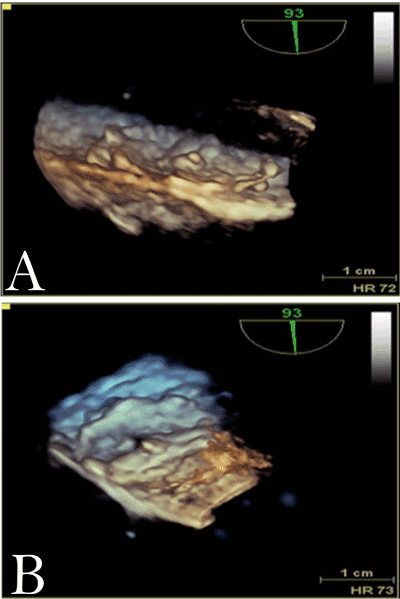
Figure 5. 3D images after full deployment of GORE CARDIOFORMâ Septal Occluder.
Discussion
PFO is the most common defect of the heart, and it has been recognized as a source for cryptogenic stroke and migraine [5-7]. A relationship between PFOs and paradoxical embolisms has been documented previously in the literature, and the cause of stroke in a patient with a PFO is believed to be paradoxical embolism [3]. In order to reduce the risk for paradoxical embolism in patients with PFO, percutaneous closure of these defects has been recommended.
Operators have typically relied on a femoral venous approach to deploy an occluder device across the PFO, and more recently a jugular venous approach has been employed [8]. An occlusion in a patient’s IVC (which may become more common in the era of IVC filters) prevents the use of the femoral venous approach, and thus the jugular venous approach is advantageous. In previously documented cases, the jugular venous approach has been used to close a PFO using the AMPLATZERä Septal Occluder (St. Jude Medical, Saint Paul, MN) device [8]. The technique for deployment of the GORE® CARDIOFORM Septal Occluder device via the right internal jugular approach differs from the transfemoral approach in terms of how to approach crossing the intratrial septum and allowing adequate support for device advancement into the left atrium. In our case, we found the Simmons 1 catheter to provide excellent angulation and support for crossing the interatrial septum. Subsequently, the wire was placed in the left ventricular apex rather than the left upper pulmonary vein to assist in our septal occluder tracking into the left atrium. Otherwise, the device and sheath tend to prolapse in the right atrium. The utilization of both these modifications to standard technique facilitated correct and safe deployment of our device.
Conclusion
These results indicate that the right internal jugular approach is a feasible alternative for patients requiring PFO closure but may suffer from IVC thrombosis. The GORE® CARDIOFORM Septal Occluder was successfully deployed in the patient, resulting in complete PFO closure with no residual shunt. PFO closure via the internal jugular venous approach has been utilized in various previously published reports, but thus far, this is the first such report using the GORE® CARDIOFORM Septal Occluder device.
Acknowledgement
Case conducted at Sharp Grossmont Hospital Catheterization Laboratory 5555 Grossmont Center Dr., La Mesa, CA 91942, USA.
References
- Webster MW, Chancel2021 Copyright OAT. All rights reserv, et al. (1988) Patent foramen ovale in young stroke patients. Lancet 2: 11-12.
- Kerut EK, Norfleet WT, Plotnick GD, Giles TD (2001) Patent foramen ovale: a review of associated conditions and the impact of physiological size. J Am Coll Cardiol 38: 613-623. [Crossref]
- Lechat P, Mas JL, Lascault G, Loron P, Theard M, et al. (1988) Prevalence of patent foramen ovale in patients with stroke. N Engl J Med 318: 1148-1152. [Crossref]
- Martin F, Sanchez PL, Doherty E, Colon-Hernandez PJ, Delgado G, et al (2002) Percutaneous transcatheter closure of patent foramen ovale in patients with paradoxical embolism. Circulation 106: 1121-1126. [Crossref]
- Ali Ebrahimi H, Hamzeaie Moghadam A, Aredestani E (2011) Evaluation of patent foramen ovale in young adults with cryptogenic stroke. ARYA Atheroscler 7: 74-77. [Crossref]
- Horton SC, Bunch TJ (2004) Patent foramen ovale and stroke. Mayo Clin Proc 79: 79-88. [Crossref]
- Wilmshurst PT (2005) The persistent foramen ovale and migraine. Rev Neurol (Paris) 161: 671-674. [Crossref]
- Sader MA, De Moor M, Pomerantsev E, Palacios IF (2003) Percutaneous transcatheter patent foramen ovale closure using the right internal jugular approach. Catheter Cardiovasc Interv 60: 536-539. [Crossref]