Introduction
We present a case of an 83-year-old male who presented with penetrating aortic ulcer, ascending aortic saccular pseudoaneurysm, and intramural hematoma. Patient history and presentation with symptoms, physical signs, labs, and CTA with images are provided. Also, management including medical and surgical contributions are discussed.
Acute aortic syndromes encompass the following life-threatening medical conditions: aortic dissection (AD), intramural hematoma (IMH) and penetrating aortic ulcer (PAU) [1]. AD is a serious condition with a tear in the inner wall of the aorta, characterized by the presence of an intimal flap separating the true and false lumens of the aorta [2]. However, IMH and PAU are defined as non-flap lesions [3]. IMH represents a hemorrhage within the aortic wall, in the absence of a tear in the intimal wall of the aorta. PAUs, while they have a different pathophysiological mechanism than AD and IMH, can lead to the formation of IMH and evolve into AD [4].
History of Presentation
83-year-old male presented to ER with shortness of breath that progressively got worse over the past 5 days. In the ED, heart rate in 120s-130s, blood pressure 110s/80s, and was started on 3 L oxygen. Patient’s past medical history was significant for COPD and was on 2L of oxygen at baseline, essential hypertension, and an ascending aortic dissection that was treated conservatively with blood pressure control.
His initial admission workup included a CTA chest to rule out PE, but it showed a penetrating ulcer with a 19 mm saccular pseudoaneurysm of the ascending aorta with intramural hematoma (Figure 1), ectatic thoracic aorta, and pulmonary artery aneurysm measuring 5.7 cm. Cardiothoracic surgery evaluated the patient in the ER, and determined he was not a surgical candidate due to end-stage respiratory failure with other comorbidities. Also, the patient did not prefer the surgical option.
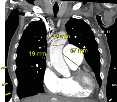
Figure 1. Day 1 Coronal CT of the Chest
Coronary angiogram with right anterior oblique view of right coronary artery showing spontaneous coronary artery dissection. Main pulmonary trunk is enlarged, measuring up to 57 mm. Ascending aorta is 40 mm. There is a 19 mm outpouching of contrast from the ascending aorta.
EKG showed paroxysmal A. fib with RVR for which he was started on amiodarone and esmolol drip with a heart rate target below 60 and blood pressure less than 120/80. With the concurrent acute/subacute pseudoaneurysm, the patient had an absolute contraindication for anticoagulation/ cardioversion. TTE showed EF of 25-30%, severe global hypokinesis of the left ventricle, aneurysmal dilatation of ascending aorta (4.8 cm), and pulmonary artery (5.1 cm).
On the first day of admission, the patient was medically stable. However, his respiratory status deteriorated through the hospital course and necessitated the use of BiPAP. Also, he started to develop AKI. CT chest was ordered on the sixth day of admission without contrast due to concurrent AKI. It showed ascending penetrating aortic ulcer with intramural hematoma, not well defined in the non-contrasted study, with progressively enlarging ascending aortic diameter with displaced atherosclerotic calcification consistent with propagation of the ascending aortic intramural hematoma with better defined high-density intramural hematoma involving the aortic arch and extending along the descending thoracic aorta to the level of the aortic hiatus (Figure 2). Palliative got consulted and spoke with the family who decided to switch the patient to comfort care. On physical exam, he developed a pulsating epigastric mass, which eventually ruptured, and the patient passed away.

Figure 2. CT Sagittal view of Chest on Days 1 and 6 of hospital admission
Left: Day 1 CT with contrast of the chest; Right: Day 6 CT non-contrast of the chest shows development of new hyperdense intramural hematoma at the distal aortic arch extending along the descending thoracic aorta to the level of the aortic hiatus measuring up to 12 mm in thickness along the posterior aortic arch.
Discussion
PAUs are often seen in elderly patients with severe atherosclerosis and require immediate medical intervention. Patients may remain asymptomatic if the ulcer remains confined to the intimal layer of the aorta. However, in about 20%– 54% of patients, symptoms such as chest pain occur when the ulcer erodes into the medial wall of aorta and forms an intramural hematoma (IMH). This hematoma could later progress to form a pseudoaneurysm and eventually rupture, causing significant mortality.
PAUs in the ascending aorta are classified as Type A, whereas those present in the descending thoracic aorta are Type B. In general, Type A patients are treated surgically, and Type B patients are managed conservatively. However, regardless of the location of the PAU, aggressive treatment is recommended for symptomatic patients because of their increased risk of rupture.
Conservative therapy with annual CT surveillance and anti-hypertensives is considered in asymptomatic patients. But surgical treatment is indicated if there’s progression of the ulcer to an aneurysm, or growth in the size of the aneurysm [5]. Thoracic Endovascular Aortic Repair (TEVAR) is indicated in patients with recurring symptoms, thoracic aorta with a diameter greater than 55mm, increases in aortic size of more than 10mm in one year and co-existence of other aortic diseases. However, these guidelines for TEVAR remain controversial.
In this case report, the patient had a Type A PAU that was complicated by the formation of IMH and saccular pseudoaneurysm. Furthermore, his ABGs showed persistent hypoxia and hypercarbia. The end-stage respiratory status of the patient contraindicated surgical treatment of PAU. Hence, a conservative approach with heart rate and rhythm control was adopted. Palliative care was also consulted for the patient. Unfortunately, the patient’s pseudoaneurysm progressively enlarged on the fifth day of his admission and it eventually ruptured resulting in the patient’s demise.
This case illustrates the challenges with medical management of PAU in a patient with severe comorbid conditions and underscores their poor prognosis. It also highlights the need for more comparative studies to evaluate the role of medical management in preventing future complications.
References
- Erbel R, Aboyans V, Boileau C, Bossone E, Di Bartolomeo R, et al. (2014) 2014 ESC Guidelines on the diagnosis and treatment of aortic diseases: Document covering acute and chronic aortic diseases of the thoracic and abdominal aorta of the adult. Eur Heart J 35: 2873-2926. [Crossref]
- Isselbacher EM, Black JH, Augoustides JG, Beck AW, Bolen MA, et al. (2022) ACC/AHA Guideline for the Diagnosis and Management of Aortic Disease. CZ. A Report of the American Heart Association/American College of Cardiology Joint Committee on Clinical Practice Guidelines. Circulation 146: e334-e482. [Crossref]
- Coady MA, Rizzo JA, Elefteriades, JA (1999) Pathologic variants of thoracic aortic dissections. Penetrating atherosclerotic ulcers and intramural hematomas. Cardiol Clin 17: 637-657. [Crossref]
- Oderich GA, Kärkkäinen JM, Reed NR, Tenorio ER, Sandri GA (2019) Penetrating Aortic Ulcer and Intramural Hematoma. Cardiovasc Intervent Radiol 42: 321-334. [Crossref]
- El-Hamamsy I, Ouzounian M, Demers P, McClure S, Hassan A, et al. (2016) State-of-the-Art Surgical Management of Acute Type a Aortic Dissection. Can J Cardiol 32: 100-109. [Crossref]