Introduction/background: It is well known that pelvic floor has multiple functions. These functions range from urination, defecation, pleasure/sexuality, parturition, and stability of pelvic organs. If the pelvic floor is not functioning correctly, these functions can be impaired. These impairments can lead to issues with quality of life and activity level.
Case description: This case reports on a 32-year-old female referred by her gynecologist to physical therapy with complaints of pelvic heaviness after her first vaginal delivery. The patient was concerned that she would not be able to return to her normal active lifestyle. The patient lived in a rural area with limited access to care. The patient was seen for evaluation and three additional visits over the course of 2 months for pelvic floor strengthening as clinical findings showed a weakness in pelvic floor endurance and power as well as mild prolapse.
Outcomes: Subjective improvements were a reduction in pelvic pressure, reduced urinary leakage as well as improved ability to exercise with less fear of leakage. Objectively, the patient had improvements in pelvic floor strength as measured by the use of the PERFECT system. The mild prolapse noted on evaluation using the POP scoring system was still present.
Discussion: The patient had limitations in the amount of sessions she could attend due to her job and the nature of the rural setting, which limited progress. The patient was compliant at home, which did assist in functional improvement.
Informed Consent was given by patient to participate in this study.
pelvic heaviness, physical therapy, parturition
Women's health encompasses many facets of treatment diagnoses. One division being pelvic health, which consists of pelvic floor dysfunction (PFD) resulting in incontinence, constipation, pain, and/or sexual dysfunction. It is well known that pelvic floor has multiple functions. These functions range from urination, defecation, pleasure and sexuality, parturition, and stability of pelvic organs [1]. If the pelvic floor is not functioning correctly, these above mentioned functions can be impaired.
The patient in this case reflection is a 32-year-old female with otherwise unremarkable past medical history. She was referred by her gynecologist to physical therapy with complaints of pelvic heaviness after her first vaginal delivery 2 months prior. The patient was selected for this case reflection based on her pelvic floor complications following the delivery of her first baby, and her compliance with home program in achieving therapeutic benefit. The patient was also self-referred, meaning she asked her gynecologist for a pelvic physical therapy referral, it was not suggested to her.
During her evaluation the patient reported a heaviness in her pelvic area since the delivery. She did not report feeling anything abnormal at the vaginal opening, just the pressure. She denied any pelvic problems prior to the vaginal delivery. The patient reported that standing long periods of time caused an increase in pelvic pressure, resulting in symptoms most often being worse at the end of the day. The patient did report a long-standing history of constipation since childhood in which she managed with diet. She also reported a history of irritable bowel syndrome (IBS) but notes it did not cause her much dysfunction. At times she experienced low back pain (LBP) and sacroiliac joint (SIJ) issues; however, she has a sister who is a Physical Therapist (PT) who helped her manage those issues in the past.
The patient noted she has a fitness routine that consists of running 20 + miles a week and occasional fitness high-intensity interval training (HIIT) workouts. She hasn’t been able to return to these activities since she gave birth, so her current routine consisted of walking and swimming. She reported occasional leakage of urine during the breast stroke kick while swimming. The patient also stated that at times she may smell of urine slightly but didn’t notice significant wetness; however due to the smell, she feared she may leak without knowing. Otherwise, no real significant urinary dysfunction was reported. She had prior knowledge of Kegel exercises [2], so she was doing those along with her fitness regimen; however, the Kegel exercises were done sporadically.
As far as voiding, she reported having the sensation to void, but often didn’t produce much urine as compared to voiding prior to her vaginal delivery. Other times she had little urge, then she voided a lot. She also noted a sense of air getting stuck in the vaginal area that produced a noise. That was bothersome to her.
With the symptoms provided, the patient reported her goal was to continue to be an active mom without fear of urinary leakage or pelvic discomfort and/or heaviness.
From the initial data gathered, the patient reported a heaviness and pressure in the vaginal area. She also reported some issues with voiding. These symptoms relate highly with pelvic organ prolapse (POP) as noted in the study by Barber [3].
To determine if this was in fact a POP, other medical and musculoskeletal diagnoses needed to be excluded. Things to consider in regard to differential diagnosis of pelvic pain/ pressure are shown in Table 1 [4]. Medically, the patient was young, physically fit, and reported no medical concerns other than IBS and constipation that had not been an issue in the past and that she managed with diet. From a medical standpoint, it was not likely she had any of the medical diagnoses listed in Table 1.
Table 1. Differential diagnostics for pelvic pain**.
In terms of the musculoskeletal component of the pressure and pelvic discomfort the patient was feeling, a few diagnoses were considered. The patient had a previous history of LBP and SIJ dysfunction, both of which can cause pelvic girdle pain. Involvement of the low back (LB) and SIJ could be ruled out with special testing such as postural assessment, the active straight leg raise test, palpation, and general testing of range of motion (ROM) and strength using manual muscle testing (MMT) [5,6]. The patient also had recently started back to light exercise, which could potentially cause muscular pain/strain due to disuse during pregnancy. Muscular pain can be ruled out with movement tests such as ROM and MMT and also palpation. Muscular dysfunction/pelvic floor instability, as seen in pelvic floor dysfunction, can be tested by internal manual muscle testing as well as POP testing. POP tests can be performed to determine if there is downward descent of either the uterus (apical prolapse), bladder (cystocele or anterior prolapse), or rectum (rectocele or posterior prolapse). Upon Valsalva, if descent is positive, prolapse can be considered [7].
The patient examination started at her initial arrival. Gait and mobility was visually inspected and there were no impairments. The patient was educated regarding the evaluation process, and she gave full consent. She filled out the Pelvic Floor Distress Inventory (PFDI) for prolapse and urinary issues as well as urinary problem survey which are proven to be a valid and reliable instrument for measuring symptom inconvenience caused by pelvic organ prolapse and the health-related quality of life (QOL) [8]. Since QOL was an issue for this patient, this was an appropriate outcome measure.
The patient had a posture screen in which pelvic asymmetry was noted. In standing, the patient had a high anterior superior iliac spine (ASIS) and iliac crest (IC) height, and a low posterior superior iliac spine (PSIS) noted on the right side. She did have normal and equal leg length. The asymmetric straight leg raise test (ASLR) was negative. The patient had reported being treated in the past for SI joint asymmetry, so the abnormal postural findings were not surprising as SIJ dysfunction is common after pregnancy [9]. Spinal motion was also noted to be normal.
The ROM of bilateral hips was normal. Strength measurements of the hips were taken according to Manual Muscle Scale are seen in Table 2.
Table 2. Strength of hip using manual muscle testing.
The PERFECT [12] scoring (P representing power, E = endurance, R = repetitions, F = fast contractions, and finally ECT = every contraction timed was completed and the patient received the following: P: 3, E: 4, R: 4, F: 8, ECT: No elevation of posterior vaginal wall with contraction, appropriate co-contraction of PFM and transverse abdominal muscles and appropriate timing of PFM involuntary contraction with a cough. POP testing was performed and the patient did have mild prolapse (cystocele and rectocele) but not to the level of the introitus. She also reported heaviness in the vaginal area upon standing but no increase in prolapse while standing.
Examination results indicate that the patient is struggling from a Physical Therapy diagnosis of PFD with mild cystocele (anterior) and rectocele (posterior) prolapse as well as mild SIJ dysfunction. Although the pain was not the same pain that she had experienced in the past when she had been treated in Physical Therapy for SIJ dysfunction, the patient did present with abnormal alignment with high ASIS and IC height and a low PSIS noted on the right side. This could be a result of changes from pregnancy, or from previous issues.
According to the PFDI results noted in Figure 1, the patient had moderate self-reported pelvic floor dysfunction geared more towards prolapse than urinary problems (which was confirmed with patient report that she only had mild urinary incontinence). Perineal scarring and decreased mobility can lead to pain in the vulva; however, this patient did not report that pain was an issue in that area. In regard to the PERFECT scoring, examination results indicates the patient has weakness in the pelvic floor. The power was a 3/5 and the endurance was 4 repetitions before fatigue. According to the Modified Oxford scale (Table 3) a power of 3 indicates only moderate strength of the pelvic floor. Her POP testing revealed a mild prolapse which likely was a cause of the increase in pelvic pressure that she felt and possibly the issue with completely voiding. It was because of this increase in pressure that the patient had a fear of doing HIIT exercise and returning to running, which is her biggest complaint. Although the patient was a high-functioning patient with more mild symptoms, these symptoms were impairing her ability to function in her daily life as well as her role as a new mom.
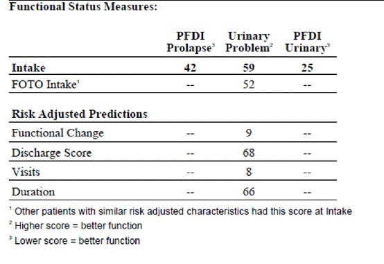
Figure 1. Pelvic floor distress inventory and urinary problem survey intake scores.
As stated, the patient is a high-functioning, active female with mild impairments in pelvic muscle strength as well as pelvic misalignment. From the history, the patient reported she was knowledgeable on the subject of pelvic floor due to her research and was willing to participate fully in a program provided by a therapist at 100% compliance. She appeared highly motivated due to her prenatal fitness regimen and lifestyle, as well as her drive to research the diagnosis and start Kegel exercises directly after delivery of her baby. A pelvic physical therapy plan of care can vary given the type of diagnosis, the severity, the motivation of the patient as well as any comorbidities they may have [13]. This patient had great health, great motivation, no comorbidities and a willingness to be 100% compliant to a program, which gave her a good prognosis for recovery.
It was recommended to start pelvic floor therapy at 2 x/week initially for up to 6 weeks. This would allow for starting exercise, making changes to a home exercise program (HEP), ensuring compliance and correctness of exercise implementation in her HEP. It was only anticipated the patient would need to be seen for 6 weeks as that duration of time would have allowed the patient to incorporate a lot of exercises into her home routine and see changes in muscle strength and function. We had discussed she would be doing most of her program at home with HEP addition/ changes during follow up visits as needed.
The patient’s plan of care included neuromuscular reduction and therapeutic exercises, such as pelvic muscle strengthening, core strengthening, biofeedback, manual therapy and self care training. These elements relate to pelvic floor dysfunction as they allow the patient to improve muscle weakness and neuromuscular control which can reduce prolapse, pain and pelvic floor stability.
Patient specific goals were as follows:
Short Term (STG) (3 weeks)
- The patient will be independent and compliant with home exercise program in order to achieve return to fitness without reported pelvic discomfort
- The patient will verbally report at least 25% reduction in pelvic heaviness/pressure and urinary leakage episodes
Long Term Goal (LTG) (6 weeks)
- The patient will score at least 68% on urinary problem survey noting improved function of the pelvic floor for return to activity.
- The patient will have pelvic muscle endurance on PERFECT test of 6-7 second holds at least 5-6 reps improving pelvic control to decrease leakage/reduce prolapse.
- The patient will verbally report she has been able to return to light exercise routine without urinary leakage
With the patient’s knowledge level, desire to improve and willingness to participate, patient education was at the top of the priority list. A study by Essery et al. [14] confirmed that self -motivation and drive leads to adherence to therapy and home exercises, so it was essential that she was educated on her dysfunction and treatment plan and given exercises to do at home to improve her success. Since the literature also shows that higher education levels show improved knowledge, attitude towards and practice of pelvic floor exercise, it was likely she would do well given her college level education [15]. This is likely due to better communication and understanding between therapist and patient, thus leading the patient to be more compliant than one who did not fully understand.
Evidence in the literature also notes that individuals with an athletic background and who remain physically active also have higher compliance to home exercise program [16]. Being that my patient was an athlete and still does recreational athletics, her rate of perceived compliance at home would be higher than an individual who was not active. She expressed high motivation to get better and was willing to do whatever was recommended, but if given the opportunity to do a home program that would be preferred.
Most research on pelvic floor disorders notes that adherence is essential to positive outcomes. Because the patient's physical exam and outcome measures indicated weakness, it needed to be addressed. Pelvic floor exercises need to be done correctly to be effective. After physical examination, it was indicated that the patient could contract the muscles correctly but struggled with power and endurance. This led to the decision that the patient would need to be consistent with routine exercises, which could be managed well from at home, with occasional check-ups to monitor and progress her program.
Initially, she received education on pelvic floor anatomy, pelvic floor conditions such as prolapse and incontinence, as well as exercises including Kegels and the importance of adherence to a home exercise program. Subsequently she returned after the evaluation for 3 sessions, which can be seen in Figure 2.
Figure 2: Treatment Sessions/Intervention
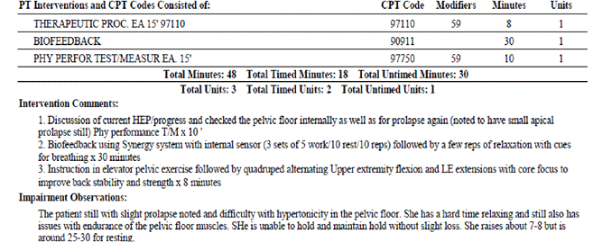
Treatment 1
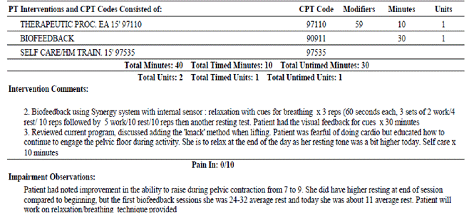
Treatment 2
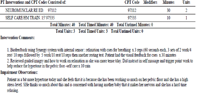
Treatment 3
The first visit after evaluation started with a reviewing the HEP and adherence. Once it was established how she was progressing, biofeedback was used. Biofeedback is noted to be an effective treatment when combined with PF therapy for motivated females [17]. The patient used pelvic floor contractions in combination with biofeedback using the Pathway MR-20 Dual Channel Surface EMG. The goal was to improve the patient’s ability to feel the contraction and maintain a long hold in order to improve power and endurance.
The second visit was shifted to do biofeedback first followed by patient education and therapeutic exercise instruction to work on relaxing the pelvic floor after working on strengthening using biofeedback. This was due to the fact that the patient had experienced some issues with relaxation after starting her pelvic floor treatment. This could be due in part to starting a focused exercise program, leading to increased muscle activity. She was instructed in diaphragmatic breathing techniques, which are shown to reduce muscle pain and tension [18,19].
The patient’s third and final treatment session consisted of another bout of biofeedback in combination with PFM exercises. We continued to use this method as it is effective in motivated patients, and the patient reported it helped her to contract the muscles correctly and effectively. After completion of biofeedback, the patient was educated on use of guided imagery (via use of applications on a mobile device at home) along with her current program to assist in stress reduction. She had some increases in pelvic floor activity since starting therapy as well as reported stress at home. Guided imagery is a cheap, accessible, and simple form of exercise that can promote relaxation and stress reduction.
The patient was only seen for a few visits in the clinic due to her schedule and ability to complete a HEP. She was seen for the initial evaluation and three treatment sessions. In the course of that time the patient did show improvements in her function both subjectively and objectively. The patient phoned after her third visit and was scheduled to see a new provider for a second opinion on her prolapse. She opted to continue with her HEP, as she felt confident with the practice, she had using biofeedback and the instruction given.
Subjective improvements were a reduction in pelvic pressure, reduced urinary leakage as well as improved ability to exercise with less fear of leakage.
Objective measurements included the use of the PERFECT system. Initially the patient scored: P: 3, E: 4, R: 4, F: 8, ECT: No elevation of posterior vaginal wall with contraction, appropriate co-contraction of PFM and transverse abdominal muscles and appropriate timing of PFM involuntary contraction with a cough. Upon completion of the 3rd visit the patient scored: P: 4 E: 7: R: 5, F: 9, ECT: elevation of posterior vaginal wall with contraction, appropriate co-contraction of PFM and transverse abdominal muscles and appropriate timing of PFM involuntary contraction with a cough.
POP testing was also performed initially which indicated and the patient did have mild prolapse (cystocele and rectocele) but not to the level of the introitus. The mild prolapse was still indicated during her last session leading her to seek out additional medical evaluation.
Further assessment of the SIJ and pelvis was not completed as the patient did not return after her third session, and she did not feel this was a primary complaint to address.
The patient completed PFDI (Prolapse and Urinary) as well as the Urinary Problem Survey using the FOTO outcome measures tool at initial evaluation and after discharge (Figure 3). Improvements are noted in PFDI prolapse and urinary (which a lower score indicates improved function) as well as Urinary survey (where higher score indicates better function).
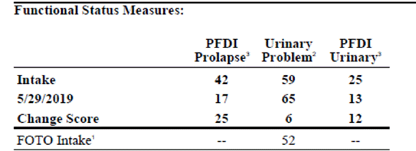
Figure 3. Pelvic Floor Distress Inventory and Urinary Problem Survey Discharge scores.
The goals created at initial evaluation were met with exception of LTG 1: The patient will score at least 68% on urinary problem survey noting improved function of the pelvic floor for return to activity. The patient had reported a 65% on the urinary problem survey with a goal set at 68%. She had made improvements in the short time frame and likely would have met this goal had she continued therapy.
Given reported compliance with the HEP and the achieved goals, the patient successfully was developing strength and functional gains in the pelvic floor. She was experiencing less pressure in the pelvic region, less episodes of incontinence and was slowly returning to activity including cardiovascular exercise and core/low back strengthening. Although she had hoped to return to her prenatal exercise routine, she had not by the time she opted for discharge. Her plan was to continue to progress with pelvic exercise at home and slowly add in jogging and running to tolerance as discussed, monitoring her pelvic discomfort; however, she wanted to follow up with a medical provider regarding her POP first.
The case patient presented for a total of 1 evaluation (including treatment) and 3 treatment sessions. As previously indicated, the patient had limitations in the amount of sessions she could attend. That limited our progress some; however, the patient was compliant at home, which did assist in her functional improvement. If given the chance to make changes, it would have been nice to see the patient for additional visits to progress into more advanced exercise routine while monitoring her progress of pelvic discomfort and incontinence as well as work on some of her SIJ issues. Although, that being said, the patient was very aware of her symptoms and felt comfortable with progressing at home.
Barriers to treatment were the distance to the clinic, busy work schedule, and a young infant to care for at home. Also, the therapist was only Per Diem in the clinic and only available three hours twice a week, which limited availability of appointments. The clinician was one of only to two pelvic floor therapists within an hour drive, so availability of appointments was limited on that aspect as well.
This patient case confirms the literature that supports that patient education level improves knowledge of, attitude towards and practice of pelvic floor muscle exercises. Also, as previously stated, literature suggests higher education levels have a positive correlation to adherence to HEP’s. Given the fact this patient had a college level degree and high self-drive, it was likely she would be more willing and open to pelvic treatment, which held true.
From the case, further research should be done on the area of access to women’s pelvic physical therapy. This case reports on the difficulty of one patient in a rural area, with limited access, which likely is a larger issue. Another potential area for further research would be referral to women’s health physical therapy following delivery of a child. This patient indicated that a referral was not suggested to her, she requested it.
- Faubion SS, Shuster LT, Bharucha AE (2012) Recognition and management of nonrelaxing pelvic floor dysfunction. Mayo Clin Proc 87: 187-193. [Crossref]
- Kegel AH (1948) Progressive resistance exercise in the functional restoration of the perineal muscles. Am J Obstet Gynecol 56: 238-248. [Crossref]
- Barber MD (2005) Symptoms and outcome measures of pelvic organ prolapse. Clin Obstet Gynecol 48: 648-661. [Crossref]
- Gunter J (2003) Chronic pelvic pain: An integrated approach to diagnosis and treatment. Obstet Gynecol Surv 58: 615-623. [Crossref]
- Robertson JA, Kendall FP, McCreary EK (1984) Muscles, Testing and Function. (3rd edn), Br J Sports Med 18: 25.
- Prather H, Dugan S, Fitzgerald C, Hunt D (2009) Review of anatomy, evaluation, and treatment of musculoskeletal pelvic floor pain in women. PM R 1: 346-358. [Crossref]
- Persu C, Chapple CR, Cauni V, Gutue S, Geavlete P (2011) Pelvic Organ Prolapse Quantification System (POP-Q) - a new era in pelvic prolapse staging. J Med Life 4: 75-81. [Crossref]
- Mattsson NK, Nieminen K, Heikkinen AM, Jalkanen J, Koivurova S, et al. (2017) Validation of the short forms of the Pelvic Floor Distress Inventory (PFDI-20), Pelvic Floor Impact Questionnaire (PFIQ-7), and Pelvic Organ Prolapse/Urinary Incontinence Sexual Questionnaire (PISQ-12) in Finnish. Health Qual Life Outcomes 15: 88.
- Mens JM, Vleeming A, Snijders CJ, Koes BW, Stam HJ (2002) Validity of the active straight leg raise test for measuring disease severity in patients with posterior pelvic pain after pregnancy. Spine 27: 196-200. [Crossref]
- Sartori DV, Gameiro MO, Yamamoto HA, Kawano PR, Guerra R, et al. (2015) Reliability of pelvic floor muscle strength assessment in healthy continent women. BMC Urol 15: 29. [Crossref]
- Laycock J (1994) Clinical evaluation of the pelvic floor. Pelvic Floor Re-education. : Springer-Verlag, London, UK. pp: 42-48.
- Laycock J, Brown JC, Cusack C, Green S, Jerwood D, et al. (2001) Pelvic floor reeducation for stress incontinence: comparing three methods. Br J Community Nurs 6: 230-237. [Crossref]
- Bø K (2012) Pelvic floor muscle training in treatment of female stress urinary incontinence, pelvic organ prolapse and sexual dysfunction. World J Urol 30: 437-443. [Crossref]
- Essery R, Geraghty AW, Kirby S, Yardley L (2017) Predictors of adherence to home-based physical therapies: a systematic review. Disabil Rehabil 39: 519-534. [Crossref]
- Muhammad J, Muhamad R, Husain NRN, Daud N (2019) Pelvic floor muscle exercise education and factors associated with implementation among antenatal women in hospital universiti Sains Malaysia. Korean J Fam Med 40: 45-52. [Crossref]
- Jack K, McLean SM, Moffett JK, Gardiner E (2010) Barriers to treatment adherence in physiotherapy outpatient clinics: a systematic review. Man Ther 15: 220-228. [Crossref]
- Halski T, Slupska L, Dymarek R, Bartnicki J, Halska U, et al. (2014) Evaluation of bioelectrical activity of pelvic floor muscles and synergistic muscles depending on orientation of pelvis in menopausal women with symptoms of stress urinary incontinence: a preliminary observational study. Biomed Res Int: 274938. [Crossref]
- Baird CL, Sands L (2004) A pilot study of the effectiveness of guided imagery with progressive muscle relaxation to reduce chronic pain and mobility difficulties of osteoarthritis. Pain Manag Nurs 5: 97-104. [Crossref]
- Arnouk A, De E, Rehfuss A, Cappadocia C, Dickson S, Lian F (2017) Physical, complementary, and alternative medicine in the treatment of pelvic floor disorders. Curr Urol Rep 18: 47. [Crossref]