Objective: to demonstrate the importance of using cone beam computed tomography (CBCT) in the surgical treatment of impacted teeth and dental anomalies.
Methodology: four cases were reported in which the CBCT provided some information that was not possible to be seen on digital panoramic radiographs. Based on this, a literature review was performed considering diferente situations in which a dental surgeon can request a CBCT after previous evaluation of a digital radiography.
Results: the presence of overlap and distortions in two-dimensional radiographs can sometimes confuse the dental surgeon in choosing an appropriate surgical technique. Therefore, some situations in which the precise tooth location by means of radiographs are not possible, or when a radiography suggests close contact with the adjacent structures and generates questions for possibles root resorption are essential to request a CBCT.
Conclusion: In view of the exposed clinical cases and the scientific basis performed in our literature review, a CBCT developed for dental use, presents an advantage for the dentist to plan the correct surgical treatment in cases where an initial radiograph generates doubts in the diagnosis.
radiography, panoramic, cone-beam cimputed tomofraphy, sugery, oral and ambulatory sugical procedures
The supernumerary tooth (extra tooth) is an anomaly in number development characterized by the presence of one or more teeth than in normal dentition. In the permanent dentition, the prevalence rate in humans varies between 0.5% and 3.8%, depending on the population. In the primary dentition the prevalence is lower, in the range of 0.3-0.6% [1-2].
Generally, the discovery of such teeth occurs during panoramic radiography performed for diagnostic purposes [2]. In some cases the position of supernumeraries and of unerupted (impacted) teeth can only be identified using location methods based on Clark, Miller-Winter, Donovan, Parma, and Le Master which require the combination of two radiographic techniques, such as for example; periapical and occlusal [3].
Clark's method is one of the most widely used and requires two periapical radiographs, ortho-radial and mesio-angulated. The method is based on the principle of parallax, which proposes that for two objects observed in a straight line, the closest to the observer will block out the most distant, yet, when the observer moves to one side, this closer object will dislocate the most, making it possible to determine the locations of the teeth [3].
In terms of locating teeth and pathologies, such localization methods are useful. However, there are limitations since a radiographic examination reveals a 3D image in a 2D plane, such that anteroposterior distances are impossible to measure [4]. In addition, image overlay impairs assessment of the case, being unreliable in terms of information involving relationships between the tooth or pathology and the adjacent anatomical regions. To locate impacted teeth and pathologies, conical beam computed tomography (CBCT) is considered the gold standard and provides three-dimensional visualizations of tooth structure, position, angulation, relationship with adjacent noble structures, distances between structures, and the lengths of impacted teeth (all in good resolution). This facilitates surgical planning with access to a better synthesis of the patient’s physical, psychological, and aesthetic well-being [3]. The purpose of this article is to demonstrate the importance, in certain cases, of the use of conical beam computed tomography (CBCT) in surgical planning for impacted teeth and dental anomalies, as well as provide information on when (for greater surgical safety) it might be necessary to request a CBCT.
Case 1
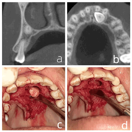
A male patient, dark-skinned, 16 years old, was referred to the Maxillofacial Anatomy and Traumatology League (LATIUM) of the Federal University of Ceará - Sobral Campus (UFC) for extraction of an included tooth (23). The included tooth was initially visualized using panoramic radiography, performed for orthodontic study. However, its vestibule-palatine positioning remained elusive, and cone beam computed tomography (CBCT) was requested. When evaluating the CBCT exam in detail, the proximity of the root apex to the nasal fossa was observed, parallel to the para-sagittal cut in the region of tooth 24 (Figure 1a), an axial cut in the region of the crown of tooth 23 (Figure 1b) its location near the palate, and the intimate contact of this crown with the roots of teeth 21 and 22, were all observed; requiring careful care in the region. Anesthesia was performed at the bottom of the buccal groove in the region of tooth 24 to anesthetize the tooth 23 nerve bundle, and, in addition anesthetizing the incisive foramen region of the palate in order to reach the mucosa of the region. The envelope incision, with a #15 blade, involved the entire palatal groove from the mesial of tooth 14 to the mesial of tooth 24. Osteotomy with a #6 spherical drill was performed at high rotation to expose the crown of tooth 23, (being in close contact with tooth 21), which was followed by a high-rotation odontosection with a #703 tapered cone drill to preserve maxillary bone. Figure 1c presents removal of the last fragment after the odontosections, managing to preserve the thin layer of bone that separated the crown of tooth 23 from the roots of teeth 21 and 22. After extraction, osteoplasty and abundant irrigation with saline were performed (Figure 1d), followed by transpapillary suture of all anterior teeth.

Figure 1. a. parasagittal section showing the location of the root apex of 23. b. axial section showing the position of the crown of the tooth 23. c. last fragment removed. d. surgical surface after osteoplasty and abundant irrigation
Case 2
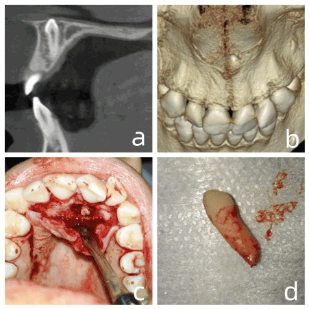
A female patient, melanoderm, 16 years old, and presenting no systemic changes was referred for extraction of an included tooth. Through panoramic radiography, an inverted mesiodens was observed between tooth 11 and 21. Being impossible to determine the vestibulo-palatal position of the referred tooth, a CBCT was requested for better evaluation. Evaluating the CBCT in detail, the inverted mesiodens and palate location were confirmed Figure 2a, and in the 3d reconstruction (Figure 2b) we noticed intimate contact with the nasal fossa. Anesthesia was performed both in the bottom of the buccal groove of tooth 21, and in the incisor foramen. An envelope incision was performed (#15 blade) involving the gingival sulcus from tooth 23 to tooth 13, followed by high-speed osteotomy with a #6 spherical drill to expose the root. The use of elevators to avoid wedge movement, rupture of the nasal membrane, and deviation of the tooth towards the nasal cavity is noted. After the osteotomy (Figure 2c), the root of the tooth was revealed, where #150 forceps were used to manage removal without odontosection (Figure 2d). Finalizing, osteoplasty and abundant washing with saline were then performed, followed by transpapillary suture in the entire anterior region.

Figure 2. a. Sagittal cut showing the position of the supernumerary tooth. b. 3D reconstruction showing proximity to the nasal cavity. c. root portion of the supernumerary exposed after odontosection. d. intact mesiodens
Case 3
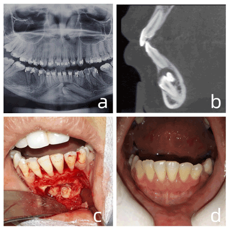
A female patient, 25 years old, leukoderm, was referred for evaluation of possible supernumerary teeth and revealed the presence of 3 supernumerary teeth in the mandibular symphysis region figure 3a. However, a panoramic examination did not provide information concerning the vestibulo-palatal positioning of the teeth, a CBCT was requested for better evaluation. Analyzing the 3D results in detail, the presence of a thick hair follicle associated with the supernumeraries was evidenced, which indicated the need for extraction. In the parasagittal section (Figure 3b) the presence of all supernumeraries by the buccal was revealed, in addition, external root resorption of tooth 31 was observed. Anesthesia was performed in the bottom of the buccal sulcus in the region of the mental foramen, bilaterally, and to the lingual gingiva in the lower incisor region. A #15 blade incision was made in the sucular region; mesial of tooth 33 to mesial of tooth 43, followed by two relaxing incisions in mesial of tooth 33 and mesial of tooth 43, featuring a quadrangular flap. Following the CBCT, the initial osteotomy was performed at 20 mm from the bony crest in the region of tooth 41, at high rotation with a spherical #6 drill. Upon exposing the crown, odontosections were started with a #703 tapered cone drill to minimize bone wear (Figure 3c). When the first supernumerary was extracted, it was possible to remove the enlarged hair follicle, again suggested by CBCT. The extraction of the other two supernumeraries followed the same protocol as the first, minimizing osteotomies and maximizing odontosections. At the end of the extractions, abundant irrigation with saline was performed, followed by suture with absorbable suture thread, the suture initially at the angles of the relaxants to avoid dehiscence of the suture, and continuing in the interdental papillae, and ending on the long relaxants axis. The postoperative period, 1 month after the surgery passed without periodontal gingival sequelae (Figure 3d).

Figure 3. a. initial panoramic radiograph. b. parasagittal section showing external root resorption of 31. c. sequential odontosections to minimize bone wear. d. one month after surgery
Case 4
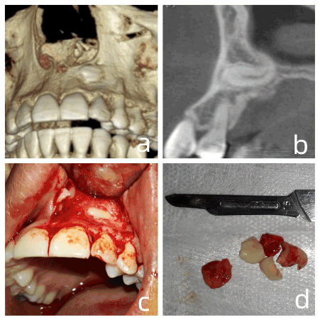
A 23-year-old female patient was referred for extraction of an included tooth (23). The initial diagnosis was performed using a panoramic radiographic examination; however, this did not provide information on vestibulo-palatal positioning, and a CBCT was requested for better assessment. When analyzing the details it was evidenced in the 3D reconstruction (Figure 4a) that the crown of the tooth was positioned by the vestibular, generating a slight bulging of the bone, and the root went towards the palatal direction, showing accentuated laceration of the apical third from intimate contact with the nasal fossa, as suggested in the parasagittal section (Figure 4b). Anesthesia was performed at the bottom of the buccal groove in the region of tooth 22 and tooth 24, and by the palate in the region of the incisor foramen. An incision (#15 blade) was carefully made in the gingival sulcus from tooth 11 to tooth 24, in association with a relaxing incision in tooth 24, and featuring a triangular flap (Figure 4c). Osteotomy was performed with a #6 spherical drill to expose the entire crown, followed by odontosections (Figure 4d), to preserve the jawbone. At the end of the surgical procedure, abundant washing with saline was performed, followed by an initial suture at the angle of the relaxant to avoid suture dehiscence, and finally, suture between the interdental papillae and along the relaxing incision.

Figure 4. a. 3D reconstruction with vestibular bone bulging. b. parasagittal section with root characteristics. c. surgical flap. d. dental fragments after several odontosections
Sometimes, in clinical practice the dental surgeon comes across cases of included teeth that whether supernumerary or permanent, for some reason did not erode in the oral cavity. And which are discovered through imaging tests that were performed for other reasons, for example evaluating the extent of caries lesion [5-7].
CBCT has been recommended in patients with supernumerary teeth in both the maxilla and mandible: to help locate them and diagnose the need for extraction, to reveal relationships with adjacent structures, to plan extractions without damaging the surrounding teeth, to apply an appropriate surgical approach, and to avoid tissue trauma [5-8].
Panoramic and periapical radiographs can enlarge and distort images in a way that results in diagnostic uncertainty [8]. In addition, reports in the literature argue that cases of included canines, especially those found close to the palate, and located above the central incisors, can be distorted in panoramic radiographs, sine the exam does not have a focal area that covers this region [5-7].
Localizing supernumerary teeth is very important for many reasons, whether for orthodontic diagnosis or for planning surgical removals. In the literature you can currently find several studies on methods of locating supernumerary teeth. The present study focused on supernumerary canines.
When it comes to conventional radiography, we are faced with several factors that hinder precise diagnosis and consequent knowledge of the exact location of the supernumerary canine. This influences the surgical plan. Factors such as overlapping structures, distortion, and two-dimensional images, require using techniques for verification in the anteroposterior position [4]. For this, the correct diagnosis of the positioning of supernumerary teeth requires complementary imaging tests with greater precision [9].
The exam of choice in the face of such situations is cone beam computed tomography (CBCT), which provides three-dimensional analysis of the area to be evaluated, with reduced distortion and without overlapping structures [10]. However, in public health it is a costly and often inaccessible test, this makes conventional radiographic examinations such as panoramic, periapical, and occlusal radiographic techniques more often chosen (Miller & Winter method) [11].
Thus, the requests for tomographic examinations in cases 1 and 2 are justified, as they allow better visualization of the region to be operated, as well as a safer and more comfortable surgery for the patient. In case 1, we mention visualization of the root apex of tooth 23, located in the tooth 24 root apex region, which enabled more effective anesthesia through a puncture point located at the bottom of the buccal groove of the apex of tooth 24.
Case 2 presented a tooth located entirely in the palate, in addition to being inverted. Evaluating only a panoramic exam, we might make the mistake of beginning access at the vestibular. However, using tomography the impossibility of carrying such access out is evident, since access by the vestibular, would require very extensive osteotomy. Thus, the CBCT made it possible to plan the extraction with the use of forceps securing the root portion of the supernumerary, and not the crown as is done conventionally.
The CBCT performed for case 3 was also decisive for locating the supernumerary teeth, since it was possible to reveal their location by the vestibular, in addition to the exact point to begin osteotomy and prevent unnecessary bone wear.
In case 4, when analyzing the details provided using CBCT, it was evidenced that the crown of the tooth was positioned by the vestibular, discreetly perforating the bone plate while its root went in the palatal direction, presenting a sharp laceration in the apical third, this proved fundamental for the surgical planning. Thus, the request for 3D exams is justified in cases where the overlapping of structures prevents precise localization and impairs the planning and safety of the surgery (10-12).
Root resorption
A systematic review carried out in 2017 [12], sought to evaluate the efficiency of CBCT as compared to conventional radiographs for location of included canines, that is, the precision of the results of both exams. Not satisfied, they also sought to assess the difference in surgical planning using both tests [12]. Their results revealed that in discrepant cases of severe root resorption, both exams are effective, yet CBCT is much more accurate in cases of initial and/or discreet external resorption [12].
Intra- and inter-exam agreement was also much greater when using CBCT as compared to two-dimensional radiographs. However, this systematic review did not report testing the effectiveness of the proposed treatment after each exam, and the most difficult cases were the ones that found the greatest disagreement [12].
In view of this, CBCT, to clearly visualize the extent and degree of external resorption present in tooth 31, was essential in Case 3, likely caused by the peri-coronary dental follicle of one of the supernumerary teeth. This type of advantage was confirmed in a study by Carolina S. Lai et al. [13], where 134 included canines were evaluated by CBCT, and it was shown that the case agreements regarding the presence and degree of resorption were greater when the evaluators used CBCT in comparison with conventional radiographic methods.
Relationship with other anatomical structures
We emphasize that in some cases, details provided by CBCT allow visualization of possible intimate contact of the included tooth with important anatomical structures, such as the maxillary sinus, nasal cavity, and even with the root of an erupted tooth [14-15].
Citing case 1, we can see intimate contact between the crown of tooth 23 and the root of tooth 21. Our surgery avoided wear in the region close to the buccal region of tooth 23 and damaging the root of tooth 21.
Contrary to case 1, in case 2 we noticed that the supernumerary crown was in close contact with the nasal fossa, and in case 4 the root of the included canine was also in close contact with the nasal fossa. This information allowed surgical planning to minimize any intrusion, and avoid wedging effect from any instrument, which would make buco-nasal communication more likely. In addition, a fibrin sponge was included at the operating table to resolve any surgical accident involving bucal-nasal communication.
When to indicate a CBCT?
An extremely important factor to consider is the radiation dose involved in the CBCT exam. For this alone, the CBCT exam must be requested only in well analyzed and, above all, well justified cases. Given this precaution, the CBCT exam will decisively contribute to reducing surgical time and avoiding possible intra- and postoperative complications [16].
In view of these factors, it is important to establish criteria for a correct CT scan referral request. The use of CBCT is not indicated for all cases, especially when surgery can be performed using conventional periapical and occlusal radiographs.
The CBCT can be indicated when:
1. Locating an unerupted tooth with suspected absorption of adjacent tooth.
2. Locating an unerupted tooth, suspected of being in intimate contact with some anatomical structure;
3. Diagnosis is not possible using radiographic methods and conventional radiographs;
4. Pre-surgical evaluation fails when using conventional radiographs.
In view of the clinical cases herein related and the scientific basis of the cited bibliographic review; in cases of included teeth located in regions where conventional two-dimensional radiography might present distortions as to the precise position of the tooth, and where accentuated overlap precludes precise pre-surgical diagnosis, CBCT (as developed for dental use), presents advantages that assist the dental surgeon in correct surgical planning.
No funding received.
Carlos Eduardo Nogueira Nunes declares that he/she has no conflict of interest.
Samuel Rocha França declares that he/she has no conflict of interest.
Renan Ribeiro Benevides declares that he/she has no conflict of interest.
Filipe Nobre Chaves declares that he/she has no conflict of interest.
Denise Helen Imaculada Pereira de Oliveira declares that he/she has no conflict of interest.
Marcelo Bonifácio da Silva Sampieri declares that he/she has no conflict of interest.
All procedures performed in studies involving human participants were in accordance with the ethical standards of the institutional and national research committee and with the 1964 Helsinki declaration and its later amendments or comparable ethical standards.
Informed consent was obtained from all individual participants included in the study.
- Sulabha AN, Sameer C (2012) Association of mesiodentes and dens invaginatus in a child: A rare entity. Case Rep Dent 9: 198032.
- Celikoglu M, Kamak H, Oktay H (2010) Prevalence and characteristics of supernumerary teeth in a non-syndrome turkish population: Associated pathologies and proposed treatment. Med Oral Patol Oral Cir Bucal 15: e575-578.
- Tsolakis AI, Kalavritinos M, Bitsanis E, Sanoudos M, Benetou V, et al. (2018) Reliability of different radiographic methods for the localization of displaced maxillary canines. American Journal of Orthodontics and Dentofacial Orthopedics 153: 308-314.
- Botticelli S, Verna C, Cattaneo PM, Heidmann J, Melsen B, et al. (2011) Two- versus three-dimensional imaging in subjects with unerupted maxillary canines. Eur J Orthod 33: 344-349.
- Merrett SJ, Drage N, Siphahi SD (2013) The use of cone bean computed tomography in planning supernumerary cases. J Orthod 40: 38-46.
- Chen KC, Huang JS, Chen MY, Cheng KH, Wong TY, et al. (2019) Unusual supernumerary teeth and treatment outcomes analyzed for developing improved diagnosis and management plans. J Oral Maxillofac Surg 77: 920-931.
- Gurler G, Delilbaşı C, Delilbaşı E (2017) Investigation of impacted supernumerary teeth: A cone beam computed tomograph (cbct) study. J Istanbul Univ Fac Dent 1: 18-24.
- Jeremias F, Fragelli CMB, Mastrantonio SDS, Dos Santos-Pinto L, Santos-Pinto A Dos, et al. (2016) Cone-beam computed tomography as a surgical guide to impacted anterior teeth. Dent Res J (Isfahan) 13: 85-89.
- Castro-Silva II, Vasconcelos JLA, Alves AD, Basilio SR, Siebra AKA, et al. (2018) Distribuição de anomalias dentárias em cidades do Norte e Nordeste do Brasil. Braz J Surg Clin Res 22: 49-53.
- Mossaz J, Kloukos D, Pandis N, Suter VG, Katsaros C, et al. (2014) Morphologic characteristics, location and associated complication of maxilar and mandibular supranumerary teeth as evaluated using cone beam computed tomography. Eur J Orthod 36: 708-718
- Khalaf K (2016) Supernumerary teeth-review of aetiology, sequelae, diagnosis and management. Part II. Int J of Adv Res 4: 1363-1375.
- Eslami E, Barkhordar H, Abramovitch K, Kim J, Masoud MI, et al. (2017) Cone-beam computed tomography vs conventional radiography in visualization of maxillary impacted-canine localization: A systematic review of comparative studies. Am J Orthod Dentofac Orthop 151: 248-258.
- Lai CS, Bornstein MM, Mock L, Heuberger BM, Dietrich T, et al. (2013) Impacted maxillary canines and root resorptions of neighbouring teeth: A radiographic analysis using cone-beam computed tomography 35: 529-538.
- Valente, Alencar N (2016) The importance of CBCT in supernumerary teeth diagnosis and location. Rev Bras Odontol Rio de Janeiro 73: 55-59.
- White SC, Pharoah MJ (2014) Oral radiology: principles and interpretation: Elsevier HEALTH SCIENCES; 2014.
- Winstanley KL, Otway LM, Thompson L, Brook ZH, King N, et al. (2018) Inferior alveolar nerve injury: Correlation between indicators of risk on panoramic radiographs and the incidence of tooth and mandibular canal contact on cone-beam computed tomography scans in Western Australian population. J Invest Clin Dent 9: 1-6.


