Introduction
Socket preservation has shown to be a very valuable adjunct procedure in implant dentistry. Preserving labial cortical bone during tooth extraction is mandatory to obtain a wider alveolar bone leading to a long-term stability of dental implants [1-8].
Several dental malocclusions, regardless of the improvement of orthodontic therapy in the last years, can still benefit from dental extractions. Severe crowding, thin periodontal tissues and dental protrusion are common conditions that can lead to teeth extractions for orthodontic treatment success [9-11]. (Figure 1)
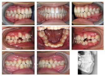
Figure 1. From top to bottom: Thin periodontal biotype, severe crowding, dental protrusion exceeding the anterior limit of denture
Oral surgery standard extraction procedures have always advocated that, due to the labial position of the dentition in the alveolar bone, any tooth should be extracted following a pathway towards the thin buccal cortical bone, because lingual or palatal cortical bone is always wider and harder to allow root dislocation during an extraction procedure [9]. (Figure 2)
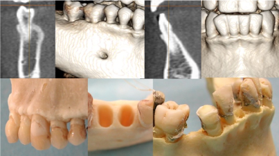
Figure 2. Labial position of teeth in the alveolar bone from CBCT scans and from human skulls. Labial cortical bone is always less represented compared to the lingual or palatal bone
Dislocation, iatrogenic disruption or removal of the labial cortical bone is the main reason responsible for progressive bone volume reduction and soft tissue adaptation during wound healing. The bone loss in the vertical dimension, is always followed by a reduction of the amount of adherent gum [5].
Socket preservation procedures have been introduced in order to reduce the amount of bone and adherent gum loss in the three dimensions.
The purpose of this paper is to show how a modified extraction technique focused in preserving the alveolar socket can improve tooth movement during space closure and can result in a healthier periodontum at the end of the treatment. Additional considerations regarding root torque setup in the orthodontic appliance must be kept in mind and will be focused later when space closure has to be achieved. (Figure 3)
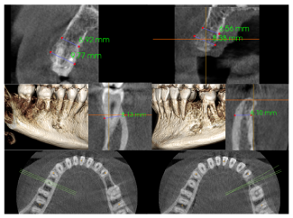
Figure 3. Bone modeling after extraction: in the first case a socket preservation surgical procedure has been performed resulting in a minimal bone loss, while in the second case, conventional extraction technique has been used
Surgical clinical procedure
Gentle tooth removal associated to root sectioning has become the gold standard when immediate implant placement after tooth extraction has been planned.
On the other hand, when implant placement must be delayed a socket preservation technique should be considered associated to the use of bone substitutes or soft tissue augmentation procedures [7].
In both cases the key factor relies on preserving the thin labial cortical bone of the socket during tooth extraction.
A modified surgical approach can help achieve this goal and should be applied to any extraction for an orthodontic purpose.
A wider alveolar bone allows safer movement of the roots far away from the cortical plates, speeding up space closure, limiting the amount of root resorption and a better periodontal adaptation at the end.
In case of a bicuspid extraction, the first step consists of removing the mesial and distal contact point of the bicuspid.
Secondly, an elevator is placed in the distal contact point of the bicuspid in order to loosen the tooth in a forward anterior direction.
Only after the tooth has shown some root dislocation and mobility, the tooth can be loosened in backward posterior direction, limiting in this way the amount of reaction force applied to the front teeth. (Figure 4)
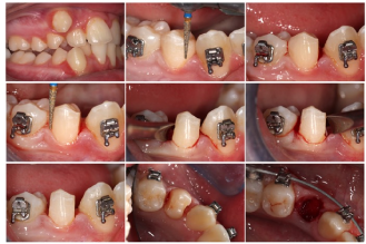
Figure 4. Extraction technique: socket preservation avoiding periodontal trauma
In case of multi rooted tooth extraction, separation of the roots is performed, while in case of single root, as a lower bicuspid, the tooth is ready to be extracted.
Special care at this point, is required to not dislocate roots towards the labial bone. Clockwise alternated to counter clockwise movements gradually lead to root removal. (Figure 5)
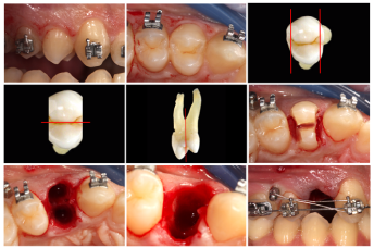
Figure 5. Additional procedures when removing teeth with multiple roots; close detail of the thin preserved labial cortical plate
Orthodontic socket preservation does not contemplate the use of bone substitutes, alveolar socket is filled only with collagen (Gingistat, Gaba Vebas) to promote the stability of the wound clot. Soft tissue augmentation might be indicated especially in presence of thin periodontal biotype or a buccal cortical bone loss.
Orthodontic Appliance Setup
Space closure and sliding mechanics normally follow teeth extractions. The labial position of the dentition is often a true limit in achieving a fast space closure and can often lead to a gingival recession in the involved teeth. When using a straight wire appliance, a correct torque selection should be assessed to move roots towards the center of the alveolar bone. If torque prescription of the teeth that are going to close the space is negative, it should be changed into a neutral or a positive torque. On the other hand, a third order information could be added to the stainless-steel wires before space closure mechanics are started without any change to the bracket prescription. (Figure 6)
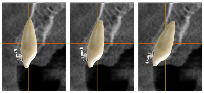
Figure 6. Different torque prescription from negative to neutral and, in the end positive, affects root position disengaging it from the labial cortical bone allowing more efficient movement through the medullar bone. Periodontum in this instance is not stressed from tooth movement
When using clear aligners, information for torque correction has always to be inserted in the first aligners before attempting space closure.
Discussion
This surgical technique adopted from implant dentistry has been routinely applicated in the treatment of any extraction case resulting in a smooth space closure, absence of gum recession and long-term stability of the periodontum, especially when a thin periodontal biotype is associated to a dental protrusion as shown in the cases below. (Figure 7-9)
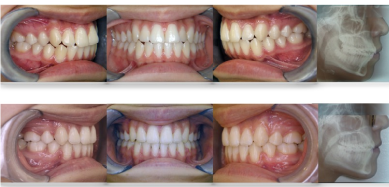
Figure 7. Thin periodontal biotype associated to dental protrusion. Treatment included four bicuspid extractions, neutral torque selection for cuspids e second bicuspids
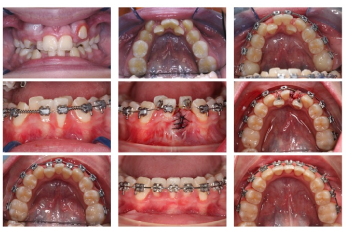
Figure 8. Severe anterior crowding associated to a thin periodontum. A lower incisor extraction has been required to solve the crowding. Hard and soft tissue management during the surgical phase allowed healty periodontal tissues. Incisors torque has been controlled through third order archwire bends using a TMA wire

Figure 9. Healthy periodontal tissues after 8 years since the end of the treatment of a case of class III camouflage. Treatment included extractions of lower first bicuspids, neutral torque selection for lower cuspids
References
- Buser D (2009) 20 years of guided bone regeneration in implant dentistry (2nd Edn), Quintessence Pub C0.
- Misch CE (2007) Contemporary implant dentistry (3nd Edn), Mosby.
2021 Copyright OAT. All rights reserv
- Newman MG, Takei H, Klokkevold PR, Carranza FA (2014) Carranza’s clinical periodontology (12nd Edn), Sanuders.
- Fee L (2017) Socket preservation. Br Dent J 222: 579-582. [Crossref]
- Van der Weijden F, Dell'Acqua F, Slot DE (2009) Alveolar bone dimensional changes of post-extraction sockets in humans: a systematic review. J Clin Periodontol 36: 1048-1058. [Crossref]
- Tan WL, Wong TL, Wong MC, Lang NP (2012) A systematic review of post-extractional alveolar hard and soft tissue dimensional changes in humans. Clin Oral Implants Res 23: 1-21. [Crossref]
- Vignoletti F, Matesanz P, Rodrigo D, Figuero E, Martin C, et al. (2012) Surgical protocols for ridge preservation after tooth extraction: a systematic review. Clin Oral Implants Res 23: 22-38. [Crossref]
- Buser D, Chen ST, Weber HP, Belser UC (2008) Early implant placement following single-tooth extraction in the esthetic zone: biologic rationale and surgical procedures. Int J Periodontics Restorative Dent 28: 441-451. [Crossref]
- Hupp JR, Tucker MR, Ellis E (2003) III: Contemporary oral and maxillofacial surgery (6th Edn), Mosby.
- Proffit WR, Fields HW, Sarver DM (2012) Contemporary orthodontics (5th Edn), Mosby.
- Graber LW, Vanarsdall RL, Vig KWL (2016) Orthodontics current principles and technique (6th Edn) Mosby.