"Off label" use of the ReMotionTM total wrist for treatment of advanced stage of post-traumatic pancarpal wrist osteoarthritis: Report of two cases and review of literature
Ingo Schmidt
Medical Centre Wutha-Farnroda (Department of Orthopaedics / Traumatology / Hand Surgery), Ringstr. 20, 99848 Wutha-Farnroda, Germany
Hospital Bad Salzungen GmbH (Department of Orthopaedics and Traumatology, Teaching Hospital of the Friedrich-Schiller-University Jena, Germany), Lindigallee 3, 36433 Bad Salzungen, Germany
Hospital Bad Salzungen GmbH (Department of Orthopaedics and Traumatology, Teaching Hospital of the Friedrich-Schiller-University Jena, Germany), Lindigallee 3, 36433 Bad Salzungen, Germany
The ReMotion total wrist is one 3rd generation implant which is current in use. Mid- to long-term outcomes regarding its survivorship revealed satisfying results. However, this implant has a design-related issue resulting in development of painful radial-side impingement. We present two cases involving two modified implantation techniques ((1) complete removal of scaphoid accompanied by partial cement augmentation of the radial-side fixation screw for carpal component, and (2) positioning of both fixation screws for carpal component into the scaphoid adjacent to the capitate peg without crossing the carpometacarpal joins) which could be able to avoid this specific complication. However, such an “off label” use in the absence of a larger number of patients and further biomechanical investigations can only be used in single cases currently.
Keywords
wrist, osteoarthritis, total wrist arthroplasty, ReMotion total wrist, “off label” use
If technically possible a total wrist arthroplasty (TWA) utilizing the 3rd generation types (ReMotion, Universal 2 / Freedom, Maestro) is the motion-preserving alternative to total wrist fusion (TWF) for treatment of pancarpal primary and post- or non-traumatic wrist osteoarthritis (OA) (Figures 1A-E, Figures 2A-D), and revision TWA or conversion to TWF are reliable options for a failed TWA [1-11].
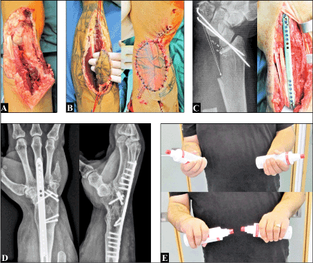
Figure 1 (35-year old male, right severe open wrist injury). (A) Initial soft tissue situation. (B) Wound coverage with a distally pedicled radial artery flap. (C) After uneventful flap healing planning and performing the TWF utilizing two corticocancellous iliac crest bone grafts, a 2,7 mm locking wrist spanning plate, and five headless compression screws. Due to the extended carpal bony defects a TWA was technically not possible. (D) Unchanged uneventful bony fusion in the absence of any implant complications at the 11-year follow-up. (E) Good restoration of forearm supination-pronation motion arc compared to the contralateral forearm
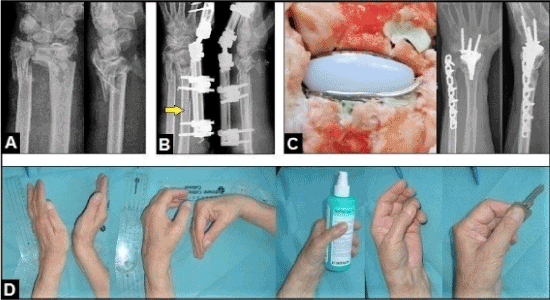
Figure 2 (71-year old female, left severe closed distal forearm injury associated with poor osteoporotic bone stock). (A) Radiographs in both planes demonstrating the initial findings, note the highly comminution of distal radius metaphysis. (B) The fracture was primarily closed reducted by external fixation. (C) After stabilization of the closed soft tissue damages an internal fixation of the distal ulna fracture utilizing a 3,5 mm titanium locking reconstruction plate was done 10 days after injury, and four weeks later the ReMotion total wrist was inserted with cementation due to the poor osteoporotic bone stock. (D) At the 2-year follow-up there was an very satisfying functional outcome for the patient
Recent evidence suggests that the complication rate of all 3rd generation TWAs is significantly lower than older generation types (p=0.002, range 0.1-2.9% vs.0.2-8.1%) [12], and its complication rate with 7% is slightly lower as well to those with 10% in patients undergoing a TWF, and TWF is associated with a higher percentage of perioperative devicerelated complications (6 vs. 3%, p < 0.001) and respiratory complications (0.54 vs. 0%, p < 0.05) potentially leading to higher costs of hospitalization than TWA in 2010 [13]. Noted that mid- to long-term outcomes of primary TWF revealed a complication rate ranging from 33 to 60.5% associated with a required re-operation rate ranging from 19 to 63% with a reported predominance in females suffered from inflammatory disease, respectively [14-17]. From all 3rd generation TWAs the Maestro was superior both in terms of longevity and functional outcome over the other types, however, this implant was withdrawn from the marketplace because it was no longer profitable for the company, and now surgeons have explanation misery to their patients [18-22].
The non-constrained ReMotion total wrist (stryker, Kalamazoo MI, USA) with it ball and socket design was introduced in 2002 and is one of the 3rd generation types which is current in use. It is a radial surface replacement, thus, the minimal bony resection allows revision TWA or conversion to TWF without greater problems. Another advantage is that the distal radioulnar joint (DRUJ) is not compromised allowing an additional ulnar head replacement (UHR) [23,24]. It can be used both in a cemented or in a non-cemented manner (Figures 2A-D) [25]. However, this type has design-related issues. We present two possible options in order to avoid these problems.
Technical note to the implant and problems
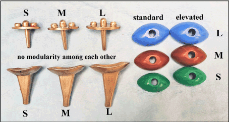
The ReMotion implant has an ellipsoid metal (cobalt-chromium)-on-ultrahigh density polyethylene articulation and consists of three components: (1) the anatomically shaped radial component which is inserted pressfit with it titanium coated stem into the distal radius metaphysis without required bony resection at the articular surface, (2) the carpal component with it horizontal straight design which is inserted pressfit with it titanium coated peg into the capitate, and (3) the intercalated polyethylene ball. The radial and carpal components are available in four sizes (extra small, small, medium, large) and the intercalated polyethylene ball in a standard or elevated (“plus”, adding 1 mm to the standard height) manner, however, the disadvantage is that there is no modularity among each other (Figure 3). The implant is designed to provide 40° of flexion, 40° of extension, and 30° arc of radial/ulnar deviation. The intercalated carpal ball can rotate 10° relative to the carpal plate in order to minimize torque transmission to the metal carpus component, and this feature could be able to preserve the complex “dart thrower’s” motion of the wrist with relative axial rotation between the intercalated segment and the carpal plate as believed by the developer [25].
Figure 3. Trials of the ReMotion total wrist (without size extra small)
Due to the anatomically determind higher instability in the carpometacarpal joint IV compared to the carpometacarpal joint II, the ulnar-side fixation screw of carpal component should only be inserted into the hamate after removal of the lunate and triquetrum whereas the radial-side fixation screw can be inserted into the second metacarpal after removal only a half of the scaphoid. However, this implantation technique utilizing a carpal component with a horizontal straight design (ReMotion, Universal 2) requires fusions of the hamate-capitate and the scaphoid-capitate joints in order to avoid micromotions potentially leading to loosening of the screws [25,26]. The advantage of the Maestro over the other types was the possibility of insertion a carpal component with a scaphoid augment after removal of the entire scaphoid which did not require fusions of the surrounding intercarpal joints [27]. Another disadvantage of the ReMotion compared to the Maestro or Freedom is that this implant is not available with locking screws for fixation the carpal component in order to enhance stability.
The main postoperative problem with the ReMotion as well as with Universal 2 is worsened radial deviation that can be associated with development of painful radial-side impingement between the distal scaphoid and the offset of radial component, and only removal of the entire scaphoid is able to avoid this symptomatic impingement, but this procedure can cause an insufficient bony wrapping of the proximal threads of the radial-side screw (Figures 4A-C) [23,28,29]. Of all third generation types, only the Maestro achieved improvement of radial deviation in the absence of radial-side impingement [20,30,31]. This advantage with the Maestro was based on design-related features: (1) it had a concave to distally curved carpal component design (i.e. tapered), and (2) the three available sizes of intercalated carpal heads allowed more better restoration of resection-related carpal height than the ReMotion with it two sizes only [18-20,28].
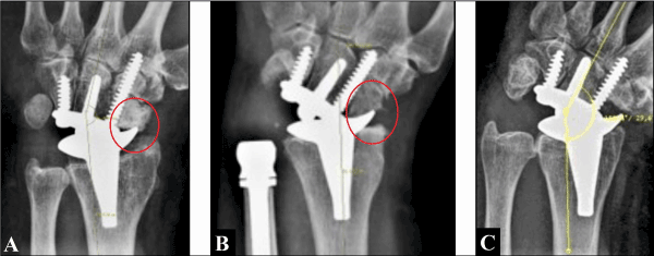
Figure 4 (Correlation of painful impingement at terminal range of radial deviation with the ReMotion [23,28]). (A) Radial-side impingement when only the proximal pole of scaphoid was removed (arrow, standard technique). (B) Radial-side impingement despite additional oblique resection at the distal pole of scaphoid (arrow). (C) No radial-side impingement between the trapez and offset of radial component when the entire scaphoid was removed. Note that only the distal threads of radial-side fixation screw are bony wrapped by the trapezoid and second metacarpal
Case presentation 1
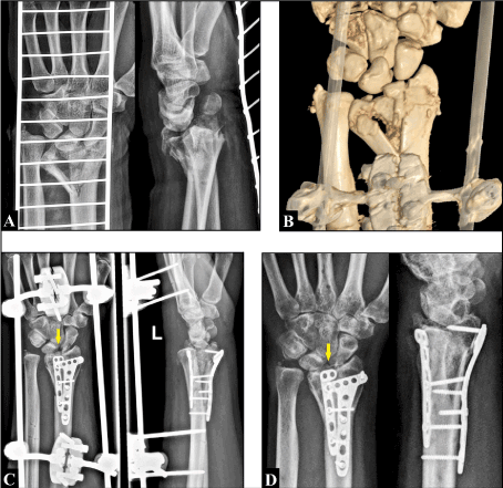
A 39-year-old male sustained a highly comminuted fracture-dislocation injury at his left wrist after a fall from height (Figures 5A-B), initially treated by combined closed and open reduction and fixation utilizing an external fixateur and two locking plates (Figure 5C). The external fixateur was removed six weeks after injury (Figure 5D), and the volar and dorsal locking plates six months after injury (Figure 6A). All these procedures were done in another two institutions. The retrospective analysis of the radiographs by us revealed a concomitant scapholunate ligament disruption (SLLD).
Figure 5 (Case presentation 1). (A) Radiographs in both planes showing the fracture-dislocation injury. (B) Three-dimensional computed tomography demonstrating the highly comminution of distal radial metaphysis. (C) Radiographs in both planes after initial treatment. Note the concomitant SLLD (arrow). (D) Radiographs in both planes after removal of the external fixateur (arrow: SLLD)
Figure 6 (Case presentation 1). (A) Radiographs in both planes showing advanced stage of post-traumatic wrist OA (arrow: SLLD). (B) Clinical photographs demonstrating loss of wrist function of approximately 50% compared to the uninjured right wrist. (C) Clinical photographs demonstrating the marked loss of forearm supination/pronation compared to the uninjured right wrist
At first presentation in our institution one year after injury the patient reported pain with 8 in visual analogue score (VAS, 0-10 points) and a marked decreased Patient-rated wrist evaluation with 69 (PRWE, 0-100 points). Radiographically, there was advanced stage of post-traumatic pancarpal wrist OA associated with scapholunate advanced collapse (SLAC) (Figure 6A). Functions of the wrist and forearm were decreased in summary of approximately 50% compared to the uninjured right wrist (Figures 6B-C). Thus, the primarily non-cemented ReMotion TWA was indicated by us. Noted that the patient has lost his job as a carpenter at this time because of the incapacity to work for one year.
TWA was done typically via the dorsal approach. The triquetrum, lunate, and scaphoid were completely removed in order to avoid postoperative painful radial impingement. After that, the trials size S of the ReMotion were placed which showed correct alignment (Figure 6A). After removal of the trials, the definitive components were inserted without cementation in standard technique but only the radial-side fixation screw could be placed intraosseous with its distal threads into the trapezoid and the base of the second metacarpal, and for fusion of the hamate-capitate joint the corresponding cartilaginous surfaces were excised (Figure 6B). Then the proximal threads of radial-side fixation screw were wrapped with cement creating a stable cement augmented “monobloc construct” with the capitate and trapezoid, and the hamate-capitate joint was fusioned with impaction of cancellous bone from the excised scaphoid (Figure 6C). At the end of surgical procedure, there was a stable TWA at passive terminal ranges of motion in the absence of radial-side impingement between the trapez and offset of radial ReMotion component (Figure 6D).
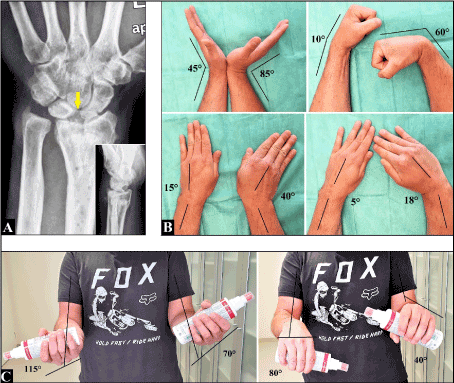
At the six-month follow-up the patient was very satisfied with his intermedium outcome. Radiographically, we observed unchanged correctly aligned placement of the implant (Figure 8A). Pain (VAS) and PRWE had improved to 2 and 27. We observed improvements of active flexion with 10°, active ulnar/radial deviation with 13°/15°, active supination/pronation with 10°/40°, and only active extension had decreased with 10° (Figures 8B-C compared to Figures 6B-C). Furthermore, we observed a very good circumduction of the left wrist compared to the uninjured right wrist (video 1 – supplementary material).
Figure 7 (Case presentation 1). (A) Intraoperative fluoroscopy demonstrating correct alignment of the trials size S in both planes. (B) Intraoperative clinical photograph after insertion of both definitive components showing the not bony wrapped proximal threads of radial-side fixation screw, and for fusion of the hamate-capitate joint the corresponding cartilaginous surfaces were excised (arrow). (C) Intraoperative clinical photograph showing the cement augmented “monobloc construct” between the proximal threads of radial-side fixation screw and the surrounding carpal bones, and the fusioned hamate-capitate joint (arrow). (D) Intraoperative fluoroscopy demonstrating the stable ReMotion TWA at passive terminal ranges of motion (black arrow: cement augmentation, green arrow: no radial-side impingement)
Figure 8 (Case presentation 1, six-month follow-up). (A) Radiographs in both planes showing unchanged correctly aligned placement of the ReMotion. (B) Clinical photographs with active terminal ranges of motion at the left wrist. (C) Clinical photographs with active terminal ranges of supination and pronation of both forearms
(1) Video to the patient in case presentation 1.
Case presentation 2
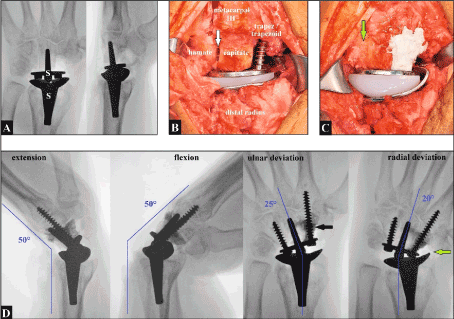
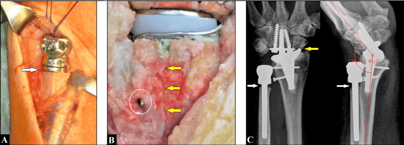
A 62-year-old male suffered from left painful post-traumatic wrist (SLAC III) and DRUJ OA and sustained primarily an UHR combined with wrist denervation. The UHR was placed in standard technique on the top of the distal ulnar stump (Figure 9A). Six months later, a TWA utilizing the ReMotion TWA (size S) became necessary because prior denervation was unsuccessful for the patient. Intraoperatively, a longitudinal periprosthetic fracture on the dorsal aspect of distal radius metaphysis occurred that required insertion of the radial component with cementation combined with internal fixation utilizing two 3,5 mm titanium compression screws (Figure 9B). In contrast to the standard technique, both screws for fixation the carpal component were placed with convergence to distal into the capitate adjacent to the capitate peg and did not cross the carpometacarpal joints, this technique required no additional fusions of the surrounding intercarpal joints (Figure 9C). One year after TWA we observed radiographically an uneventful periprosthetic fracture healing with unchanged correctly aligned placements of both implants, impingement between the distal scaphoid and the offset of radial component already in neutral position which did not allow further active radial deviation by the patient, and periprosthetic osteolysis (PPO) around the collar of UHR (Figure 9C).
Figure 9 (Case presentation 2). (A) Intraoperative clinical photograph showing the UHR inserted exactly on the top of distal ulnar stump (arrow). (B) Intraoperative clinical photograph showing the longitudinal periprosthetic fracture on the dorsal aspect of distal radius metaphysis (arrows), insertion of the radial ReMotion component with cementation, and one of the two compression screws (circle). (C) Radiographs in both planes one year after TWA demonstrating unchanged correctly aligned implants without any signs of loosening, PPO around the collar of UHR (white arrows), and evident radial impingement in neutral position between the distal scaphoid after removal of its only proximal one half regarding the standard technique and the offset of radial ReMotion component (yellow arrow)
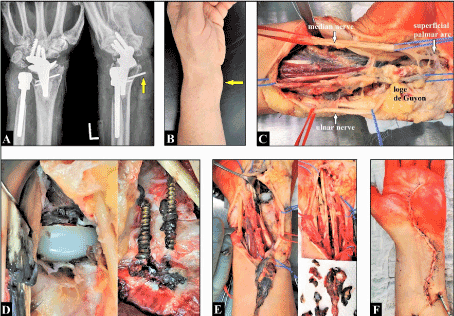
At the 10-year follow-up regarding left TWA the patient reported progressive painless swelling on the volar aspect of his wrist and sensory deficits at his all finger tips within one year in the absence of decrease in function. Radiographically, there were no signs of loosening of both implants, PPO around the collar of UHR had been stabilized, we saw firstly a new ulnar-side PPO under the offset of radial ReMotion component subsequently leading to loosening and migration of one titanium compression screw in volar direction (Figure 10A). Clinically, there was a subcutaneous giant cyst at the volar-ulnar aspect of the wrist (Figure 10B). Neurological examination revealed both carpal tunnel and loge de Guyon syndrome. Therefore, surgical revision via a volar approach became necessary. Intraoperatively, a giant metal debris induced black-colored cyst starting in all flexor tendon sheaths at the distal forearm and extending up into the palm was present which led to entrapment both of the median and ulnar nerve. We performed a radical debridement, decompression of both nerves with opening the loge de Guyon, and removal of the migrated screw. Metallosis originates around both TWA components and from the hole of the migrated screw which was broken. Similar to the preoperative radiographs no loosening of both TWA components could be observed, and the intercalated polyethylene ball did not show any signs of destruction macroscopically. Primary wound closure could be done (Figures 10C-F). Histopathological examination revealed encapsulated benign cyst formations containing collagen fibers and fibroblasts / fibrocytes, myxoidal connective soft tissue changes, and metallic wear particles out- and inside of histiocytes and macrophages (Figures 11A-B). The wound healing was uneventful.
Figure 10 (Case presentation 2). (A) Radiographs in both planes showing both implants without any signs of loosening. PPO around the collar of UHR had been stabilized. A new PPO was present under the offset of radial ReMotion component ulnar-side which led to loosening of one titanium compression screw accompanied by its migration in volar direction (arrow). Noted the bony erosions at the distal scaphoid adjacent to the radial ReMotion component. (B) Clinical photograph demonstrating the subcutaneous giant cyst formation (arrow). (C) Intraoperative clinical photograph showing the extended black colored metallosis-induced giant cyst formation after volar surgical incision. Both volar nerves were decompressed with opening the loge de Guyon. (D) Metallosis originates from the TWA, however, both components were not loosened and the polyethylene ball did not show any signs of destruction macroscopically. Metallosis also originates from the hole of the migrated and broken titanium screw. (E) A radical debridement involving of all flexor tendon sheaths and wrist synovia was done. (F) Primary wound closure was possible
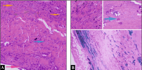
Figure 11 (Case presentation 2, histopathological findings, Courtesy of Dr. Peca Mihaly, Centrum for Clinical Pathology Eisenach, Germany). (A) Microscopic image showing the fibroblasts/fibrocytes (blue arrow) and the histiocytes/macrophages (yellow arrows) inside the connective tissue of ganglion cyst, and macrophages on the surface of the ganglion cyst membrane (pink arrow) in hematoxylin-eosin staining. (B) The two microscopic images above showing the black metallic wear particles and a calcifying deposit (blue arrow) inside the connective tissue of ganglion cyst in hematoxylin-eosin staining. The one microscopic image below showing the blue colored histiocytes/macrophages with incorporated metallic wear particles in ferritin staining
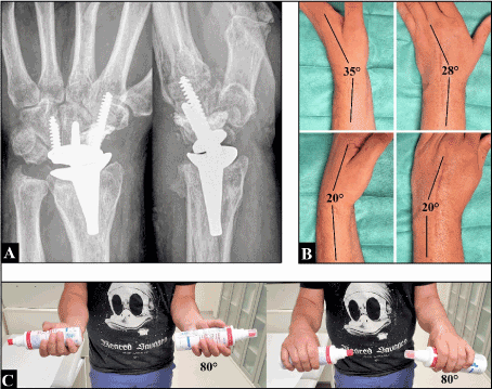
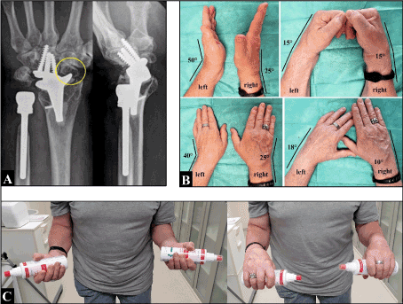
At the last 11-year follow-up regarding left TWA we observed a good outcome. Radiographically, there were unchanged well aligned implants, PPO around the collar of UHR did not show progression, PPO around the ulnar-side offset of radial ReMotion component did not show any clinical signs of loosening, and there were no adverse effects regarding adjacent placements of the capitate peg and both fixation screws into the capitate. Noted evident erosions at the distal scaphoid adjacent to the offset of radial TWA component (Figure 12A, in comparison to Figure 9C). Neurological symptoms had resolved. Pain in VAS and PRWE were 2 and 21. Functionally (compared to the right wrist and forearm in the presence of a surgically non-treated SLAC III currently), left active extension (50°) was with 25° better than right (25°), left active flexion (15°) equal to right, left active ulnar deviation (40°) with 15° better than right (25°), left active radial deviation (18°, clinically in the absence of painful radial impingement) with 8° better than right (10°), and left active supination/pronation with 90°/90° equal to right (Figures 12B-C). Wrist circumduction showed an excellent restoration (video 2 – supplementary material).
Figure 12 (Case presentation 2). (A) Radiographs in both planes showing do not show progressive pathological changes as compared to one year previously (Figure 10A). Note the evident bony erosions at the distal scaphoid adjacent to the radial ReMotion component (arrow), however, the patient did not report painful impingement. (B) Clinical photographs demonstrating the good functional outcome for the patient as described in text. (C) Supination-pronation motion arc was equal to the contralateral right forearm
(2) Video to the patient in case presentation 2.
Discussion
The ReMotion total wrist was introduced in 2002, and first short- to medium term results were encouraging [25,32-35]. Depending on the definition for failed TWA (required implant removal vs. loosening of carpal component without required implant removal) and the varying number of patients were evaluated in uni- or multicentric studies, mid- to long-term survivorship for the ReMotion is reported to be 69 to 100% [29,36-40]. However, the main problem of all third generation TWAs is unchanged loosening of its carpal components which is probably primarily based on progressive mechanical imbalance and secondarily followed by pronounced polyethylene and/or metal wear [5,22,41-43]. However, histopathological evaluation of in vivo taken samples from patients received a ReMotion TWA revealed that these wear particles were commonly found even in patients without any signs of radiographical or clinical loosening [44].
Metallosis as an evident cause for loosening was mainly observed with older TWA generation types utilizing a metal-on-metal articulation [45]. This specific complication could be reduced significantly since introducing the 3rd TWAs with its metal-on-polyethylene articulations, however, it is unchanged observed in single cases due to pronounced polyethylene wear that can result secondarily in a metal-on-metal impingement between both metallic TWA components [46]. Impingement between a TWA component and the surrounding bones can be a concern as well. It has been described for the Universal 2 that led to bony erosions followed by metallosis and/or metallosis-induced carpal tunnel syndrome, and revision surgery became inevitably necessary [47,48]. But noted, it is also be presumed that metallosis and/or development of metallosis-induced pseudotumor following a 3rd generation TWA can caused by fretting at the metal interface between the non-locking screws and its holes of carpal component [22,42,49-51]. Interestingly with our case, despite a massive metallosis was found, both implants were not loosened radiographically as well as clinically (no pain), and that was confirmed intraoperatively. However, it must be emphasized by us that removal of the ReMotion would become necessary if such a severe metallosis should recur in future.
Painful impingement utilizing the ReMotion between the distal scaphoid and the offset of radial component is a design-related issue when placement was done in the standard technique, and it can only be avoided by removal of the entire scaphoid (i.e. “off label” use) [20,23,28,29]. However, when the entire scaphoid is removed then the radial-side screw for fixation the carpal component cannot be completely placed intraosseous which could negatively affect long-term stability. Froschauer et al. [29] did not observe loosening of the radial-side fixation screws when its proximal threads were not bony wrapped at a mean follow-up of four years, however, loosening of all TWAs is mostly observed eight years after its insertion [3]. Insertion of cement-augmented screws (i.e. “implant-to-bone monobloc construct”) in order to improve its pullout strength especially in patients with poor bone stock has proven to be an useful and reliable concept, and it is increasingly in use at the spine, pelvis, around the hip / knee and shoulder, and the distal radius [52-55]. To our knowledge, our presented patient is the first report in the literature which describes cement augmentation of an intercarpal screw with the use of the ReMotion total wrist (i.e. “off label” use). However, it is only one case in an “ultrashort-term” follow-up, and further studies are needed to validate this concept.
With our second patient we presented another “off label” use of the ReMotion total wrist, and we could not find any similar references in the literature which described such a modified surgical technique with a 3rd generation TWA. Placement of both screws into the capitate directly adjacent to the capitate peg does not require additional fusion of the hamate-capitate as well as the scaphoid-capitate joint, and offers an option for complete intraosseous placement of the radial-side fixation screw if the entire scaphoid is removed. Interestingly with our case, the longstanding pressure-related erosions associated with bone loss at the distal scaphoid led to marked improvement of primary radial impingement. Furthermore, we observed a functional outcome which was marked better than function in the affected opposite wrist. A newest TWA study revealed that the center of motion for extension-flexion in TWA is localized in the proximal third of capitate similar to a healthy wrist, whereas the center of motion for radial-ulnar deviation in TWA is localized in contrast to a healthy wrist at the distal tip of capitate [56]. The question is: Does placement of both screws for fixation a carpal TWA component with convergence to distal into the capitate directly aligned to the two centers of motion in the absence of rigid fixation the carpal plate along the entire width of carpus and without crossing a carpometacarpal joint improve function and decrease the risk of loosening? However, although a long-term follow-up was presented, it was only one patient; hence, it is not the intention by us to advocate for general use of this modified technique. Further biomechanical investigations comparing the standard with our described modified technique are needed to validate this concept.
Occurrence of PPO after UHR and TWA, observed with our second patient, is a well-known phenomenon and remains unpredictable. Boeckstyns and Herzberg reported in 2014 about 44 patients received a TWA with the ReMotion total wrist and evaluated at a mean follow-up of 3.7 years, significant periprosthetic radiolucency (more than two mm in width) were found juxta-articularly around the prosthetic components in 36,4% at the radial component and in 15,9% at the carpal component, whereas clinical manifestation of loosening (defined as progressive angulation or subsidence) was present in 14% of all cases only; and radiolucency seemed to be stabilize within three years after surgery [57]. These observations were confirmed with the use of the Motec type in which PPO juxta-articularly around the radial component seemed to be stabilize within eight years after surgery without any clinical signs of loosening [58]. This discrepancy between the amounts of occurrence of PPO and its real clinical manifestation of loosening is also observed in patients receiving an UHR in which PPO around the collar of implant are observed in nearly all cases and followed by its stabilization within three years after surgery, and that phenomenon is discussed as a result of "stress-shielding" [59]. Hence, PPOs in the absence of any clinical signs of loosening do not require surgical intervention. That phenomenon is probably not new. Julius Wolff, a german orthopaedic surgeon (†1902), first described in 1892 that cortical bones primarily became atrophic in lesser loaded regions whereas it became hyperthrophic in higher loaded regions, and resulted secondarily in a steady-state over time ("Wolff's law") [60].
Periprosthetic fracture, observed with our second patient, can be a concern. Occurrence has been reported in approximately 2% of all TWAs independently of its intra- or postoperative occurrence observed at an average of 7.9 years after TWA, and for intraoperative occurrence only age was found to be a risk factor [61]. Non-displaced fractures can be treated by splinting, however, in order to achieve early stability for movement of the wrist, cementation and/or internal fixation are the methods of choice [62,63]. Noted that sufficiently treated intraoperative periprosthetic fractures are not associated with fracture development and do not appear to affect long-term implant survival [61].
Funding
None.
Conflicts of interest
The authors declare no conflicts of interest, financial or otherwise.
Adams BD (2013) Wrist arthroplasty: partial and total. Hand Clin 29: 79-89.
Boeckstyns ME (2014) Wrist arthroplasty--a systematic review. Dan Med J 61: A4834.
Reigstad O, Røkkum M (2014) Wrist arthroplasty: where do we stand today? A review of historic and contemporary designs. Hand Surg 19: 311-322. [Crossref]
Halim A, Weiss AC (2017) Total Wrist Arthroplasty. J Hand Surg Am 42: 198-209.
Boeckstyns MEH, Herzberg G (2017) Current European Practice in Wrist Arthroplasty. Hand Clin 33: 521-528. [Crossref]
Srnec JJ, Wagner ER, Rizzo M (2018) Total Wrist Arthroplasty. JBJS Reviews 6: e9.
Adams BD, Kleinhenz BP, Guan JJ (2016) Wrist Arthrodesis for Failed Total Wrist Arthroplasty. J Hand Surg Am 41: 673-679. [Crossref]
Reigstad O, Holm-Glad T, Thorkildsen R, Grimsgaard C, Røkkum M (2016) Successful conversion of wrist prosthesis to arthrodesis in 11 patients. J Hand Surg Eur Vol 42: 84-89. [Crossref]
Sargazi N, Philpott M, Malik A, Waseem M (2017) Ulna Autograft for Wrist Arthrodesis: A Novel Approach in Failed Wrist Arthoplasty. Open Orthop J 11: 768-776. [Crossref]
11.Pinder EM, Chee KG, Hayton M, Murali SR, Talwalkar SC, Trail IA (2018) Survivorship of Revision Wrist Replacement. J Wrist Surg 7: 18-23.
Berber O, Garagnani L, Gidwani S (2018) Systematic review of total wrist arthroplasty and arthrodesis in wrist arthritis. J Wrist Surg 7: 424-440. [Crossref]
Melamed E, Marascalchi B, Hinds RM, Rizzo M, Capo JT (2016) Trends in the utilization of total wrist arthroplasty versus wrist fusion for treatment of advanced wrist arthritis. J Wrist Surg 5: 211-216.
Hazewinkel MHI, Lans J, Lunn KN, Garg, R, Eberlin KR, Chen NC (2020) Complications and Factors Associated with Reoperation following Total Wrist Fusion. J Wrist Surg.
Field J, Herbert TJ, Prosser R (1996) Total wrist fusion. A functional assessment. J Hand Surg Br 21: 429-431.
Reigstad O, Holm-Glad T, Korslund J, Grimsgaard C, Thorkildsen R, Røkkum M (2019) High re-operation and complication rates 11 years after arthrodesis of the wrist for non-inflammatory arthritis. Bone Joint J 101-B: 852-859. [Crossref]
Owen DH, Agius PA, Nair A, Perriman DM, Smith PN, Roberts CJ (2016) Factors predicting outcome following total wrist arthrodesis. Bone Joint J 98-B: 647-653.
Schmidt I (2018) A critical appraisal to the decision by the company Zimmer Biomet to withdraw the MaestroTM Wrist Reconstructive System from the marketplace. Trauma Emerg Care 3.
Schmidt I (2018) All surgeons who are planning a Total Wrist Arthroplasty with the MaestroTM implants should be aware. Recent Adv Arthroplast 2: 69-74.
Schmidt I (2019) Functional Outcomes After Salvage Procedures for Wrist Trauma and Arthritis (Four-Corner Fusion, Proximal Row Carpectomy, Total Wrist Arthroplasty, Total Wrist Fusion, Wrist Denervation): A Review of Literature. Open Orthop J 13: 217-231.
Fischer P, Sagerfors M, Jakobsson H, Pettersson K (2020) Total Wrist Arthroplasty: A 10-Year Follow-Up. J Hand Surg Am 45: 780.e1-780.e10.
Kandemir G, Smith S, Schmidt I, Joyce TJ (2020) Explant analysis of a Maestro™ wrist prosthesis and calculation of its lubrication regime. J Mech Behav Biomed Mat 110: 103933. doi.org/10.1016/j.jmbbm.2020.103933.
Schmidt I (2014) Primary combined replacements for treatment of distal radius physeal arrest. J Wrist Surg 3: 203-205. [Crossref]
Gvozdenovic R, Boeckstyns M, Merser S (2020) Ulnar Head or Total Distal Radioulnar Joint Replacement, Isolated and Combined with Total Wrist Arthroplasty: Midterm Results. J Wrist Surg 9: 411-416.
Gupta A (2008) Total wrist arthroplasty. Am J Orthop (Belle Mead NJ) 37: 12-16.
Adams BD (2004) Total wrist arthroplasty. Orthopedics 27: 278-284.
Dellacqua D (2009) Total wrist arthroplasty. Tech Orthop 24: 49-57.
Schmidt I (2017) RE-MOTIONTM total wrist arthroplasty for treatment of advanced stage of scaphoid non-union advanced collapse. Does excision of the entire scaphoid bone prevent impingement at terminal range of radial deviation? Trauma Emerg Care 2.
Froschauer SM, Zaussinger M, Hager D, Behawy M, Kwasny O, Duscher D (2019) Re-motion total wrist arthroplasty: 39 non-rheumatoid cases with a mean follow-up of 7 years. J Hand Surg Eur Vol 44: 946-950.
Nydick JA, Greenberg SM, Stone JD, Williams B, Polikandriotis JA, Hess AV (2012) Clinical outcomes of total wrist arthroplasty. J Hand Surg Am 37: 1580-1584.
Sagerfors M, Gupta A, Brus O, Rizzo M, Pettersson K (2015) Patient related functional outcome after total wrist arthroplasty: A single center study of 206 cases. Hand Surg 20: 81-87. [Crossref]
Herzberg G (2011) Prospective study of a new total wrist arthroplasty: short term results. Chir Main 30: 20-25.
Herzberg G, Boeckstyns M, Ibsen Sorensen A, Axelsson P, Kroener K, Liverneaux P, Obert L, Merser S (2012) "Remotion" total wrist arthroplasty: preliminary results of a prospective international multicenter study of 215 cases. J Wrist Surg 1: 17-22.
Ogunro S, Ahmed I, Tan V (2013) Current indications and outcomes of total wrist arthroplasty. Orthop Clin North Am 44: 371-379.
Bidwai AS, Cashin F, Richards A, Brown DJ (2013) Short to medium results using the remotion total wrist replacement for rheumatoid arthritis. Hand Surg 18: 175-178.
Cooney W, Manuel J, Froelich J, Rizzo M (2012) Total wrist replacement: a retrospective comparative study. J Wrist Surg 1: 165-172. [Crossref]
Boeckstyns ME, Herzberg G, Merser S (2013) Favorable results after total wrist arthroplasty: 65 wrists in 60 patients followed 5-9 years. Acta Orthop 84: 415-419. [Crossref]
Chevrollier J, Strugarek-Lecoanet C, Dap F, Dautel G (2016) Results of a unicentric series of 15 wrist prosthesis implantations at a 5.2 year follow-up. Acta Orthop Belg 82: 31-42. [Crossref]
Honecker S, Igeta Y, Al Hefzi A, Pizza C, Facca S, Liverneaux PA (2019) Survival Rate on a 10-Year Follow-Up of Total Wrist Replacement Implants: A 23-Patient Case Series. J Wrist Surg 8: 24-29.
Fischer P, Sagerfors M, Jakobsson H, Pettersson K (2020) Total Wrist Arthroplasty: A 10-Year Follow-Up. J Hand Surg Am 45: 780.e1-780.e10.
U.S. Food & Drug Administration, 2009. MAUDE Adverse Event Report: BIOMET ORTHOPEDICS, MAESTRO TOTAL WRIST DISTAL RADIAL BODY 7.5 MM LEFT PROSTHESIS, WRIST.
U.S. Food & Drug Administration, 2012. MAUDE Adverse Event Report: SMALL BONE INNOVATIONS, INC. REMOTION TOTAL WRIST WRIST PROSTHESIS.
Boeckstyns ME, Toxvaerd A, Bansal M, Vadstrup LS (2014) Wear particles and osteolysis in patients with total wrist arthroplasty. J Hand Surg Am 39: 2396-2404.
Radmer S, Andresen R, Sparmann M (2003) Total wrist arthroplasty in patients with rheumatoid arthritis. J Hand Surg Am 28: 789-794.
Marinello PG, Peers S, Bafus BT, Evans PJ (2015) Modified brachioradialis wrap for stabilizing the distal radioulnar joint: case report. Hand (N Y) 10: 802-806.
Johnson ST, Patel A, Calfee RP, Weiss APC (2007) Pisiform impingement after total wrist arthroplasty. J Hand Surg Am 32: 334-336.
Nunez Jr FA, Wright L, Kilpatrick SE, Seitz Jr WH (2020) Revision Total Wrist Arthroplasty Due to Polyethylene Wear, Metallosis-Induced Carpal Tunnel Syndrome, Distal Ulnar Impingement, and Fourth Carpometacarpal Joint Pain: Case Report and Pitfalls to Avoid. Hand (N Y) 15: NP1-NP6. [Crossref]
Day CS, Lee AH, Ahmed I (2013) Acute carpal tunnel syndrome secondary to metallosis after total wrist arthroplasty. J Hand Surg Eur Vol 38: 80-81. [Crossref]
Taha R, Roushdi I, Williams C (2015) Pseudotumor secondary to metallosis following total wrist arthroplasty. J Hand Surg Eur Vol 40: 995-996.
Heyes GJ, Julian HS, Mawhinney I (2018) Metallosis and carpal tunnel syndrome following total wrist arthroplasty. J Hand Surg Eur Vol 43: 448-450. [Crossref]
Shea TM, Laun J, Gonzalez-Blohm SA, Doulgeris JJ, Lee WE 3rd, Aghayev K, Vrionis FD (2014) Designs and techniques that improve the pullout strength of pedicle screws in osteoporotic vertebrae: current status. Biomed Res Int 2014: 748393. [Crossref]
Höch A, Pieroh P, Henkelmann R, Josten C, Böhme J (2017) In-screw polymethylmethacrylate-augmented sacroiliac screw for the treatment of fragility fractures of the pelvis: a prospective, observational study with 1-year follow-up. BMC Surg 17: 132. [Crossref]
Rai AK, Goel R, Bhatia C, Singh S, Thalanki S, Gondane A (2018) Cement Augmentation of Dynamic Hip Screw to Prevent Screw Cut Out in Osteoporotic Patients with Intertrochanteric Fractures: A Case Series. Hip Pelvis 30: 269-275.
Kammerlander C, Neuerburg C, Verlaan JJ, Schmoelz W, Miclau T, Larsson S (2016) The use of augmentation techniques in osteoporotic fracture fixation. Injury 47 (Suppl 2): 236-243. [Crossref]
Akhbari B, Morton AM, Shah KN, Molino J, Moore DC, et al. (2020) Proximal-distal shift of the center of rotation in a total wrist arthroplasty is more than twice of the healthy wrist. J Orthop Res 38: 1575-1586.
Boeckstyns ME, Herzberg G (2014) Periprosthetic osteolysis after total wrist arthroplasty. J Wrist Surg 3: 101-106. [Crossref]
Reigstad O, Holm-Glad T, Bolstad B, Grimsgaard C, Thorkildsen R, Røkkum M (2017) Five- to 10-Year Prospective Follow-Up of Wrist Arthroplasty in 56 Nonrheumatoid Patients. J Hand Surg Am 42: 788-796. [Crossref]
Herzberg G (2010) Periprosthetic bone resorption and sigmoid notch erosion around ulnar head implants: a concern? Hand Clin 573-577.
Wolff J (1862) Das Gesetz von der Transformation der Knochen (August Hirschwald, Berlin).
Wagner ER, Srnec JJ, Mehrotra K, Rizzo M (2017) What Are the Risk Factors and Complications Associated With Intraoperative and Postoperative Fractures in Total Wrist Arthroplasty? Clin Orthop Relat Res 475: 2694-2700. [Crossref]
Dawson WJ (1989) Radius fracture after total wrist arthroplasty. J Hand Surg Am 14: 630-634.
Barrera-Ochoa S, Muñetón D, Mir X (2014) Periprosthetic proximal fracture in total wrist arthroplasty. J Hand Surg Am 39: 399-400. [Crossref]
Editorial Information
Editor-in-Chief
Guo-Gang Xing
Perking University
Article Type
Case Series Article
Publication history
Received: October 21, 2020
Accepted: November 16, 2020
Published: November 19, 2020
Schmidt I, Beylich T (2020) “Off label” use of the ReMotionTM total wrist for treatment of advanced stage of post-traumatic pancarpal wrist osteoarthritis: Report of two cases and review of literature. Trauma Emerg Care 5: DOI: 10.15761/TEC.1000203
Corresponding author
Dr. med. Ingo Schmidt
Medical Centre Wutha-Farnroda, Ringstr. 20, 99848 Wutha-Farnroda, Germany
Figure 1 (35-year old male, right severe open wrist injury). (A) Initial soft tissue situation. (B) Wound coverage with a distally pedicled radial artery flap. (C) After uneventful flap healing planning and performing the TWF utilizing two corticocancellous iliac crest bone grafts, a 2,7 mm locking wrist spanning plate, and five headless compression screws. Due to the extended carpal bony defects a TWA was technically not possible. (D) Unchanged uneventful bony fusion in the absence of any implant complications at the 11-year follow-up. (E) Good restoration of forearm supination-pronation motion arc compared to the contralateral forearm
Figure 2 (71-year old female, left severe closed distal forearm injury associated with poor osteoporotic bone stock). (A) Radiographs in both planes demonstrating the initial findings, note the highly comminution of distal radius metaphysis. (B) The fracture was primarily closed reducted by external fixation. (C) After stabilization of the closed soft tissue damages an internal fixation of the distal ulna fracture utilizing a 3,5 mm titanium locking reconstruction plate was done 10 days after injury, and four weeks later the ReMotion total wrist was inserted with cementation due to the poor osteoporotic bone stock. (D) At the 2-year follow-up there was an very satisfying functional outcome for the patient
Figure 3. Trials of the ReMotion total wrist (without size extra small)
Figure 4 (Correlation of painful impingement at terminal range of radial deviation with the ReMotion [23,28]). (A) Radial-side impingement when only the proximal pole of scaphoid was removed (arrow, standard technique). (B) Radial-side impingement despite additional oblique resection at the distal pole of scaphoid (arrow). (C) No radial-side impingement between the trapez and offset of radial component when the entire scaphoid was removed. Note that only the distal threads of radial-side fixation screw are bony wrapped by the trapezoid and second metacarpal
Figure 5 (Case presentation 1). (A) Radiographs in both planes showing the fracture-dislocation injury. (B) Three-dimensional computed tomography demonstrating the highly comminution of distal radial metaphysis. (C) Radiographs in both planes after initial treatment. Note the concomitant SLLD (arrow). (D) Radiographs in both planes after removal of the external fixateur (arrow: SLLD)
Figure 6 (Case presentation 1). (A) Radiographs in both planes showing advanced stage of post-traumatic wrist OA (arrow: SLLD). (B) Clinical photographs demonstrating loss of wrist function of approximately 50% compared to the uninjured right wrist. (C) Clinical photographs demonstrating the marked loss of forearm supination/pronation compared to the uninjured right wrist
Figure 7 (Case presentation 1). (A) Intraoperative fluoroscopy demonstrating correct alignment of the trials size S in both planes. (B) Intraoperative clinical photograph after insertion of both definitive components showing the not bony wrapped proximal threads of radial-side fixation screw, and for fusion of the hamate-capitate joint the corresponding cartilaginous surfaces were excised (arrow). (C) Intraoperative clinical photograph showing the cement augmented “monobloc construct” between the proximal threads of radial-side fixation screw and the surrounding carpal bones, and the fusioned hamate-capitate joint (arrow). (D) Intraoperative fluoroscopy demonstrating the stable ReMotion TWA at passive terminal ranges of motion (black arrow: cement augmentation, green arrow: no radial-side impingement)
Figure 8 (Case presentation 1, six-month follow-up). (A) Radiographs in both planes showing unchanged correctly aligned placement of the ReMotion. (B) Clinical photographs with active terminal ranges of motion at the left wrist. (C) Clinical photographs with active terminal ranges of supination and pronation of both forearms
Figure 9 (Case presentation 2). (A) Intraoperative clinical photograph showing the UHR inserted exactly on the top of distal ulnar stump (arrow). (B) Intraoperative clinical photograph showing the longitudinal periprosthetic fracture on the dorsal aspect of distal radius metaphysis (arrows), insertion of the radial ReMotion component with cementation, and one of the two compression screws (circle). (C) Radiographs in both planes one year after TWA demonstrating unchanged correctly aligned implants without any signs of loosening, PPO around the collar of UHR (white arrows), and evident radial impingement in neutral position between the distal scaphoid after removal of its only proximal one half regarding the standard technique and the offset of radial ReMotion component (yellow arrow)
Figure 10 (Case presentation 2). (A) Radiographs in both planes showing both implants without any signs of loosening. PPO around the collar of UHR had been stabilized. A new PPO was present under the offset of radial ReMotion component ulnar-side which led to loosening of one titanium compression screw accompanied by its migration in volar direction (arrow). Noted the bony erosions at the distal scaphoid adjacent to the radial ReMotion component. (B) Clinical photograph demonstrating the subcutaneous giant cyst formation (arrow). (C) Intraoperative clinical photograph showing the extended black colored metallosis-induced giant cyst formation after volar surgical incision. Both volar nerves were decompressed with opening the loge de Guyon. (D) Metallosis originates from the TWA, however, both components were not loosened and the polyethylene ball did not show any signs of destruction macroscopically. Metallosis also originates from the hole of the migrated and broken titanium screw. (E) A radical debridement involving of all flexor tendon sheaths and wrist synovia was done. (F) Primary wound closure was possible
Figure 11 (Case presentation 2, histopathological findings, Courtesy of Dr. Peca Mihaly, Centrum for Clinical Pathology Eisenach, Germany). (A) Microscopic image showing the fibroblasts/fibrocytes (blue arrow) and the histiocytes/macrophages (yellow arrows) inside the connective tissue of ganglion cyst, and macrophages on the surface of the ganglion cyst membrane (pink arrow) in hematoxylin-eosin staining. (B) The two microscopic images above showing the black metallic wear particles and a calcifying deposit (blue arrow) inside the connective tissue of ganglion cyst in hematoxylin-eosin staining. The one microscopic image below showing the blue colored histiocytes/macrophages with incorporated metallic wear particles in ferritin staining
Figure 12 (Case presentation 2). (A) Radiographs in both planes showing do not show progressive pathological changes as compared to one year previously (Figure 10A). Note the evident bony erosions at the distal scaphoid adjacent to the radial ReMotion component (arrow), however, the patient did not report painful impingement. (B) Clinical photographs demonstrating the good functional outcome for the patient as described in text. (C) Supination-pronation motion arc was equal to the contralateral right forearm