Although the relationship between aural symptoms and dental occlusion, as well as the improvement of tinnitus using occlusal position correcting therapy have been reported, the treatment of low-frequency hearing loss and ear fullness using occlusal position correcting therapy have not yet been reported. Occlusal improvement was achieved using the occlusal position correcting therapy on a patient with low frequency hearing loss and ear fullness. Occlusal analysis was performed by mounting dental models on an articulator and identifying a premature occlusal contact on the right second molars and a deviation of the habitual occlusal position from the muscular reference position. Occlusal modification was performed to make the habitual occlusal position coincide with the muscular reference position. The low- frequency hearing loss and fullness on the right ear disappeared and have not recurred at 2-year follow-up. The low frequency hearing loss and ear fullness were thought to be caused due to the tensor tympani and stapedius tension synchronously produced with the masticatory muscle contraction.
low frequency hearing loss, ear fullness, habitual occlusal position, muscular position, occlusal position correcting therapy
The relationship between temporomandibular disorders and aural symptoms (tinnitus, low-frequency hearing loss and ear fullness) have been reported [1-8]. However, to be the best of our knowledge the relationship between these aural symptoms (low-frequency hearing loss and ear fullness) and dental occlusion has not yet been reported. Various symptoms have reportedly been caused by the occlusal discrepancy between the habitual occlusal position (HOP) and the muscular position (MP) [9]. Some masticatory muscle tension might be caused due to the occlusal discrepancy, which may affect tensor tympani and stapedius [10,11]. The present case study might be useful to understand one of the mechanisms of low-frequency hearing loss and ear fullness.
A 66-year-old woman presented with a chief complaint of pain of the lower right third molar. She had right tinnitus, difficulty in hearing low-frequency sound, and right ear fullness. She initially visited an ENT clinic. After various tests, she was diagnosed with sudden deafness and low-frequency hearing loss. She was treated with oral steroids, such as isosorbide, diphenidol and domperidone without any improvement of the symptoms [12]. Her medical history was unremarkable, except for the above-mentioned aural symptoms. She reported sounds and pain of the temporomandibular joints (TMJs) during jaw movements, a right stiff shoulder, right tinnitus, low-frequency hearing loss and right ear fullness. Mouth opening was 40 mm and the opening pass deviated laterally to the right. There was no TMJ tenderness; however, tenderness of the right temporal and medial pterygoid muscle on palpation was observed. Dental occlusion was anatomically normal (Figure 1). The TMJs bilaterally appeared normal on the computed tomography images taken in the HOP at the initial consultation (Figure 2).
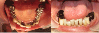
Figure 1. Upper and lower dental arches after the extraction of the right lower third molar.
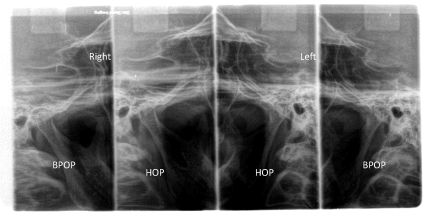
Figure 2. Bilateral tomographic images of TMJ in the HOP and the BPOP.
The lower right third molar was extracted at the initial visit. At the second visit (5 days later), an HOP record was obtained using a vinyl polysiloxane bite registration material (Exabiite, GC, Tokyo, Japan), while the patient was seated upright with her jaw voluntarily closed. Subsequently, the upper and lower jaw impressions were obtained, and dental models were fabricated. An anterior flat bite plate was fabricated on the upper cast using a self-curing acrylic resin (Ortho-fast, GC, Tokyo, Japan). At the third visit (3 days after the second visit), she wore the bite plate for 5 min and the bite plate-induced occlusal position (BPOP) record was obtained, using the same material as used for obtaining the HOP record (Figure 3). A BPOP wax record was obtained using the registration wax material (Bite Wafer, Kerr USA, Romulus, MI, U.S.A.) in the previously described manner. The upper and lower models were mounted on an articulator with the BPOP wax record. A premature occlusal contact was recognized on the right second molars (Figure 4). To examine the difference between HOP and BPOP, two-dimensional measurements were performed with the modified articulator using previous records (Figure 5). Her mandible deviated 2 mm posterior from the BPOP on both sides (this is the physiological MP) (Figure 6). Occlusal adjustments were performed on the dental models mounted on the articulator until the posterior teeth bilaterally made contacts according to the occlusal position correcting therapy (Figure 7) [13]. Subsequently, the occlusal adjustments in the mouth were performed following the aforementioned procedure. The adjustments were performed at the third and fourth visits (the fourth visit was 7 days after the third visit). At the fifth visit (4 days after the fourth visit), she reported no sounds in the TMJs, right tinnitus, right stiff shoulder, low-frequency hearing loss, and right ear fullness (Figure 4). She had complained of cheek biting at the fourth visit; however, it disappeared at the fifth visit. The low-frequency hearing loss and fullness in the right ear have not recurred at 2-year follow-up.
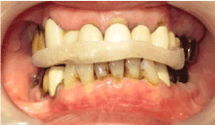
Figure 3. Wearing the bite plate.
Figure 4. Examination record
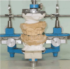
Figure 5. Mandibular position analyzer
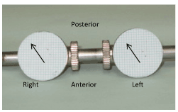
Figure 6. The HOP and BPOP records. The arrows indicate the shift from the BPOP to the HOP
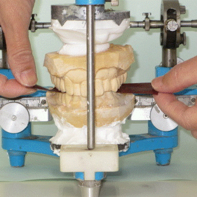
Figure 7. Confirmation of occlusal contacts on both sides in the muscular contact position
Pharmacological therapies have been reported for low-frequency hearing loss. In a previous study, out of all the patients treated with oral and intratympanic steroid, 39% of patients showed complete recovery and 77% of patients showed audiometric improvement [12]. Conversely, another study reported on 9 cases of unilateral tinnitus to have been completely resolved with occlusal adjustments without recurrence [11]. This means that unilateral deviation of the mandible might cause the tension of the masticatory muscles and the tension in turn might cause tinnitus with the spasmodic synkinesis of the tensor tympani and the stapedius [11]. In the present case, the premature occlusal contact on the right second molars retracted the mandible backward with the contraction of the right temporal muscle, making the teeth meet together and causing the tensor tympani and the stapedius tension to be synchronously produced with the temporal muscle contraction (Figure 8) [10]. The tensor tympani and stapedius tension restricted the movement of auditory ossicles (malleus, incus, and stapes), causing low-frequency hearing loss. Moreover, the tensor tympani tension caused the contraction of the semicanalis musculi tensoris tympani that resulted in ear fullness.
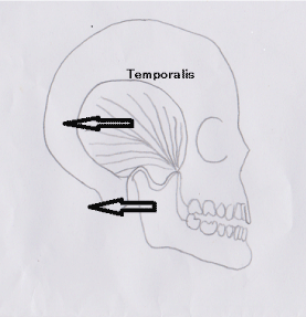
Figure 8. The temporalis strongly pulls the mandible backward for the teeth to meet together
Written consent was obtained from the patient for publication of the study.
The author declares no competing interests.
- Torii K (2014) Various symptoms of temporomandibular disorders. In: Torii K. Evidence-based occlusal management for temporomandibular disorders. Sharjah: Bentham Science Publishers 6: 172-185.
- Nicolakis P, Nicolakis M, Piehslinger E, Ebenbichler G, Vachuda M, et al. (2000) Relationship between craniomandibular disorders and poor posture. Cranio 18: 106-112. [Crossref]
- Stack B, Sims A (2009) The relationship between posture and equilibrium and the auriculotemporal nerve in patients with disturbed gait and balance. Cranio 27: 248-260. [Crossref]
- Torii K, Chiwata I (2010) Occlusal adjustment using the bite plate-induced occlusal position as a reference position for temporomandibular disorders: A pilot study. Head & Face Medicine 6: 1-5
- Torii K, Chiwata I (2010) A case report of the symptom-relieving action of an anterior flat plane bite plate for temporomandibular disorder. The Open Dentistry Journal 4: 218-222.
- Torii K (2015) Causal relationship between occlusal factors and temporomandibular disorders. J Dent Res 2: 1-25.
- Myrhaug H (1964) The incidence of ear symptoms in cases of malocclusion and temporo-mandibular joint disturbances. Br J Oral Surg 2: 28-32. [Crossref]
- Maciel LFO, Landim FS, Vasconcelos BC (2018) Otological findings and other symptoms related to temporomandibular disorders in young people. Br J Oral Maxillofac Surg 56: 739-743.
- Torii K (2018) Occlusal disease. J Den Health Res 1: 1-10.
- Watanabe I, Kumagami H, Tsuda Y (1974) Tinitus due to abnormal contraction of stapedial muscle. J Otorhinolaryngol 36: 217-226.
- Torii K (2011) Tinnitus and temporomandibular disorders. In: Up to Datebon Tinnitus, Fayez B j red. Rijeka, Intech 1: 15-32.
- Torii K (2018) Occlusal position correcting therapy for temporomandibular disorders. EC Dental Science 17: 168-176.
- Jung AR, Kim MG, Kim SS, Kim SH, Yeo SG (2016) Clinical characteristics and prognosis of low frequency sensorineural hearing loss without vertigo. Acta Otolaryngol 136: 159-163. [Crossref]