Critical limb ischemia (CLI) is a severe blockage in the arteries of the lower extremities, which significantly reduces blood-flow. Often multisegmental atherosclerosis is detected in patients who have the critical limb ischemia (CLI). Usually there are two or three levels of occluded arterial lesions of the lower extremities. Typically, these are the patients who underwent multiply revascularization procedures aiming to salvage the limb. The main limiting factor of this procedure is absent or poor outflow in the arteries. If revascularization is unsuccessful or not possible to perform, it can lead to progression of critical limb ischemia, which most likely will result in amputation. We present two cases of successful treatment of patients with CLI, who had history of thrombosis of aortobifemoral bypass (ABF) branches in the absence of distal arterial outflow. We successfully performed hybrid revascularization procedure in these patients after multiple attempts of open interventions.
critical limb ischemia, hybrid procedures, outflow disease
Critical limb ischemia (CLI) is a severe blockage in the arteries of the lower extremities, which markedly reduces blood-flow. It is a serious form of peripheral arterial disease, or PAD, but less common than claudication. PAD is caused by atherosclerosis, the hardening and narrowing of the arteries over time due to the buildup of fatty deposits called plaque [1,2].
CLI is a chronic condition that results in severe pain in the feet or toes, even while resting. Complications of poor circulation can include sores and wounds that won't heal in the legs and feet. Left untreated, the complications of CLI will result in amputation of the affected limb [1-4].
Prevalence of this disease is increasing as life expectancy and incidence of diabetes continue to increase worldwide [1,2]. Lower extremity PAD and CLI are associated with significantly worse clinical outcomes, including higher rate of mortality, major adverse limb events and significant peri-operative morbidity and mortality as well [3,4].
Often, multisegmental atherosclerosis is detected in patients who have the CLI. Usually there are two or three levels of occluded arterial lesions of the lower extremities [5]. Typically, these are the patients who underwent multiple revascularization procedures aiming to salvage the limb. The main limiting factor of this procedure is absent or poor outflow in the arteries. If revascularization is unsuccessful or not possible to perform, it can lead to progression of critical limb ischemia, which most likely will result in amputation. We present two cases of successful treatment of patients with CLI, which have had history of thrombosis of branches of aortobifemoral bypass (ABF) with the absence of distal arterial outflow. We successfully performed hybrid revascularization procedure in these patients after multiply attempts of open interventions.
Sixty three years old patient was brought to the vascular surgery department with medical complaints of rest pain, which were poorly treated with opioid pain relieve medication and discoloration of the skin on the left foot.
This patient has history of short-distance claudication, which has been present during past 15 years. The patient has noticed the pain at rest in both lower limbs. The following symptoms were noted: stenosis of the right ileal-femoral segment arteries, 75% stenosis of the left common ileum artery and 70% stenosis of left external iliac arteries, occlusion of the left common, profunda and superficial femoral arteries as detected by computed tomography angiography (CTA). Aortobifemoral bypass was performed with a GORE-TEX 16-8-8 mm graft. At that time, we recorded a poor state of the left deep femoral artery, which raised doubts about the possibility of an arterial reconstruction of the left lower extremity. Nevertheless, the left distal anastomosis was performed with profunda femoral artery at the level of fourth and fifth order branch, which allowed to stop the critical ischemia of the left lower extremity. Notably, patient reported that there was no limit of walking distance after surgical procedure.
Four years later after initial surgery, patient had reported acute pain in the left lower extremity, which was attributed to thrombosis of the left branch of the aortobifemoral bypass. The thrombectomy was performed from the left branch of the aortobifemoral bypass at the beginning of January in 2014. The procedure included the reconstruction of the left distal anastomosis using the 8-mm GORE-TEX extension to the 4th order branch of the profunda artery and the autologous venous femoral (from the left branch of the aortobifemoral bypass) above-the-knee popliteal bypass grafting. The re-thrombosis of the left branch of the aortobifemoral bypass and femoral (above-the-knee) - popliteal bypass with the restoration of rest pain in the limb was noted the day after surgery. Re-thrombectomy was done from the grafts on the following day. In addition, reconstruction of the left distal anastomosis of the aortobifemoral bypass by direct anastomosing of the aortobifemoral bypass branch to the autologous venous femoral(above-the-knee)- popliteal bypass was conducted with offloading of the aortobifemoral bypass branch to the profunda femoral artery through the 8 mm GORE-TEX graft. It was noted that the cause of thrombosis, most likely, was precisely the poor condition of the left profunda femoral artery. The “poor” condition of the left profunda femoral artery was the reason for the formation of the main outflow route to the autologous venous femoral (above-the-knee) - popliteal bypass and creation of offloading of this “long”, actually “aorto-popliteal bypass”, by the graft extension to the “poor” profunda femoral artery. After surgery, the rest pain in the left lower extremity ceased.
In June of 2018, the patient again noted the appearance of rest pain in the left lower extremity. During further examination, there were two new sites of thrombosis detected: at the aortobifemoral bypass left branch and venous femoral (above-the-knee) popliteal bypass (Figure 1A-1C). There were no left profunda femoral artery and its branches detected on CTA (Figure 1A-1C).
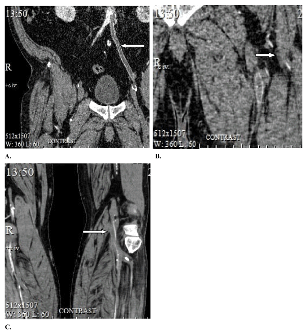
Figure 1. Pre-operative imaging finding obtained by computed tomography angiography (CTA). A. Thrombosis of the aortobifemoral bypass of the left branch as detected by CTA before the last surgery (indicated by an arrow). B. The left common, profunda and superficial femoral arteries were not detectable by CTA before the last surgery. Branches of the lateral circumflex artery were detected by CTA (indicated by an arrow). C. Significant stenosis was detected in the left popliteal artery (indicated by an arrow) and inflow in the left anterior tibial artery and distal parts of the left peroneal artery were noted on CTA examination before the last surgery (indicated by an arrow)
On June 15th of 2018, the re-thrombectomy and the bypass were performed from the branch of the aortobifemoral graft to the peroneal artery. However, there was no possibility to “offload” the branch, therefore, the thrombosis of these grafts was diagnosed and confirmed by CTA imaging after the procedure was completed. The surgical procedure was considered ineffective at that time. Post operatively this patient took a course of drug treatment with Vasaprostan (Alprostadil) 60 mkg for 10 days. The pain in the left lower extremity decreased, and the patient was discharged with the recommendation of limb amputation, if his condition would worsen.
After his discharge from the hospital, this patient had noticed that the pain increased again in the left lower extremity. Also the cyanosis of the skin of the left foot has appeared. The patient was admitted again to the local vascular center and digital subtraction angiography (DSA) was performed to assess progression of his disease. The findings of this imaging procedure indicated multiple sites of thrombosis (Figure 2A-2C).
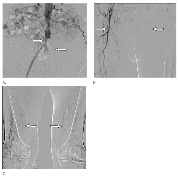
Figure 2. Findings of digital subtraction angiography (DSA) which was performed to assess progression of the disease. A. The right branch of the aortobifemoral bypass was detected on the angiogram of the abdominal aorta and pelvis’s arteries before the last surgical procedure, however the left one was found to be thrombosed by DSA. B. The right anastomosis of the aortobifemoral bypass is shown on the angiogram of the thighs, however arteries of the left thigh, including the left profunda femoral artery were not detectable. C. The arteries of the left lower extremity were not detectable on the angiogram of the thighs and popliteal areas
According to the data obtained by angiography, the only chance to salvage the limb was to perform a hybrid procedure. On July 17th of 2018, two surgical teams (endovascular and vascular surgeons) performed the mechanical retrograde recanalization and balloon angioplasty of the left anterior tibial artery, the left popliteal artery and the left superficial femoral arteries. Also, the thrombectomy and the reconstruction of the distal anastomosis of the left branch of the aortobifemoral bypass graft were performed. The new distal anastomosis of ABF branch was formed to the left superficial femoral artery by the extension of 6 mm PTFE graft (Figure 3A-3E).
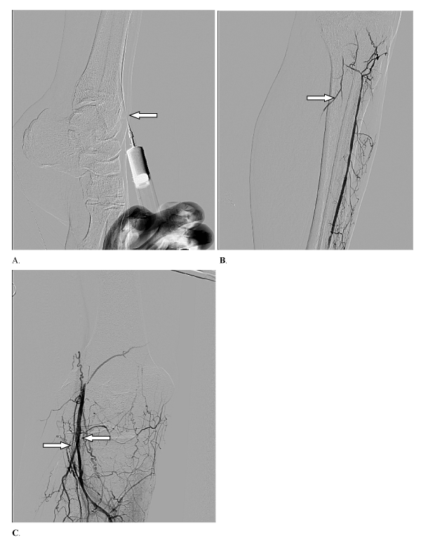
Figure 3. Angiogram of the thigh prior and after the retrograde recanalization and balloon angioplasty. A. Retrograde puncture of the left anterior tibial artery visualized on the angiogram. B. The left anterior tibial artery is contrast enhanced prior to the procedure. The left popliteal artery, the left posterior tibial artery and the left peroneal artery are occluded. C. An angiogram after retrograde recanalization and balloon angioplasty of the left popliteal artery showed restored flow
The anterior tibial artery puncture was performed in the ankle area under ultrasound guidance and the spinal anesthesia. The mechanical retrograde recanalization and balloon angioplasty of the left anterior tibial artery, the popliteal artery and the superficial femoral arteries was performed. In addition, the left superficial femoral artery was recanalized sub-intimally and retrogradely. It was not possible to lead the guidewire into the left external iliac artery. Then the surgical access to the superficial femoral artery was performed in the upper third of the left thigh artery. After arteriotomy the subintimal space with the guidewire was visualized (Figure 3A). Later, after the guidewire was extracted, the antegrade angioplasty of the left anterior tibial artery was performed by utilizing the balloon catheter 2.5-200 mm and the left popliteal artery by the balloon catheter 4-80 mm and the left superficial femoral artery by balloon catheter 7-200 mm. In the area of the inguinal ligament, the thrombosed left branch of the aortobifemoral graft was isolated, crossed and the thrombectomy was performed. The hyaline thrombus was removed until the central pulsating blood flow was obtained. Then the anastomosis of the end-to-end type was formed between the extension of the reinforced Ecoflon 6 mm graft and the left branch of the aortobifemoral graft. After this, the extension (the reinforced Ecoflon 6 mm graft) was anastomosed with the subintimal lumen of the left superficial femoral artery with fixation of detached intima to the walls of the anastomosis. The blood flow was restored along the branch of the prosthesis.
The Ecoflon 6 mm graft extension was punctured, the introducer was installed and control angiography was performed. The stenosis of “between-graft” anastomosis and the extended stenosis of the recanalized left superficial femoral artery were revealed. Thus, balloon angioplasty of the stenosis was performed and visualized by angiography (Figures 3B,3C). The introducer was removed as the defect of the graft was fixed. The wound was drained and closed.
The early post-procedural period was uneventful, the wounds healed well during post-operative period. In addition, post procedural imaging revealed restored blood flow in the left superficial femoral and left popliteal arteries (Figure 4D,4E). After the surgery, the pulsation on the left popliteal artery and the left dorsalis pedis artery was restored. The rest pain in the limb diminished and the foot cyanosis disappeared. In the hospital, the patient received triple antiplatelet and anticoagulation therapy (Acetylsalicylic Acid 100 mg, Clopidogrel 75 mg, low molecular weight heparins in prophylactic doses). After discharge, triple therapy in the form of Acetylsalicylic Acid 100 mg, Clopidogrel 75 mg, Rivaroxaban 5 mg was prescribed.
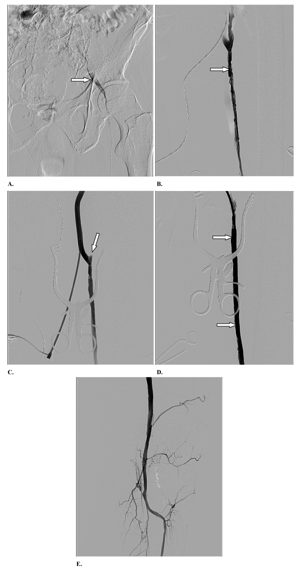
Figure 4. Imaging results representing different steps of performed recanalization and balloon angioplasty. A. The guidewire was retrogradely inserted into the left common femoral artery as shown by angiography. B. An angiogram of the left superficial femoral artery, which had been retrogradely recanalized and distal anastomosis of the extension and left superficial femoral artery was formed. C. Post procedural angiogram after antegrade balloon angioplasty of the left superficial femoral artery was performed (the arrow indicates the distal anastomosis of the extension and the left superficial femoral artery). D. Post procedural angiogram after balloon angioplasty of the left superficial femoral artery was performed and showed restored blood flow in that vessel. E. Post procedural image obtained by angiography after balloon angioplasty of the left popliteal artery showed restored blood flow in the vessel
Two months later, in September 2018, the patient was diagnosed with a para-graft infection in the area of the distal anastomosis, which was managed by the surgical treatment of the left thigh. There was no pain in the left foot reported at that time. According to the ultrasound and CTA diagnostic imaging, the inflow was detected at the zone of arteries reconstruction in the left lower extremity (Figure 5A-5C).
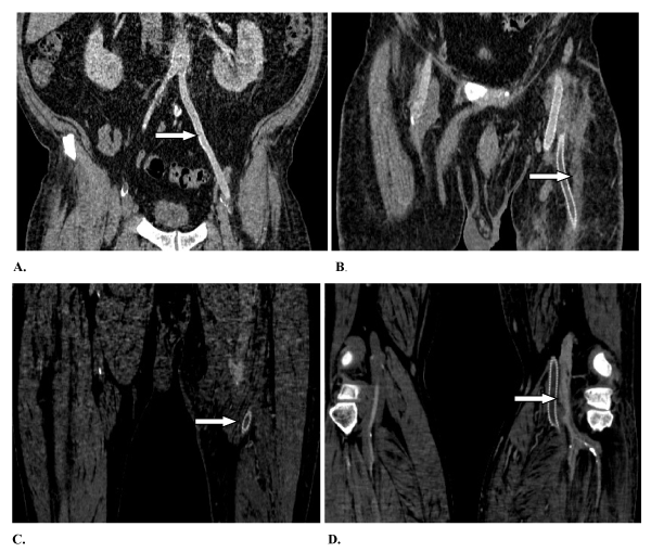
Figure 5. Post procedural computed tomography angiography (CTA) imaging at 2 months follow up. A. The left branch of the aortobifemoral bypass is shown to be intact. B. The reinforced PTFE 6mm graft is shown to be functional. C. The distal anastomosis of reinforced PTFE 6mm graft as shown is viable. D. The left popliteal artery with the elements of dissection was detected on post-surgery CTA after 2 months
In May of 2019 this patient underwent digital conventional angiography for routine follow up. The results are shown on Figure 6, indicating that foot was revascularized and well perfused.
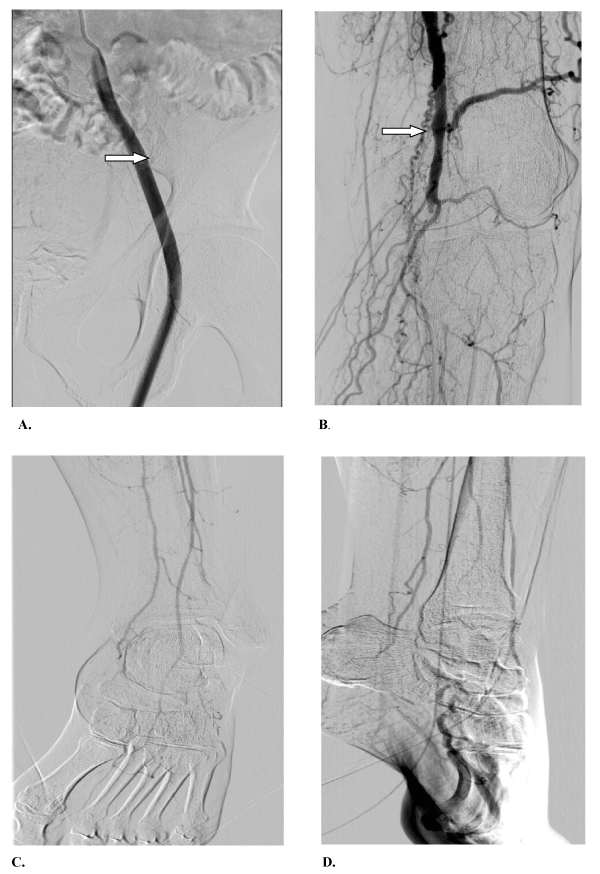
Figure 6. The left branch of the aortobifemoral bypass (A) and left popliteal artery (B) were detected by angiogram. Good vascularization in the left foot was detected by angiogram on anterior (C) and lateral (D) views of the foot
In terms of quality of life outcomes, the patient didn’t report any limitations to his walking distance during follow up visit. At the time of writing the paper (the end of September 2019, 15 months after revascularization), the patient's condition was satisfactory, there was no pain in the left lower extremity. The left foot was viable, allowing us to conclude that the limb was preserved.
Sixty three years old patient was brought to the vascular surgery department with medical complaints of rest pain, which were poorly treated with opioid pain relief medication and ischemic ulcers on the left foot in December of 2018.
This patient has a history of claudication, which has been present during past 3 years. The patient has noticed the pain at rest in the left lower limb and appearance of ischemic ulcers in 1st, 2nd, 3rd and 4th interdigitalis space of the foot in March of 2018. The following symptoms were noted at physical examination: occlusion of the left common ileum artery, left external ileum artery, left common femoral artery, left superficial and deep femoral arteries, left popliteal artery and tibioperoneal trunk on CTA (Figure 7).
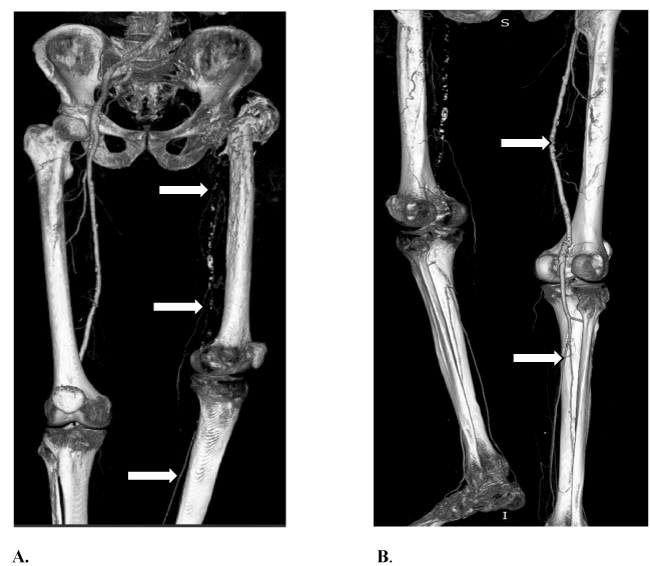
Figure 7. Pre-procedural imaging findings by utilizing computed tomography angiography (CTA) with 3D image reconstruction. A. Occlusion of the left common iliac artery, left external iliac artery, left common femoral artery, left superficial and profunda femoral arteries, left popliteal artery and tibioperoneal trunk were detected (indicated by an arrows). B. Occlusion of the left common femoral artery, left superficial and profunda femoral arteries, left popliteal artery and tibioperoneal trunk were detected. Also, left posterior tibial artery, left peroneal artery and distal part of the left anterior tibial artery were contrasted through collateral arteries (indicated by arrows)
On December 24th of 2018, two surgical teams (endovascular and vascular surgeons) have performed mechanical recanalization and balloon angioplasty of the left anterior tibial artery, left popliteal artery, left superficial femoral artery. The bypass from right common femoral artery to the left superficial femoral artery was also performed.
The antegrade puncture of the distal third of the left superficial femoral artery was performed by 21G needle under X-ray, ultrasound control and using Seldinger technic. 6F introducer was used. The guide wire for balloon catheter 3.0 – 150 mm was provided through occlusion of left anterior tibial artery, left popliteal artery, left superficial femoral artery till left common femoral artery. The balloon angioplasty of the left superficial femoral artery, left popliteal artery was performed by balloon catheter 3.0 – 150 mm and 5.0 – 200 mm on 8-10 bar pressure.
The bypass from right common femoral artery to the left superficial femoral artery was performed by reversed autologous vein. The longitudinal arteriotomy was performed; it was revealed the guide was in the subintimal lumen. Then the guide through a separate hole above the arteriotomy was moved outside the artery. The distal anastomosis was formed between the vein graft and subintimal space of the superficial femoral artery (Figure 8).
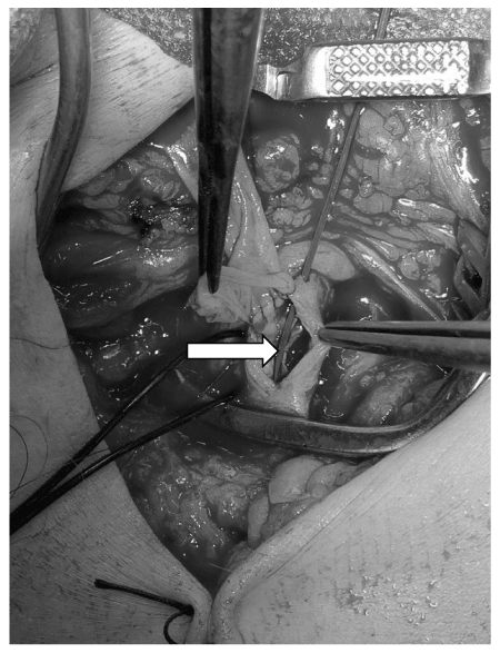
Figure 8. The picture of subintimal space of the left superficial femoral artery with the guide wire and the beginning of formation of a distal anastomosis of the vein graft with the subintimal space (indicated by an arrow)
According to angiogram which was performed immediately post – operatively, there were no signs of dissection, thrombosis, the blood flow in the vein-graft was re-established. Also the blood flow from the left anterior tibial artery and left posterior tibial artery was restored to the plantar arch of the foot. The patient has received 10,000 units of heparin during the procedure.
The early post-surgical period was uneventful. The wounds healed by primary intention. After the surgery, the left popliteal artery pulsation and left dorsalis pedis artery pulsation were recovered. The rest pain ceased in the left lower extremity as well. In the hospital, the patient had been receiving triple antiplatelet and anticoagulation therapy (Aspirin 100 mg, Clopidogrel 75 mg, low molecular weight heparins in prophylactic doses). After discharge, triple therapy (Aspirin 100 mg, Clopidogrel 75 and Rivaroxaban 5 mg) was also continued. In June of 2019 the patient underwent digital subtraction angiography (DSA) as part of scheduled follow up visit. The results are shown in Figure 9, indicating femoral-femoral bypass was functional and blood flow was re-established in the popliteal artery.
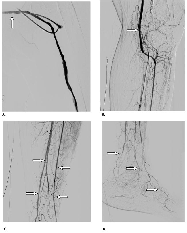
Figure 9. Results of angiogram performed at 6 months follow up. The femoral-femoral bypass (A) and left popliteal artery (B) were detected by angiogram. Arteries of the left thigh (C) and left foot (D) were detected by angiogram, indicating that left limb was vascularized and blood flow established
At the time of writing the paper (the end of September 2019, 9 months after revascularization), the patient's condition was satisfactory, there was no pain in the left lower extremity. The left foot was viable, allowing us to conclude that foot was preserved. At the point of last follow up, the patient didn’t report any limitation to the walking distance.
For many years, arterial bypass has been the traditional “gold standard” therapy for aortopopliteal arterial disease; however, the field of endovascular treatment has rapidly expanded in the recent years. With the expansion of the indication for endoluminal treatment and the development of techniques or devices in endovascular treatment, more complex multisegmental lesions are being treated currently. In addition, iliac arterial lesions and below-the-knee arterial lesions combined with femoropopliteal lesions are common problems in the treatment of patients with low extremity occlusive disease [6].
Patients with thrombosis of the aorta-femoral bypasses are special cohort, because often the reason of the thrombosis of the graft branch is the poor runoff score due to arteries occlusion (most often the profunda femoral artery). The long obstruction of the profunda femoral artery "closes" the possibility of performing thrombectomy from the graft and reconstruction of the distal anastomosis, which ultimately leads to amputation of the lower extremity. In some extremely rare cases, the blood flow is restored in the branch of the the aortobifemoral bypass and formed the bypass from the branch of the aortobifemoral graft to the popliteal or tibial arteries, with offloading into the lateral circumflex artery of the thigh, if there is one. Subintimal arterial recanalization is another option for endovascular revascularization of the lower extremity [7-9]. After thrombosis of the femoral-popliteal-distal grafts, successful rescue of an extremity is possible in virtue of those interventions [10,11]. However, as far as we know, there were no works of the combination of subintimal arterial recanalization with open procedure reported previously.
We have not found any data published in existing literature regarding similar procedures been performed as compared to these two cases we have performed and described. From our perspective, the most interesting aspect is the concept of restoring blood flow in the extremity in virtue of recanalization of the superficial femoral artery with occlusion of the profunda femoral artery and the formation of the distal anastomosis of the branch of the aortobifemoral graft with subintimal space of the superficial femoral artery. Such approach expands the possibilities of repeated revascularization of the extremity in the absence of outflow arteries, in particular, the profunda femoral artery. We believe, it can provide another chance for limb preservation in these severely ill patients with poor prognosis.
- Selvin E, Erlinger TP (2004) Prevalence of and risk factors for peripheral arterial disease in the United States: results from the National Health and Nutrition Examination Survey, 1999-2000.Circulation110: 738-743. [Crossref]
- Kharazov A, Kaliaev A, Isaev A (2016) PAD prevalence in Russian Federation. Khirurgiia 2016: 58-61. [Crossref]
- Armstrong EJ, Chen DC, Westin GG, Singh S, McCoach CE, et al. (2014) Adherence to guideline-recommended therapy is associated with decreased major adverse cardiovascular events and major adverse limb events among patients with peripheral arterial diseases. J Am Heart Assoc 3: e000697. [Crossref]
- Davies MG, Saad WE, Peden EK, Mohiuddin IT, Naoum JJ, et al. (2008) Impact of runoff on superficial femoral artery endoluminal interventions for rest pain and tissue loss. J Vasc Surg 48: 619-625. [Crossref]
- Veith FJ, Gupta SR, Wengerter KR, Goldsmith J, Rivers SP, et al. (1990) Changing arteriosclerotic disease patterns and management strategies in lower limb threatening ischemia. Ann Surg 212: 402-412. [Crossref]
- Balzer JO, Thalhammer A, Khan V, Zangos S, Vogl TJ, et al. (2010) Angioplasty of the pelvic and femoral arteries in PAOD: results and review of the literature.Eur J Radiol75: 48-56. [crossref]
- Sultan S, Hynes N (2014) Contemporary management of critical lower limb ischemia in TASC D lesions with subintimal angioplasty in femoro-popliteal lesions, tibial angioplasty and sequential compression biomechanical device for infra-inguinal arterial occlusion. Experience and quality of life outcome learned over 25 years. J Cardiovasc Surg 55: 813-825. [Crossref]
- Hong SJ, Ko YG, Kim JS, Hong MK, Jang Y, et al. (2013) Midterm outcomes of subintimal angioplasty supported by primary proximal stenting for chronic total occlusion of the superficial femoral artery. J Endovasc Ther 20: 782-791. [Crossref]
- Tay JS, Ching SS, Tan YK, Kum SWC (2017) Endovascular retrograde recanalization in Asian critical limb ischaemia patients. ANZ J Surg 87: E61-E64. [Crossref]
- Yin M, Wang W, Huang X, Hong B, Liu X, et al. (2015) endovascular recanalization of chronically occluded native arteries after failed bypass surgery in patients with critical ischemia. Cardiovasc Intervent Radiol 38: 1468-1476. [Crossref]
- Li Z, Feng R, Qin F, Zhao Z, Yuan L, et al. (2018) Recanalization of native superficial femoral artery chronic total occlusion after failed femoropopliteal bypass in patients with critical limb ischemia. J Interv Cardiol 31: 207-215. [Crossref]