Neuronal intranuclear inclusion disease (NIID) and acute ischemic stroke (AIS) weren’t simultaneous reported before. We report a case of adult onset sporadic NIID, and brain MRI showed AIS at tubera corporis callosi and left Wallenberg. The clinical manifestation of this patient presented dysarthria and ataxia. Laboratory analyses revealed hyperlipemia in this case. We supposed NIID may be induced metabolic disturbance, it may have relationship with AIS.
stroke, intranuclear inclusion, skin biopsy, diffusion weighted image, ataxia, hyperlipemia
Neuronal intranuclear inclusion disease (NIID) is a progressive and degenerative disease that can involve multiple systems, especially involves the nervous system. The pathologic characters of NIID is eosinophilic hyaline intranuclear inclusions in cells in the central, peripheral, and autonomic nervous system and visceral organs [1]. The typical eosinophilic, p62-positive, and ubiquitin-positive intranuclear inclusions from skin biopsy is an effective and less invasive diagnostic tool for NIID [2,3]. The clinical manifestations of this disease are varie, such as seizure, coma, migraine, tremors and so on. Here we report a case of NIID with acute ischemic stroke (AIS).
A 56-year-old women was taken to the Third Hospital of Zhangzhou in Fujian, China and admitted to the Stroke Unit for walking unsteadily one day, with slurred speech for 10 hours suddenly. She had numbness of right limb and tired. The neurologic evaluation showed dysarthria, ataxic finger-nose test right sides, and a positive Romberg sign. Further clinical history revealed a hypertension for 6 years. There was no past family history of cardiovascular diseases, diabetes or epilepsy.
On the same day a brain computed tomography (CT) scan showed there wasn't obvious abnormality (Figure 1, A-B). Computed tomography angiography (CTA) of the head and neck showed severe stenosis of the left internal carotid artery (ICA)-C6 segment (Figure 1C), and mild stenosis of the right middle cerebral artery (MCA)-M1 segment (Figure 1D). No obvious abnormality was observed in computed tomography perfusion (Figure 1E).
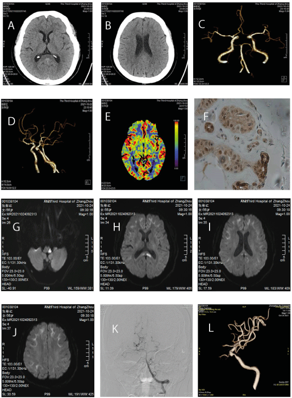
Figure 1. Brain CT scan: A-B, CTA: C-D, CTP: E, Skin biopsy: F, brain MRI: G-J, DSA: K-L
Laboratory analyses revealed blood platelet 351×109/L, low density lipoprotein (LDL) 8.47mmol/L, triglyceride 3.41mmol/L, cholesterol 13.44 mmol/L, hemoglobin A1c 6.05%, fasting blood-glucose 5.21 mmol/L. Other results including C reactive protein, D-Dimer, renal, liver and thyroid function, HIV, syphilis, hepatitis B, coagulation function was in the normal ranges.
On the third day, brain MRI showed AIS at tubera corporis callosi and left Wallenberg (Figure 1, G-H), and hyperintensity in the corticomedullary junction on diffusion weighted image (DWI) (Figure 1, I-J). The electrocardiogram and echocardiography were normal. Ultrasound examination revealed fatty liver.
One week after hospitalization, digital subtraction angiography (DSA) examination revealed bilateral posterior communicating arteries are absent (Figure 1K), and severe stenosis of the left internal carotid artery (ICA)-C6 segment (Figure 1L).
Skin biopsy showed eosinophilic ubiquitin-positive and P62-positive intranuclear inclusions in adipocytes, fibroblasts and sweat gland cells (Figure 1F).
NIID with AIS were diagnosed in this case. Aspirin (Bayer S.p.A) 100mg qd, clopidogrel bisulfate tablets (Sanofi Winthrop Industrie) 75mg qd, atorvastatin calcium tablets (Pfizer Inc.) 40mg qd were given in this case for 8 weeks.
As no one in her family had similar symptoms,this patient was an adult-onset sporadic NIID case. Previous study had reported repeated cerebellar ataxia in NIID case, but it had resolved immediately [4]. In this case, the characterized of ataxia wasn't repeated, we consider it was caused by AIS. What is the relationship between NIID and AIS, more literature is needed. Approximately 20% of NIID cases showed elevated glycated haemoglobin [5]. Elevated uric acid was found during follow-up. The elevation of hemoglobin A1c, uric acid and hyperlipemia in this case, we think the intranuclear inclusions may be induced metabolic disturbance. The hyperintensity in the corticomedullary junction on DWI and high DWI signals on the splenium of corpus callosum help to eatly diagnose NIID [6] and confirmed by skin biopsy.
There was deficiency in our study. No electromyography or electroencephalogram was taken in this case. Epileptic seizure, muscle weakness and sensory disturbance had been reported in NIID [5,7,8]. We will continue to follow up this patient.
- Lu X, Hong D (2021) Neuronal intranuclear inclusion disease: recognition and update. J Neural Transm 128: 295-303. [Crossref]
- Sone J, Tanaka F, Koike H, Inukai A, Katsuno M, et al. (2011) Skin biopsy is useful for the antemortem diagnosis of neuronal intranuclear inclusion disease. Neurology 76: 1372-1376. [Crossref]
- Sone J, Kitagawa N, Sugawara E, Iguchi M, Nakamura R, et al. (2014) Neuronal intranuclear inclusion disease cases with leukoencephalopathy diagnosed via skin biopsy. J Neurol Neurosurg Psychiatry 85: 354-356. [Crossref]
- Sakurai T, Harada S, Wakida K, Yoshida M, Nishida H (2016) Sporadic adult-onset neuronal intranuclear inclusion disease with the main presentation of repeated cerebellar ataxia: a case study. Rinsho Shinkeigaku 56: 439-443. [Crossref]
- Sone J, Mori K, Inagaki T, Katsumata R, Takagi S, et al. (2016) Clinicopathological features of adult-onset neuronal intranuclear inclusion disease. Brain 139: 3170-3186. [Crossref]
- Wang Y, Wang B, Wang L, Yao S, Zhao J, et al. (2020) Diagnostic indicators for adult-onset neuronal intranuclear inclusion disease. Clin Neuropathol 39: 7-18. [Crossref]
- Fujita K, Osaki Y, Miyamoto R, Shimatani Y, Abe T, et al. (2017) Neurologic attack and dynamic perfusion abnormality in neuronal intranuclear inclusion disease. Neurol Clin Pract 7: e39-e42. [Crossref]
- Shindo K, Tsuchiya M, Hata T, Ichinose Y, Koh K, et al. (2019) Non-convulsive status epilepticus associated with neuronal intranuclear inclusion disease: A case report and literature review. Epilepsy Behav Case Rep 11: 103-106.