Meaningful and scientific cardiotocography (CTG) interpretation is vital to minimise intrapartum fetal asphyxia and its serious consequences. This paper analyses myths at the heart of cardiotocography viz. the majority ‘rapid’ fetal heart rate (FHR) decelerations are variable (from cord-compression), early decelerations must be ‘gradual’, shoulders are indicative of cord-compression, true early decelerations are extremely rare etc. The burgeoning scientific knowledge about human cognitive biases in the last few decades helps us to understand these myths and to find a way forward. Importantly, this paper demonstrates that the current unscientific untruthful framework of pattern-recognition of FHR decelerations based on misconceived ideology (rapid vs gradual) is not just semantics, but is posing a serious risk to the unborn babies because of dysfunctional interpretation. The commonest cause of fetal hypoxemia in labor is contraction induced drop in uteroplacental perfusion and not cord-compression at all. There is ‘false-alarm fatigue’ due to overburden of atypical variable decelerations. Concurrently, deep late decelerations are increasingly misinterpreted as ‘variable’ in UK, leading to confusion, complacency and dangerous procrastination. Nobel prize winning Prospect theory of decision making informs us that the British obstetricians and British guideline-groups are in the best position to discard these myths and put CTG interpretation on a sound scientific basis. This is because they have the ‘reference point’ of primarily timing-based scientific categorization of FHR decelerations (majority early) for four decades before 2007. Without this reform, the cardiotocography and its computerized interpretation will remain blighted. Obstetricians, midwives and guideline-groups need to overcome the inertia and urgently address these unsafe myths.
cardiotocography, intrapartum fetal monitoring, fetal heart rate decelerations, backfire effect, electronic fetal monitoring
Intrapartum cardiotocography (CTG) is besieged by controversies and myths, but will remain widely practiced. There is plenty of space for controversies and unknowns in science but none for myths or post-truths. The “Each Baby Counts” 2018 progress report by the Royal College of Obstetricians and Gynaecologists (RCOG, London) stated that out of 700,000 babies born in 2016, there were 124 stillbirths, 145 babies died early and 854 babies sustained severe hypoxic brain injuries (and many more mild-moderate injuries) during labor at term [1]. It seems important to critically look at how to maximise the potential of CTG even if it may not reach perfection. Fetal heart rate (FHR) baseline, variability and accelerations need consideration, but there is little controversy about them. On the other hand, FHR decelerations have been rightly described as most important or center-stage [2,3] but continue to be surrounded by much controversy. A baby healthy at the onset of labor will almost never develop hypoxic-ischemic encephalopathy (HIE) without displaying FHR decelerations. This article critically examines some common myths about FHR decelerations which are at the very heart of CTG interpretation and debates how to resolve them. It borrows from the recent burgeoning knowledge of the scientifically proven principles from human cognitive and behavioural psychology (not simply philosophy). Moreover, there is no such thing as philosophy-free science [4]. A somewhat adversarial approach of challenging cognitive biases becomes necessary when there is a significant scientific principle or a major health issue or serious patient harm at stake as in the case.
Human beings have a potential to be rational which is essential in science. There is no good quality empirical evidence about CTG interpretation (sometimes called evidence-free zone). This commentary, particularly in the discussion of myth 4, points to a substantial proof that a false ideology is already posing a major risk to the unborn babies in the UK [5].
Myth No. 1
The most common FHR decelerations with ‘rapid’ descent (majority with trough corresponding to the peak of contraction) are due to cord-compression and hence should be called “variable”. An ideological myth.
This myth seems to have arisen following experiments in sheep fetus to study fetal response to hypoxemia mimicked by artificial cord clamping which produced rapid FHR decelerations. Unfortunately, the finding that the recovery of FHR decelerations in these sheep experiments started only after complete relief of cord occlusion [6-11] was overlooked. This recovery certainly cannot start at peak of contraction where the hypothetical cord-compression would be at its maximum remain unrelieved (Figures 1A and 1B). Moreover, the proposition that "Cord-compression causes rapid decelerations, hence all rapid decelerations are due to cord-compression (hence should be called variable)" is a fundamental "formal" fallacy of logic called "affirming the consequent" [3]. Studies of umbilical artery blood flow velocity waveform (UABVW) using Doppler during FHR deceleration in labor and amnioinfusion trials also do not support cord-compression theory [3].
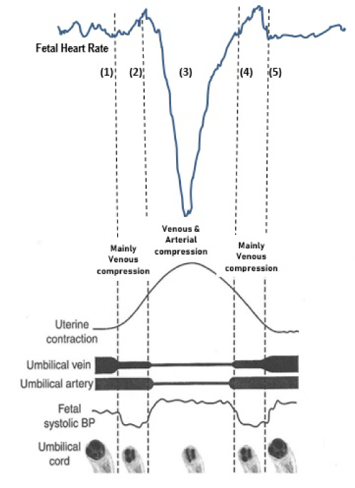
Figure 1A. Schematic drawing of the popular but flawed hypothesis of cord-compression [12-16]. For CTG speed 1cm/min. (1) UV occlusion hypothesized - fetus loses blood causing small acceleration. (2) UA occlusion hypothesized claiming to cause BP rise and rapid FHR drop via baroreceptor stimulation. But, fetal blood volume stabilising at a lower level than before the contraction would not lead to significant rise in BP at all. (3) Relief of UA occlusion is proposed to cause loss of blood volume and hypotension causing recovery of FHR ending with a small overshoot. But UA opens at point (4), where the recovery of deceleration should start rather than at point (3). Thus, the baroreceptor mechanism does not explain the drop of FHR or the commonly seen recovery of FHR close to the peak of contraction. In fact, chemoreflex from hypoxemia is accepted to be the main mechanism in cord-compression FHR deceleration as shown in figure1B. The deceleration depicted in figure 1A seems consistent with 'direct' or 'pure' vagal reflex (head-compression rather than cord-occlusion) [18].
FHR: Fetal heart rate, UV: Umbilical Venous, UA: Umbilical Arterial, BP: Blood Pressure
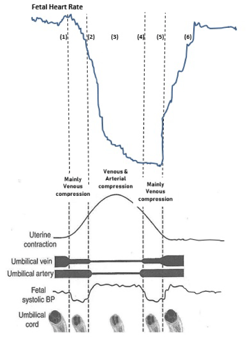
Figure 1B. The schematic drawing of the truthful (pathophysiological) cord-compression deceleration: CTG speed 1cm/min. UV compression occurring at point (1) will produce progressive fetal hypoxemia with a slight delay and a chemoreflex mediated FHR drop. Immediately after the peak of contraction (point 3), the FHR drop will slow down but continue until UV compression is released at point (5), where the recovery of FHR will start. Recovery will be complete late after the contraction. There may be small accelerations before and after the deceleration simply as a reflection of longer but smaller sympathetic response. Thus cord-compression tends to cause deep late FHR decelerations (Figure 2) as seen in practice during cord-prolapse [2], but implausible to cause common benign decelerations in labor. Marked hypoxemia from drop in uteroplacental oxygenation during contractions would also result in similar rapid deep late decelerations on British CTG. Thus, gradual late decelerations will become rapid and deep as hypoxemia becomes severe. It has become a common pitfall in British practice to underestimate the seriousness of these rapid late deceleration [3,5].
FHR: Fetal heart rate, UV: Umbilical Venous, UA: Umbilical Arterial, BP: Blood Pressure
Following are the main unsurmountable objections against this myth (Figures 1A and 1B).
- The popular baroreceptor mechanism posits “complete cord-compression” (venous and arterial occlusion) responsible for vast majority of “variable” FHR decelerations starting in the early part of contraction phase [12-17]. This is not plausible given that we perform crash caesareans for cord-compression during cord-prolapse as it leads to severe hypoxemia.
- The baroreceptor mechanism does not explain the fall of FHR [2,3,6-10] (Figure 1A). Animal experiments show that with umbilical cord clamping the rise in mean arterial pressure is only about 10 – 20 mm of Hg [2,6,17], not enough to account for the substantial fall in FHR. Most importantly, the FHR drop and its trough precedes any rise in blood pressure thus disproving the baroreceptor hypothesis [6,17].
- Both baroreceptor and chemoreceptor mechanisms completely fail to explain the recovery of most common FHR decelerations occurring at the peak of contraction [3,18]. Because, neither umbilical arteries nor vein which are postulated to get occluded in the early part of contraction would open around the height of the contraction [3,18]. Hence, only non-hypoxic reflex mechanisms like head compression (and placental compression?) can explain these decelerations which therefore should be called ‘early’ [3].
- Chemoreceptor stimulation (hypoxemia) is the main proven mechanism underlying cord-compression [2,6-11], thus the recovery of resultant decelerations will start only after relief of umbilical venous occlusion which occurs towards the end of the contraction [3,18]. These decelerations will be late in timing (Figures 1b and 2).
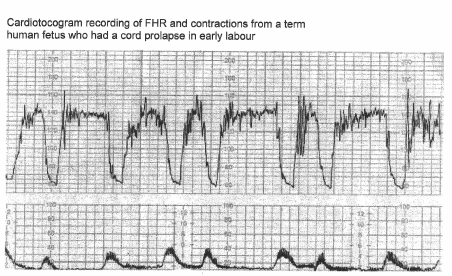
Figure 2. CTG in a case of cord prolapse showing late decelerations (reproduced with thanks from Westgate et al, AJOG, 2007) [2]. Although these decelerations ‘look’ rapid (paper speed 1cm/min), the ‘descent-time’ is well over 60 seconds (hence late by even American definition). Most importantly, they are consistently “late” in timing
The etiolgoy of FHR decelerations remains presumptive, most likely multifactorial with one of the factors predominant [19]. The basic distinction in Hon’s and Caletyro-Barcia’s pioneering timing-based categorization was not etiological but pathophysiological viz. “reflex” (early in timing) and “hypoxemic” (late in timing) (Figures 3a and 3b) [20-22]. In contrast, the rate of descent is not discriminatory of the causative agent, underlying pathophysiological mechanism or presence / absence/ degree of fetal hypoxemia [3,5]
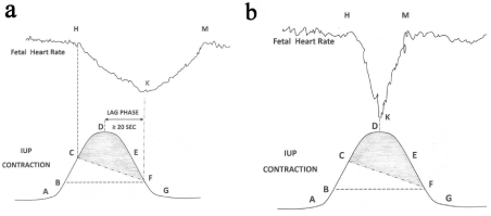
Figure 3. 3(a) Schematic drawing of FHR deceleration resulting from peripheral chemoreflex due to hypoxemia based on scientific rationale. Hypoxemic trigger is very likely to produce a classical “late deceleration”[3]. A: Contraction commences. B: IUP enough to commence fetal hypoxemia. C: Worsening fetal hypoxemia enough to start FHR deceleration. D: Peak of contraction where speed of worsening of hypoxemia will slow down but will continue to worsen (PaO2 continues to drop). E: Hypoxemia will continue to worsen. F: Hypoxia will start recovering because IUP equivalent to point B. Thus, chemoreflex induced FHR deceleration will start recovering at point F and recovery will extend beyond the end of contraction.
Shaded area: Level of IUP where fetal PaO2 will continue to drop during deceleration. FHR- Fetal heart rate, IUP - Intrauterine pressure, PaO2- Fetal partial pressure of Oxygen
3(b). Schematic drawing of common rapid short-lasting FHR decelerations with early ‘timing’ due to non-hypoxic mechanisms. There is probably some transient drop in fetal PaO2 (but not tissue anoxia) during many contractions. But it is erroneous to conclude that all decelerations must be because of hypoxemia [6]. The FHR deceleration above can be best explained by benign non-hypoxemic reflex mechanisms including head-compression [23-27]. Note that the recovery of decelerations starting at the peak of contraction makes hypoxemic cause improbable because the degree of hypoxemia will continue to worsen until point F.
Shaded area: Level of IUP where mild transient drop in PaO2 may occur, but not enough to lead to a hypoxemic deceleration
Myth No. 2
Myth: “True” early decelerations are very rare
The National Institute for Health and Clinical Excellence (NICE) guidelines in 2007 declared that “true” early decelerations are very rare implying that the British birth attendants were mistaken previously [28]. These “true” early decelerations have been variously and inconsistently described as “truly uniform”, “gradual” or “true mirror image of contractions” which are all ideological fantasies on British as well as American CTG [3,18]. This emperor has no clothes. These amount to post-truth concepts (blue lies).
How can one explain the anomaly that currently there no FHR decelerations due to head-compression at all in labor? This is because, the benign early decelerations (commonly because of head compression) are postulated / defined to be gradual (contrasting to rapid variables) as an arbitrary ‘convention’ despite many clinical studies in human labor showing that head compressions cause rapid decelerations and no studies showing gradual ones [29-32]. Head compression can occur at any stage of labor although more pronounced in the second stage. It has become a fashionable to opine that early decelerations before late first stage of labor cannot be benign [13,14]. This is not consistent with the observation of clinicians who had experience of the British system for almost four decades before 2007 (Figures 4-6) [3,24-27]. Head-compression decelerations are not associated with cerebral ischemia or anoxia and hence are due to benign reflex mechanisms [33]. The claim that early decelerations must be “gradual” has been shown to be post-truth ideology [3]. The well documented British traditional practice for four decades from 1970 to 2007 with majority early benign decelerations seems truthful, scientific and correct (Figures 4,5,6) [19,23-27].
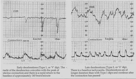
Figure 4. Early and late decelerations in traditional British practice (Reproduced from Textbook of Obstetrics by Bryan Hibbard,1988) [27]. Note rapid (descent less than 30 seconds) rather than gradual descent of early as well as some of the late FHR decelerations. Illustrations by other British authorities before 2007 were also very similar [24-26]. Many other old as well as recent illustrations [15,16] show that even late decelerations on British CTG can look more like ‘V’ than ‘U’ especially when they are deep (more serious). Hence there is a danger of miscategorising late decelerations [5] if ‘gradual’ shape is included in the British definition of late decelerations
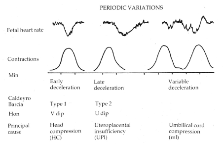
Figure 5. Diagrammatic representation of early, late and variable decelerations as practiced in British Obstetrics for four decades before 2007 (Reproduced with permission from ‘Principles of Obstetrics’ by Bryan Hibbard, 1988) [27]. Etiology is not a primary defining feature. Late decelerations are described as ‘U’-shaped only in comparison to ‘V’-shaped (rapid) early decelerations. Severe deep late decelerations from serious hypoxemia will have rapid descent and recovery on British CTG [5]
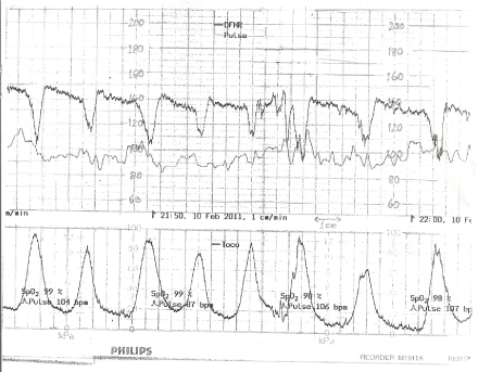
Figure 6. Cardiotocograph (1cm/min) in a primigravida in spontaneous early labor (4 cm dilatation). These FHR decelerations were quietly rightly called “early” (non-hypoxemic reflex) by a very senior expert obstetrician in 2011 in UK, based on previous four decades of practice [24-27]. Others were concerned regarding lack of the mythological “shoulders”. After a few hours of these decelerations (no oxytocin) a cesarean was performed for non-progress (4 cm) delivering a baby in an excellent condition and normal cord blood gases. These decelerations seem to reflect dystocia (head-compression) but not hypoxemia or cord-compression
Myth No. 3
Myth: The shoulders (small accelerations) on one or both sides are a confirmation that the FHR deceleration is likely to be because of cord-compression – An alluring story but a fiction
This hypothesis is not widely accepted in the USA for very good reasons. But in UK, the ideological hold of this myth is so strong that the shoulders have become a byword for the so-called benign cord-compression (typical variable) decelerations. The first shoulder is owed to the highly hypothetical (but implausible) complete umbilical arterial (cord) occlusion (Figure 1A) [12-17]. The secondary shoulder (small acceleration) is hypothesized to indicate relief of umbilical venous occlusion [12-17]. But, the chemoreceptor mechanism (triggered by hypoxemia) is the proven predominant mechanism in cord-compression FHR deceleration [2,6-11], which refutes the highly hypothetical explanation of the second shoulder (Figure 1B). Thus, the current ideology of both shoulders is a by-product of a disproven baroreceptor hypothesis and should be abandoned. Hence, a conclusion behoves that both the “shoulders” have nothing to do with cord-compression at all, but simply represent sympathetic response before and after the FHR drop due to a much stronger parasympathetic response [3]. Thus, the shoulders could be seen with any type of decelerations. Krebs et al who first described the concept atypical features in 1983, considered the lack of shoulders to be a common but benign feature [34]. Moreover, the lack of one or both shoulders (atypical feature) has been shown to have no correlation to presence of fetal hypoxemia in contrast to the ideological belief [35]. The myth of shoulders simply leads to frequent false-alarms, resultant fatigue and clouding of judgement.
Myth No. 4
Myth: The precise nomenclature of the FHR decelerations (truthful pattern recognition) does not matter in clinical practice (a matter of semantics?)
Cardiotocography is said to be a combination of science and art. It is underpinned primarily by meaningful pattern-recognition of FHR parameters (decelerations being the most crucial or center-stage). It is indeed a common belief (or resignation) on both sides of Atlantic that the precise nomenclature of decelerations does not matter provided everyone accepts a particular system. But, is a post-truth ideological categorization of FHR decelerations [3] acceptable in scientific clinical practice? This is clearly not a matter of semantics (just difference in definitions). “Relativism” in science cannot be overstretched to mean that any theory or scientific framework is as good as another even if one of them is completely outlandish. Enforcing wrong pattern-recognition is like giving wrong tools for the job or giving wrongly labelled colors to paint a picture. Teaching myths to impressionable trainees amounts to priming and burdening them with false ‘intuition-pumps’. In the UK, the obstetric Registrars are increasingly failing to recognise late decelerations because of the dysfunctional pattern-recognition. The flawed categorization of FHR decelerations can be seen to lead to a primary focus on discrimination of majority (mistaken) variable decelerations using abstract, random and variable criteria to decide which of these “cord-compression” decelerations cause fetal asphyxia, while the majority of cases of fetal acidemia in labor are due to reduction in uteroplacental perfusion (not cord-compression at all) [3]. The flawed categorization creates a lot of “noise” (artefacts, false alarm fatigue, confounding) in the visual /computerized interpretation of CTG by grass-root clinicians [3]. This can explain why they are unable to take appropriate action on the real subset of “abnormal” CTGs which had led to poor fetal outcome in practice and in RCTs like INFANT [36]. Unnecessary complexity reduces validity in science (Occam’s razor) [37]. The errors in CTG interpretations account for the biggest medicolegal expense, professional stress and increasingly performance-investigations and disciplinary measures. Many senior obstetricians are still instinctively and preferentially using their knowledge and experience of the pre-2007 traditional British CTG interpretation system [23-27]. The dysfunction is also abundantly evident from a recent voluntary comment by a young midwife with six years of experience (many others agreed) that while interpreting CTG currently, midwives quite often first decide what action feels necessary in that particular patient, then classify the CTG as normal / suspicious/ pathological tailored to justify that action. Then lastly, they decide the FHR deceleration categorization tailored to achieve that predetermined outcome (personal communications). This is CTG interpretation in “reverse gear”. Many midwives with much longer experience have also expressed serious worries about the unreliability of the current system (personal communication.) In marked contrast, such comments were unheard before 2007 and the midwives commented that the previous British scientific physiological categorization of decelerations before 2007 [24-27] was indeed very useful in clinical practice [23].
A set of fundamental contradictions / fallacies leads to a constant need of corrections or amendments lurching from one crisis to another on a road to nowhere. Thus, British national (NICE) guidelines [38] in 2014 disbanded typical / atypical terminology because of lack of usefulness emphasizing that, “the terms atypical and typical variable decelerations should not be used because they cause confusion” [38]. NICE then did a ‘U’ turn by reinstating the very same terminology in 2016 after consultation amongst its ‘stakeholders’. The burden of this confusion is being borne by the grass-root clinicians. There seems renewed speculation that the NICE may again do another U-turn (personal correspondence). Instead NICE should rather adopt the truth i.e. the categorization of FHR decelerations prior to 2007 [23-27] (Figure 7).
In the USA, there are good clinical studies showing evidence of “loss of meaning” from of the unscientific and untruthful categorization of FHR decelerations which amounts to going down a wrong road. Cahill et al (2012) in an Editor’s choice paper presented a very good clinical study which proved that there was no correlation between incidence of fetal acidemia and different types of FHR decelerations [35]. They attributed this lack of correlation to the “peculiar” (read inexplicable) definitions / categorization of decelerations. This raises the risk of abandonment of the categorization of FHR decelerations altogether because of its current untruthful and dysfunctional nature. That would be like throwing the baby out with the dirty bathwater because the early/late/variable categorization was the “low lying fruit” quickly picked up by the pioneers which would have strong correlation with fetal condition if applied correctly. Another important clinical study by Clark et al (2017) showed that under the actual practice conditions, the detection rate of fetal acidemia would be as low as 30 - 40% even with additional expert-algorithm to elucidate FHR decelerations [39]. The futility of current British categorization of decelerations led a group of prominent British experts [14] to teach abandonment of early/late/variable categorization with potential disastrous consequences for patient safety [5].
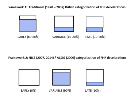
Figure 7. Schematic representation of the two frameworks of FHR categorization. The marked difference is not a matter of semantics. Only one of the above two frameworks must be meaningful, correct and scientific. The current framework 2 (with no early benign head-compression decelerations) seems invalid based on the principle of “reductio ad absurdum”. In addition, framework 2 has several irreconcilable fallacies and is dysfunctional in practice [3]
This important hypothesis is described previously in detail [5] and is a cautionary tell. Rapid decelerations early in timing (benign reflex) can often be seen in oxytocin stimulated as well as spontaneous labors. But when they become late in timing (shift to right), they may indicate onset of significant and worsening of hypoxemia [5]. It has become a common risk to assume them to be continuation of the same (mistaken) variable cord-compression decelerations engendering complacency. If cord-compression decelerations (hypoxemia) were to worsen during labor, these would commence and reach nadir even earlier and become more prolonged. In contrast, the ‘shift to right’ of the onset and nadir seems to suggest that one type of decelerations - early in timing - are replaced by a different variety – late in timing. This hypothesis proposes that hypoxemic late decelerations would overshadow and suppress early benign reflex (e.g. head-compression) decelerations as the fetus responds to the worsening hypoxemia / acidemia [5]. This could explain how multiple causative factors could produce one predominant pattern.
Reductio ad absurdum
The principle of “reductio ad absurdum” has been described as the crowbar of rational inquiry and the lever that enforces consistency [4]. One should check the hypothesis for any contradictions and take the hypothesis to its natural consequences and conclusions. If this leads to absurdity then the hypothesis must be discarded or sent back to the shop for retooling [4]. The current NICE, American Congress of Obstetricians and Gynecologists (ACOG) and International Federation of Gynecology and Obstetrics (FIGO) definitions of FHR decelerations lead to complete extinction of “early benign” decelerations [38,40,41]. This is absurd considering that head compressions have been demonstrated to cause benign decelerations [29-32] and there is plenty of head-compression during labor. The peculiar American definition of early decelerations (descent time > 30 seconds) leads to a paradox that all early benign decelerations will be more than 60 seconds in duration (which itself is an unfavourable feature) [3,18]. Moreover, the uterine contractions that last for less than 60 seconds cannot be associated with early benign decelerations by the accident of the unscientific definition, clearly an absurdity [3,18].
Why did the British framework change in 2007?
This change was simply an accident. There was no dissatisfaction with the previous categorization of FHR decelerations in fact quite the contrary [3,19,23]. Unfortunately, in 2007, the NICE included the guidelines on intrapartum fetal monitoring as a small subsection in a much larger guideline entitled “Intrapartum Care - Care of healthy women and their babies during childbirth” [28]. The intrapartum fetal monitoring is a highly specialised major subject. In future to avoid unintended consequence, the NICE should issue a separate guideline for it (just like other developed countries). This will facilitate optimum subject-specific specialized expertise on the guideline panel including obstetricians and midwives with substantial experience dating back well before 2007. An internationally renowned scientist Richard Dawkins in his acclaimed 1976 book “The Selfish Gene” proposed a term “meme” (analogous to a “gene”) as a unit of culture (an idea, belief, narrative, pattern of behaviour etc.) [42]. The memes generally undergo progressive evolution [42]. But the discontinuation of Hon’s categorization of FHR decelerations [20,21] in the British obstetrics in 2007 [28] seems to represent the rare “deleterious meme mutation” where the clinical practice was propelled significantly backwards. Many obstetricians and midwives including the author raised objections to this unwelcome change but these were sidestepped without satisfactory explanation (personal correspondence with NICE). With the enforcement of national guidelines, yearly mandatory training and the strong regulatory environment (with very good intentions); the birth-attendants had to comply, despite knowing that the new FHR deceleration categorization is irrational. Most of us are busy, our work and lives are complicated, and we can’t spend all our time thinking and analysing everything. Fortunately, this deleterious meme-mutation could be and must be reversed before time and experience runs out.
Status quo bias and Prospect theory
For lots of reasons, there is a tendency to stick with the current situation (inertia). This collective conservatism or status quo bias can occur even when stakes are very high [43]. One reason stated is the lack of attention (to detail) described by Thaler and Sunstein as “yeah, whatever” heuristic [43]. In 2002, Daniel Kahneman and Amos Tversky shared the Nobel Prize for research into cognitive biases leading to systematic errors and the “Prospect Theory” explaining how people make choices [37]. In decision making there is a strong desire of loss-aversion i.e. the fear of losses looms much larger than hope of any gains, about 2:1 or sometime more [37]. Even more importantly, these potential losses and gains are evaluated relative to the earlier state or the “reference point” rather than the absolute outcomes [37]. For the American obstetricians, the current “rapid versus gradual” categorization of decelerations (vast majority variable) has always been the default and they probably don’t have the practical experience of the alternative primarily “timing based” categorization (majority early). Hence, they would be greatly worried about what they may lose by changing (better the devil you know). Add to that a very natural and common bias of backfire effect i.e. when people are confronted with rational arguments that contradict their long held deep-seated beliefs, they become even more committed to them [37,44]. An ‘affective tipping point’ needs to be reached in terms of volume / wide knowledge of valid counter-arguments before the opinion changes [44]. In contrast, the situation is reverse for the British obstetricians in that they do have alternative “reference point” of the traditional primarily timing based categorization of FHR decelerations (majority early) practiced for four decades from 1970 to 2007 [24-27] which was found to be far more satisfactory than the current system [19,23] in the UK, Australia, New Zealand and Canada. The majority of British obstetricians with long experience have no attachment to the current system, instead they are sceptical of it. They know that there are hardly any losses but mainly gains. Hence, the ball is firmly in the court of NICE and the British obstetricians to reform to a scientific and truthful categorization, and set an example for the rest.
CTG redundant? Will a new technology or Artificial Intelligence (AI) make visual CTG interpretation redundant?
The difficulties with computer-aided CTG analysis are multiplicity of softwares, need for providing good quality evidence of its reliability or superiority over visual CTG interpretation and medicolegal liabilities. Different computer algorithms will be required during first and the second stage to achieve good predictive capability [18]. The author is not aware of such an approach being tried. Even fetal ECG (STAN) is contingent on prior CTG interpretation. Moreover, any computerized CTG interpretation will be built upon (to mimic) the frameworks of visual CTG interpretation and will be compared to it as well. Much is expected from the rapid progress in AI and machine learning. For example, computer programmes have been comprehensively beating the human Chess masters for a few years. In 2017, Google’s “AlphaZero” Chess program reigned supreme after self-learning chess by playing against itself in a matter of hours without any human guide. But the game of chess has a specified definitive structure and rules. In contrast, the CTG interpretation has innumerable variables of labor, biological variability of FHR patterns, different patient characteristics and outcomes that are changed by intervention. Hence, the machine self-learning in CTG interpretation seems a distant dream at present.
This commentary analyzes important common myths at the heart of the current CTG interpretation. Truly uniform, gradual or true mirror image (of contraction) early decelerations are an ideological fantasy. The most common rapid decelerations with trough corresponding to peak of contraction cannot be explained by cord-compression hypothesis, hence should be called ‘early’ rather than ‘variable’. The current misconceived ideological framework (majority variable and no early decelerations) is not only meaningless but places clinicians at the risk of misjudgements and repercussions thereof. It also poses a significant risk to the patient safety [5]. Disproven hypotheses and the myths described above must be discarded (Sir Karl Popper). As a result, the British guidelines must revert to the primarily timing-based categorization of FHR decelerations (majority early) for the sake of patient safety and future progress in visual as well as computerized interpretation of CTG. The preferred unambiguous scientific definitions of FHR decelerations are described in details in previous publications [3,45].
The author conceived and wrote this paper.
Additional illustrations will be made available after publication on the Researchgate webpage for further reading and clarification.
The author is thankful to the obstetricians and midwives who participated in previous studies. He is also grateful to many experts for their contribution to the field of intrapartum monitoring.
No funding was received for this paper.
The author has no conflict of interest to declare. All the concepts presented are the author’s personal views only and do not necessarily reflect clinical practice or teaching.
- Royal College of Obstetricians and Gynaecologists (2018) Each Baby Counts: 2018 Progress Report. London: RCOG. Available: https://www.rcog.org.uk/globalassets/documents/guidelines/research--audit/each-baby-counts/each-baby-counts-report-2018-11-12.pdf
- Westgate JA, Wibbens B, Bennet L, Wassink G, Parer JT, et al. (2007) The intrapartum deceleration in center stage: a physiologic approach to the interpretation of fetal heart rate changes in labor. Am J Obstet Gynecol 197: 236 e231-211. [Crossref]
- Sholapurkar SL (2017) Critical Imperative for the Reform of British Interpretation of Fetal Heart Rate Decelerations: Analysis of FIGO and NICE Guidelines, Post-Truth Foundations, Cognitive Fallacies, Myths and Occam's Razor. J Clin Med Res 9: 253-265. [Crossref]
- Dennet DC (2013) “By parody of reasoning”: Using Reductio ad absurdum. In: Dennett DC. editor. Intuition Pumps and other tools for thinking, 1st edition. London, UK: Allen Lane. p. 29-32.
- Sholapurkar SL (2019) Study of interpretation of cardiotocography educational illustrations by experts discarding early/late/ variable deceleration categorization – Physiology or unscientific ideology, myths and road to perdition? Ann Obstet Gynecol 2: 1010-1019.
- Lear CA, Galinsky R, Wassink G, Yamaguchi K, Davidson JO, et al. (2016) The myths and physiology surrounding intrapartum decelerations: the critical role of the peripheral chemoreflex. J Physiol 594: 4711-4725.
- Giussani DA, Unno N, Jenkins SL, Wentworth RA, Derks JB, et al. (1997) Dynamics of cardiovascular responses to repeated partial umbilical cord compression in late-gestation sheep fetus. Am J of Physiol 273: H2351- H2360. [Crossref]
- Itskovitz J, LaGamma EF, Rudolph AM (1983) Heart rate and blood pressure responses to umbilical cord compression in fetal lambs with special reference to the mechanism of variable deceleration. Am J Obstet Gynecol 147: 451-457. [Crossref]
- Yasui T, Kimura Y, Murotsuki J, Watanabe T, Koshino T, et al. (2002) V-shaped deceleration differs in the pattern of carotid blood flow from variable deceleration provoked by cord compression. J Perinat Med 30: 257-264. [Crossref]
- Bennet L, Westgate JA, Liu YC, Wassink G, Gunn AJ (2005) Fetal acidosis and hypotension during repeated umbilical cord occlusions are associated with enhanced chemoreflex responses in near-term fetal sheep. J Appl Physiol 99: 1477-1482. [Crossref]
- Mallard EC, Gunn AJ, Williams CE, Johnston BM, Gluckman PD (1992) Transient umbilical cord occlusion causes hippocampal damage in the fetal sheep. Am J Obstet Gynecol 167: 1423-1430. [Crossref]
- Lee CY, Di Loreto PC, O'Lane JM (1975) A study of fetal heart rate acceleration patterns. Obstet Gynecol 45: 142-146. [Crossref]
- Gibb D, Arulkumaran S (2007) Control of fetal heart rate and NICE guidelines. In: Gibb D, Arulkumaran S, editors. Fetal Monitoring in Practice, 3rd edition. London, UK: Churchill Livingstone. p. 27-44.
- Chandraharan E (2017) Applying fetal physiology to interpret CTG traces. In: Chandraharan E. editor. Handbook of CTG interpretation: From patterns to physiology, 1st edition. Cambridge, UK: Cambridge University Press. p. 32-44.
- eFM (2011) Fetal Heart Rate Monitoring. An e-learning resource improving the interpretation of EFM in labour and its subsequent management. Department of Health Programme in partnership with Royal College of Obstetricians and Gynaecologists and the Royal College of Midwives, United Kingdom. [Accessed April 20, 2014].
- OFSEP (2012) Online Fetal Surveillance Education Programme. The Royal Australian and New Zealand College of Obstetricians and Gynaecologists. https://ofsep.fsep.edu.au/mod/book/view.php?id=411&chapterid=305 (Accessed 24th February 2019).
- Cunningham FG, Leveno KJ, Bloom SL, Hauth JC, Rouse DJ, et al (2010) Intrapartum assessment. In: Cunningham FG, Leveno KJ, Bloom SL, Hauth JC, Rouse DJ, Spong CY. Editors. Williams Obstetrics. 23rd Edition, New York, USA: McGraw Hill Medical. p. 410-433.
- Sholapurkar SL (2013) Critical evaluation of American categorization of fetal heart rate (FHR) decelerations and three tier classification —Shortcomings, contradictions, remedies and need for debate. Open J Obstet Gynecol 3: 362-370.
- Sholapurkar SL (2012) The conundrum of vanishing early decelerations in British obstetrics, a step backwards? Detailed appraisal of British and American classifications of fetal heart rate decelerations - fallacies of emphasis on waveform and putative aetiology. J Obstet Gynaecol 32: 505-511. [Crossref]
- Hon EH (1958) The electronic evaluation of the fetal heart rate; preliminary report. Am J Obstet Gynecol 75: 1215-1230. [Crossref]
- Hon EH, Quilligan EJ (1968) Electronic evaluation of fetal heart rate. IX. Further observations on "pathologic" fetal bradycardia. Clin Obstet Gynecol 11: 145-167. [Crossref]
- Mendez-Bauer C, Poseiro JJ, Arellano-Hernandez G, Zambrana MA, Caldeyro-Barcia R (1963) Effects of atropine on the heart rate of the human fetus during labor. Am J Obstet Gynecol 85: 1033-1053. [Crossref]
- Sholapurkar SL (2013) Are 'early' and 'late' fetal heart rate decelerations extinct? A survey of British midwives and analysis of controversies, facts and fiction. Br J Midwifery 21:710-715.
- Williams J, Blanchard J (1996) Suspicious and Pathological Fetal Heart Traces. In: Electronic monitoring of fetal heart. 1st edition. Hale, Cheshire, UK; Books for midwives Press. p. 24-42.
- Ritchie JWK (1986) Fetal surveillance. In: Whitfield CR, editors, Dewhurst’s Textbook of Obstetrics and Gynaecology for Postgraduates, 4th edition. Oxford, UK: Blackwell Scientific Publications. p. 442-462.
- Malvern (1989) The clinical management of labour. In: Turnbull A, Chamberlain G, editors. Obstetrics, 1st edition. London, UK: Churchill Livingstone. p. 713-723.
- Hibbard BM (1988) Assessment of fetal condition during labour. In: Hibbard BM, editor. Principles of Obstetrics, 1st edition. London, UK: Butterworth & Co. (Publishers) Ltd. p. 472-489.
- National Collaborating Centre for Women's and Children's Health (2007) Intrpartum Care - Care of healthy women and their babies during childbirth. National Institute for Health and Clinical Excellence (NICE) clinical guideline 55, London, UK. Available: http://guidance.nice.org.uk/CG55, [Accessed 15 October 2012].
- Chung F, Hon EH (1959) The electronic evaluation of fetal heart rate. I. With pressure on the fetal skull. Obstet Gynecol 13: 633-640. [Crossref]
- Rech W (1933) Untersuchungen uber die Herztatigkeit des Fe-tus. III. Die Wirkung des Kopfdruckes auf die Frequenz des fetalen Herzchlages. Arch Gynaecol 154:47-57.
- Ball RH1, Parer JT (1992) The physiologic mechanisms of variable decelerations. Am J Obstet Gynecol 166: 1683-1688. [Crossref]
- Walker D, Grimwade J, Wood C (1973) The effects of pressure on fetal heart rate. Obstet Gynecol 41: 351-354. [Crossref]
- Heyborne KD (2017) A Systematic Review of Intrapartum Fetal Head Compression: What Is the Impact on the Fetal Brain? AJP Rep 7: e79-79e85. [Crossref]
- Krebs HB, Petres RE, Dunn LJ (1983) Intra- partum fetal heart rate monitoring. VIII. Atypical variable decelerations. Am J Obstet Gynecol 145: 297-305.
- Cahill AG, Roehl KA, Odibo AO, Macones GA (2012) Association of atypical decelerations with acidemia. Obstet Gynecol 120: 1387-1393. [Crossref]
- NFANT Collaborative Group (2017) Computerised interpretation of fetal heart rate during labour (INFANT): a randomised controlled trial. Lancet 389: 1719-1729. [Crossref]
- Kahneman D (2012) Prospect theory. In: Kahneman D, editor. Thinking, fast and slow. 1st edition. London, UK: Penguin Random House. p.278-288.
- National Institute for Health and Clinical Excellence (2014) National Collaborating Centre for Women's and Children's Health. Intrapartum care - Care of healthy women and their babies during childbirth. Guideline No 190, London (UK). [Accessed 15 March 2016]. Available: http://www.nice.org.uk/guidance/cg190/resources/guidance-intrapartum-care-care-of-healthy-women-and-their-babies-during-childbirth-pdf.
- Clark SL, Hamilton EF, Garite TJ, Timmins A, Warrick PA, et al. (2017) The limits of electronic fetal heart rate monitoring in the prevention of neonatal metabolic acidemia. Am J Obstet Gynecol 216: 163. [Crossref]
- American College of Obstetricians and Gynecologists (2009) ACOG Practice Bulletin No. 106: Intrapartum fetal heart rate monitoring: nomenclature, interpretation, and general management principles. Obstet Gynecol 114: 192-202. [Crossref]
- Ayres-de-Campos D, Spong CY, Chandraharan E; FIGO Intrapartum Fetal Monitoring Expert Consensus Panel (2015) FIGO consensus guidelines on intrapartum fetal monitoring: Cardiotocography. Int J Gynaecol Obstet 131: 13-24. [Crossref]
- Dawkins R (2016) Memes: the new replicators. In: dawkins R, editor. The Extended Selfish Gene, 40th anniversary edition. Oxford, UK: Oxford University Press. p.245-260.
- Thaler RH, Sunstein CR (2008) Biases and blunders. In: 43. Thaler RH, Sunstein CR, editors. Nudge, 1st edition. New Haven & London: Yale University Press. p. 17-39.
- Pinker S (2019) Reason. In: Pinker S. editor. Enlightenment Now: The case for reason, science, humanism and progress, 1st edition. London, UK: Penguin Books. p. 351-384.
- Sholapurkar SL (2015) Categorization of fetal heart rate decelerations in American and European practice: Importance and imperative of avoiding framing and confirmation biases. J Clin Med Res 7: 672-680. [Crossref]