Lichenoid lesions, nitidus, planus, dermatosis
A 20-year-old female presented to our dermatology department with a 3-week history of an asymptomatic diffuse papular dermatosis, previously untreated.
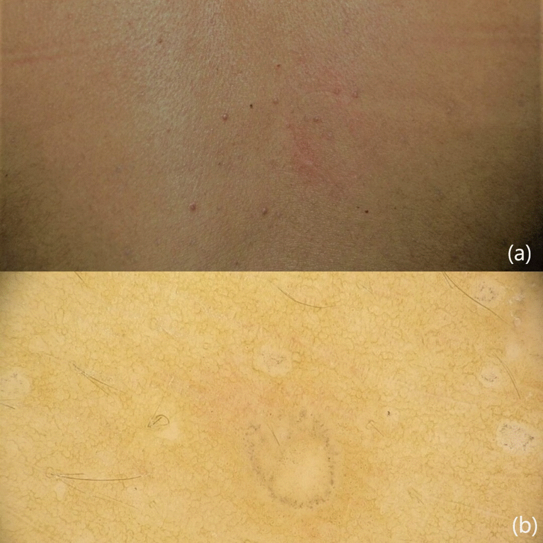
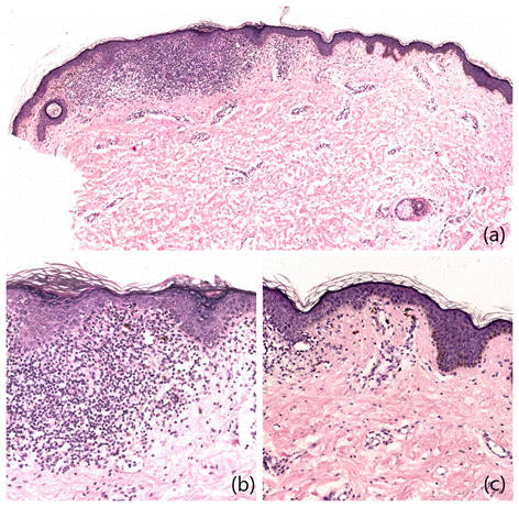
Clinical examination revealed red-brownish minute pinpoint dome-shaped monomorphic papules widespread on the trunk and upper limbs, predominantly on the flexor surfaces (Figure 1a). The remainder of physical examination and medical history were unremarkable. These lesions initially led to a diagnosis of lichen nitidus. Dermoscopy was performed and showed the presence of a brown-greyish rim and peppering with central fine scales on a structureless whitish area (Figure 1b). A biopsy was performed with an unclear final histopathological diagnosis of lichen planus/lichen nitidus (Figure 2).

Figure 1. (a) Magnification of the widespread dermatosis characterized by red-brownish dome-shaped monomorphic papules on an aphlegmasic skin (b) Dermoscopic evaluation revealed the presence of a brown-greyish rim with disseminated peppering and central fine scales in a white structureless area. This distinctive dermatoscopic feature could be described as a "jellyfish-sign".
Several types of lichen have been described, including lichen nitidus (LN) and lichen planus (LP). LP is a chronic, inflammatory, and immune-mediated dermatosis involving the skin, especially the flexural surfaces, scalp, nails and mucous membranes. Clinically it presents as small itchy flat purple papules, while dermatoscopically the pathognomonic criterion is Wickham's striae (WS). Other recognised dermoscopic features are radial capillaries, red dots, whitish streaks, diffuse brownish pattern described as hyperpigmented areas, grey-blue dots, comedo and milium-like cysts. Dermoscopy may differ in active and regressive lesions according to the developmental stage. Regressing lesions do not show pathognomonic WS, but pigment patterns could persist after treatment. In 20% of patients the dermoscopic pattern is characterised by peripheral brown dots in a circular arrangement1. The pigment pattern corresponds to the presence of pigment incontinence and dermal melanophages [1,2].
LN is a rare chronic inflammatory condition first described in 1907 by Pinkus, characterized by the presence of asymptomatic, flesh-coloured, 1-2 mm pinpoint dome-shaped papules, which usually occur on the abdomen, chest, extremities, and genitalia, and often affect children and young adults.
Only a few studies describe the dermoscopy of LN. The main dermoscopic features are defined hypopigmentation, diffuse erythema, and absence of dermatoglyphics with radial ridges. Brown shadows, as described by Malakar et al., could represent peculiar findings in LN [3].
In our case, the absence of pruritus and the presence of small-size pinpoint dome-shaped papules, suggested a diagnosis of LN. In contrast, the red-brownish colour of the papules and the dermoscopic features were not suggestive of LN but resembled the appearance of a regressing lesion of LP [1]. The brown-greyish rim on a whitish background may be reminiscent of a jellyfish head and could be described as a "jellyfish-sign".
Histology did not reveal a clear diagnosis but identified an intermediate pattern between LN and LP. The histological aspects including a preponderance of monomorphic infiltrate of lymphocytes lying on dermo-epidermal junction, corresponding to the white structureless area on dermoscopy, a vacuolar degeneration of the basal layer of the epidermis and the presence of wedge-shaped hypergranulosis, suggested a diagnosis of LP. On the other hand, the focal lymphocytic infiltrate, and the presence of histiocytes, confirmed by CD68 positivity on immunohistochemistry, suggested the diagnosis of LN (Figures 2a and 2b). The presence of dermal melanophages could correspond to the areas of peppering visible on dermoscopy (Figure 2c).

Figure 2. (a) Superficial perivascular dermatitis, interface focally lichenoid. H&E 5x. (b) Vacuolar alteration at dermoepidermal junction by an infiltrate of lymphocytes, histiocytes, melanophages in the papillary dermis. Wedge-shaped hypergranulosis, hyperplasia, and compact orthokeratosis of the epidermis. H&E 20x. (c) Fibroplasia and melanophages in the papillary dermis. Basket-woven orthokeratosis of the epidermis. H&E 20x.
In conclusion, we describe here a dermatosis presenting with minute miliary papular lesions characterised by a clinical aspect of LN, regressive dermoscopic features, previously described in LP, and a histopathological pattern straddling LP and LN. In 1961 Wilson et al. reported a similar scenario with the presence of miliary lesions clinically indistinguishable from those of LN in patients affected by widespread LP, in some cases with a histological appearance intermediate between the two entities [4].
Finally, we agree with Wilson et al. [4] that LP can sometimes appear with miliary lesions very similar to those of LN. The term LN should be reserved for those cases where the lesions are small, asymptomatic and with a specific dermoscopical and histopathological pattern. In the presence of miliary diffuse lichenoid lesions, dermoscopy can be an effective tool to establish the correct differential diagnosis between LN and LP, given that histopathologic findings are not always conclusive.
This manuscript has not been published or submitted for publication elsewhere.
The authors state that they have obtained written informed consent to publish the reported case and clinical images. The authors declare to have not conflicts of interest for the publication of this article.
None
The study was performed in line with the Declaration of Helsinki (1964) and its subsequent amendments.
- Güngör S, Topal IO, Göncü EK (2015) Dermoscopic patterns in active and regressive lichen planus and lichen planus variants: a morphological study. Dermatol Pract Concept 5: 45-53. [Crossref]
- Lallas A, Kyrgidis A, Tzellos TG, Apalla Z, Karakyriou E, et al. (2012) Accuracy of dermoscopic criteria for the diagnosis of psoriasis, dermatitis, lichen planus and pityriasis rosea. Br J Dermatol 166: 1198-1205. [Crossref]
- Malakar S, Save S, Mehta P (2018) Brown Shadow in Lichen Nitidus: A Dermoscopic Marker!. Indian Dermatol Online J 9: 479-480. [Crossref]
- Wilson HT, Bett DC (1961) Miliary lesions in lichen planus. Arch Dermatol 83: 920-923. [Crossref]


