Although the real incidence of metallosis is unknown, metallosis is described as a rare diagnosis and it seems to be more frequent in hip than in knee arthroplasty with a 5% estimated incidence in the hip prosthetic replacements.
Introduction: Metallosis is a condition that occurs when there is a high level of toxic metals in the blood. This occurs when metal particles detach from the implant and are released into the bloodstream or nearby tissues.
The symptoms of metallosis are made from a mixture of several metals that includes chromium, cobalt, nickel, titanium and molybdenum. When friction occurs between the metal components, they discharge microscopic particles into the blood and surrounding tissues.
Case presentation: The 46-year-old patient who developed postoperatively a metallosis following a total hip prosthesis. Total hip arthroplasty was performed for secondary hip osteoarthritis due to a congenital hip dysplasia of the patient.
Conclusion: Metallosis develops as these metal ions accumulate in the bone, muscles, and other tissues around the implant, and this can lead to bone death or surrounding tissues.
The particularity of this case is the fact that the trophic disorders caused by metallosis did not affect the prosthesis itself, but they affected the surrounding tissues. As a result of the disorders produced, external suppuration appeared, and the surgery was needed again.
A correct and careful debridement can bring us a favorable result with the remission of local and systemic phenomena without the need to remove and change the implant.
Metallosis, hip, arthroplasty, osteoarthritis, congenital hip dysplasia
The hip prostheses are made of a mixture of several metals including chromium, cobalt, nickel, titanium, and molybdenum. When friction occurs between the metal components, they discharge microscopic particles into the blood and surrounding tissues. Metallosis develops as these metal ions accumulate in the bone, muscles, and other tissues around the implant, and this can lead to tissue death. bone or surrounding tissues [1-5].
The medical story of this case began in 2014 while I was intern of surgery department. The patient 46- year- old female was admitted in Orthopedics and Traumatology Department of Emergency Clinical Hospital of Galati (where I am working as orthopedic surgeon specialist) for a scheduled intervention.
Processing anamnestic data, from personal pathological history she declared a congenital hip dysplasia that led to a secondary hip osteoarthritis. This hip osteoarthritis was quite advanced, a stage four with functional impotence and prolonged orthostatic deficiency, which is why we decided for a total cement less hip replacement by arthroplasty. In addition to this pathology, she declared a subtotal hysterectomy.
Following the preoperative preparation and the laboratory analyzes, we highlighted the following: Wbc 10.40 (4–10) 103/q/l; Neutrophils 7.30 (1,5–7.0)103/q/l; Glycaemia 106 (75–105) mg/dl.
Nothing pathological, so after 2 days of hospitalization the surgery was performed, and we did a total hip arthroplasty of the left hip with the endo-prosthesis ATLAS type.
Postoperative, the general and local evolution was a normal one, without fever, suppression of aspiration drainage 48 hours after the intervention.
The duration of hospitalization was 13 days, during this time she began rehabilitative physiotherapy, started to walk with a frame and full weight bearing on the left side.
The sutures were removed and discharged without any complications. She came for periodic controls like in the recommendations with a normal evolution. A quite good evolution for a total hip replacement so far, we thought the case was solved.
After 4 years from primary intervention (24.06.2018) she came into the emergency service for: pain in the operated hip; swelling on a keloid scar; in the middle of 1/3 of the wound there was a fistula that eliminates a secretion.
I was on duty that day, so I took care of the case, I did a history of her evolution. She declared that symptomatology began approximately two weeks ago with intermittent pain in the operated hip. These pains intensified, locally a regional swelling that endures the area to than fluctuate in the average 1/3 of the scar and externalize through a secretion.
The first step was to collect secretion from the fistula, along with biochemistry because I was heading for an infection. I also started broad-spectrum antibiotic therapy and waited for the result of culture.
Following the laboratory analyzes we highlighted the following: Fibrinogen 849,96 (200-400) mg/dl; Blood sugar 131 (70 – 105) dg/dl.
The culture went negative, no germs found.
After all these results we thought of a granuloma of thread, reason for which daily we cleaned the fistula chemically and mechanically we debrided very well.
The local evolution was favorable with a secondary closure of the fistula. The duration of the hospitalization was 12 days, with no fever or other symptoms.
Approximately one year after her last presentation, she returns to the emergency service (04.04.2019) with the same symptoms prior to the last presentation and locally fistula with secretion. We did the same, we collected secretion from the fistula for culture and antibiotic-gram we initiated large spectrum antibiotic therapy, associated with the following biochemistry: blood sugar 206 (70- 205) dg/dl, a newly discovered diabetes most likely, due the stress over the time, a stage II hypertension, and a thyroid node that she forgotten to mention before. PLT: 461 (150–450)103/units/liter; Wbc: 10.82 (4–9) 103/units/liter; Pct: 4.34 (0.15–0.45) %; Neutrophils: 99 (2–6.3) 103/units/liter; erythrocyte sedimentation rate (ESR) 45 (2- 20) mm/h; Fibrinogen 500 (200–400) mg/dl; PCR>70.
This time the biochemistry showed a high inflammation, we thought about a sepsis which is why we decided to have surgery because we wanted to explore the region and see what is actually there.
We found multiple black areas adhering to the tissues (metallosis) that were going with the fistula and granuloma. I removed them, I debrided very carefully the entire area, but I didn’t go through the tensor fascia-late into the prosthesis room because we didn’t find any communication washed with antiseptic solutions, and anatomically, we closed with resorbable threads at 90 days. The local evolution was a good one with the decrease of inflammatory phenomena and she was discharged at 6 days after the surgery.
Outpatient control was done to 12-14 days, where the sutures were removed.
At an interval of approx. 4 months, in august she returns with the same symptoms, secretion, and again negative culture, but increased inflammatory evidence. We underwent surgery again and debrided the black modified tissues and closed in the same session with a favorable evolution. This time we had a short successfully period because that year we didn’t saw her anymore.
In 2020 she had two presentations, first was in May and the second was in December.
Again, we proceeded standard, we harvested secretion and we waited for the result without starting antibiotic therapy which was again negative. Instead the inflammatory evidence was very high, higher than ever, such as: Wbc: 12.40 (4-9 ) 103/units/liter; Rbc: 3.60 ( 3,7-5) 106/units/liter; HGB: 10.40 (12–14) g/dl; HCT: 31.10 (35-42) %; NEU#: 10.66 ( 2-6.3) 103/UL; LYM%: 9.40 (25-40) %; urea 13 (15-44) mg/dl; ESR 55 (2-20) mm/h; fibrinogen 588.20 (200-400) mg/dl; PCR >100 (0 5) ml/l.
The pelvic radiograph didn’t show anything, the implants showed good integration without any signs of lysis or other elements that would have indicated radiologically any process at the level of the prosthesis.
I decided again for the surgical procedure and to explore the prosthesis room, eventually to remove it and to do a totally hip revision. Intraoperatively we found many areas of metallosis for that we did a very good debridement, we cleaned and removed any modified tissues, and we applied a calcium sulfate bio composite mixed with vancomycin and gentamicin.
Postoperatively, the evolution was favorable, which is why at 7 days she was discharged normally. After 3 days after she went home, I received a phone call from her, and she sent me some pictures to see the evolution of the wound.
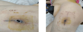
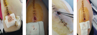
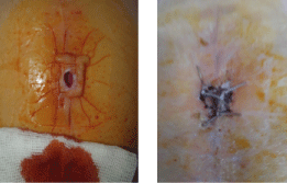
In the lower 1/3 of the scar, it removes a large amount of serous fluid that has finally penetrated the skin. By setting up a meeting she showed up in the ambulatory department where I continued the local treatment (Figures 1 to Figures 5).

Figure 1. Pelvic X-Ray of patient.

Figure 2. Before the surgery of patient.

Figure 3. It’s showed up the calcium sulfate that was eliminated true that fistula. I cleaned the area daily removing the granules exposed.

Figure 4. After that when the area was red granulated, we applied negative pressure therapy for 21 days, and we obtained this result.

Figure 5. After this 21-day cure, how the defect has diminished, which allowed me to make a final suture such in the following pictures.
Local symptoms of metallosis include hip pain, swelling, intermittent fistula, paresthesia’s, weakness, and functional impotence. In some cases, skin, heart, kidney, nervous system, or thyroid disorders may be observed before the local effects occur.
Metallosis develops as these metal ions accumulate in the bone, muscles, and other tissues around the implant, and this can lead to bone death or surrounding tissues.
A correct and careful debridement can bring us a favorable result with the remission of local and systemic phenomena without the need to remove and change the implant.
The particularity of this case is the fact that the trophic disorders caused by metallosis did not affect the prosthesis itself, but they affected the surrounding tissues. As a result of the disorders produced, external suppuration appeared, and the surgery was needed again.
Sources of Funding: No funding for the research
Ethical approval: This is not a research study. No ethical approval was necessary.
Consent: Consent for publication was obtained from the parents of the patient being a minor.
DIMOFTE Florentin collected the data; made the surgery and wrote part of surgery.
MARINA Virginia corresponding author, wrote the final form of the article, processed the pictures.
Registration of research studies: This is a case report. Is not a research study.
Guarantor: DIMOFTE Florentin
Provenance and peer review: Not commissioned externally peer- reviewed.
Declaration of Competing Interest: The author declarate that there is not conflict of interest regarding the publication of this article.
- US Food and Drug Administration (2019) Concerns About Metal-on-Metal Hip Implants.
- Oliveira CA, Candelária IS, Oliveira PB, Figueiredo A, Caseiro-Alves F (2014) Metallosis: A Diagnosis Not Only in Patients with Metal-on-Metal Prostheses. Eur J Radiol Open 2: 3-6. [Crossref]
- Pritchett JW (2012) Adverse Reaction to Metal Debris: Metallosis of the Resurfaced Hip. Current Orthopaedic Practice 23: 50-58.
- Kelmer G, Stone AH, Turcotte J, King PJ (2021) Reasons for Revision: Primary Total Hip Arthroplasty Mechanisms of Failure. J Am Acad Orthop Surg 29: 78-87. [Crossref]
- US Food and Drug Administration (2017) Implants and Prosthetics: Metal-on-Metal Hip Implants, Information for Orthopaedic Surgeons.


